. 2020 Feb 28;94(6):e01175-19.
doi: 10.1128/JVI.01175-19. Print 2020 Feb 28.

Short Direct Repeats in the 3' Untranslated Region Are Involved in Subgenomic Flaviviral RNA Production

Affiliations

Short Direct Repeats in the 3' Untranslated Region Are Involved in Subgenomic Flaviviral RNA Production

Qiu-Yan Zhang et al. J Virol. .

Abstract

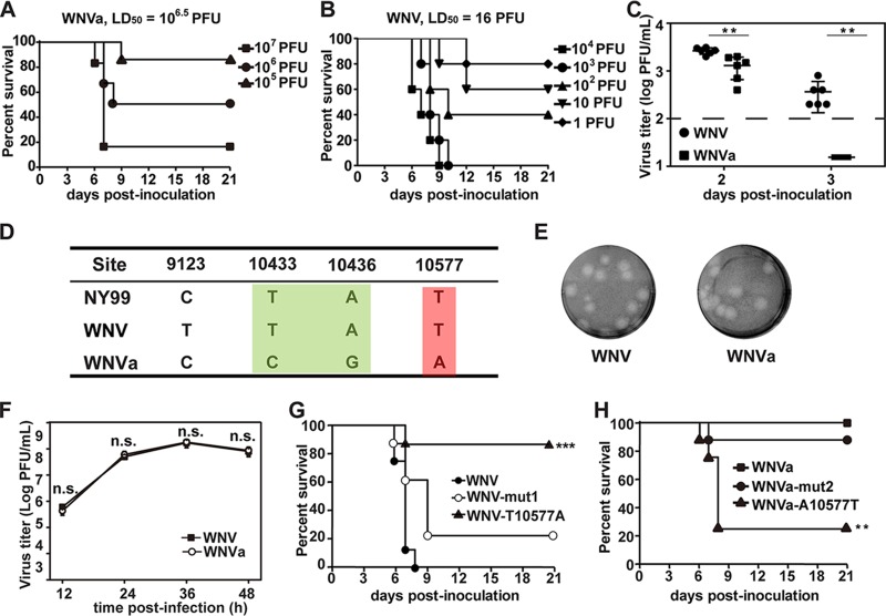
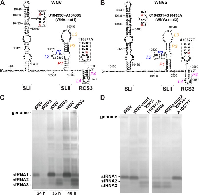
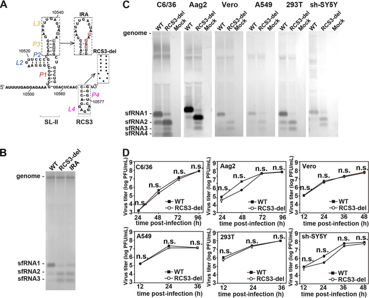
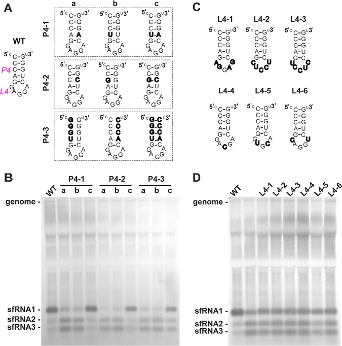
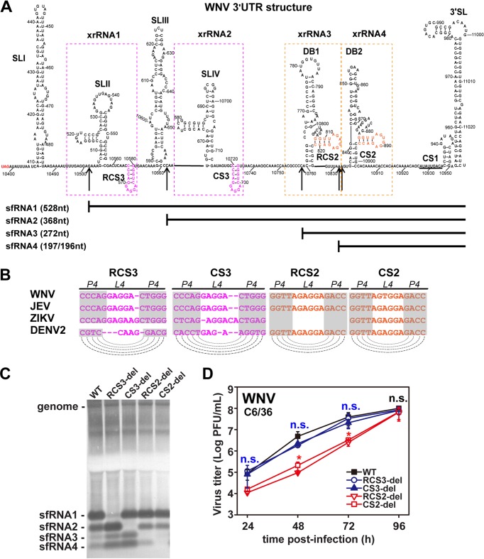
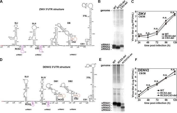
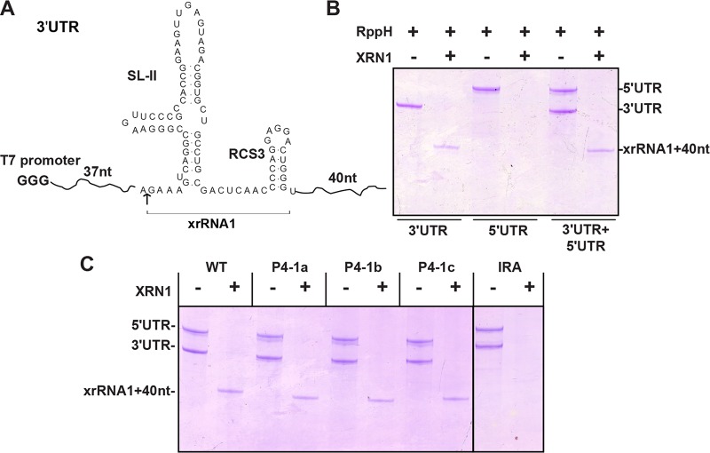
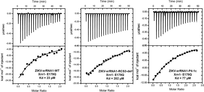
Mosquito-borne flaviviruses consist of a positive-sense genome RNA flanked by the untranslated regions (UTRs). There is a panel of highly complex RNA structures in the UTRs with critical functions. For instance, Xrn1-resistant RNAs (xrRNAs) halt Xrn1 digestion, leading to the production of subgenomic flaviviral RNA (sfRNA). Conserved short direct repeats (DRs), also known as conserved sequences (CS) and repeated conserved sequences (RCS), have been identified as being among the RNA elements locating downstream of xrRNAs, but their biological function remains unknown. In this study, we revealed that the specific DRs are involved in the production of specific sfRNAs in both mammalian and mosquito cells. Biochemical assays and structural remodeling demonstrate that the base pairings in the stem of these DRs control sfRNA formation by maintaining the binding affinity of the corresponding xrRNAs to Xrn1. On the basis of these findings, we propose that DRs functions like a bracket holding the Xrn1-xrRNA complex for sfRNA formation.IMPORTANCE Flaviviruses include many important human pathogens. The production of subgenomic flaviviral RNAs (sfRNAs) is important for viral pathogenicity as a common feature of flaviviruses. sfRNAs are formed through the incomplete degradation of viral genomic RNA by the cytoplasmic 5'-3' exoribonuclease Xrn1 halted at the Xrn1-resistant RNA (xrRNA) structures within the 3'-UTR. The 3'-UTRs of the flavivirus genome also contain distinct short direct repeats (DRs), such as RCS3, CS3, RCS2, and CS2. However, the biological functions of these ancient primary DR sequences remain largely unknown. Here, we found that DR sequences are involved in sfRNA formation and viral virulence and provide novel targets for the rational design of live attenuated flavivirus vaccine.

Keywords: Flavivirus; West Nile virus; direct repeats; sfRNA.


Figures

FIG 1
FIG 1
A single nucleotide change (T10577A) within RCS3 of the 3ʹ-UTR leads to attenuation of WNV virulence in mice. (A and B) Survival analysis of mice infected with WNV. Four-week-old BALB/c mice (n = 5 to 9) were i.p. inoculated with the indicated dose of (A) WNVa or (B) WNV and then monitored for clinical symptoms and mortality over 21 days. LD50, 50% lethal dose. (C) Viremia differences between WNV and WNVa. Four-week-old of BALB/c mice (n = 6) were intraperitoneally (i.p.) inoculated with 105 PFU indicated viruses. Blood was taken at the second and third days after infection, and virus titer was determined by plaque assay. The asterisks denote the statistical significance of differences between results from the indicated groups. **, P < 0.01. (D) Four nucleotide differences between WNV and WNVa were identified at nucleotide positions 9123, 10433, 10436, and 10577 through complete-genome sequence alignment. Corresponding nucleotides from the isolated virulent NY99 strain are also listed. (E) Plaque morphologies of WNV and WNVa in BHK-21 cells. (F) Growth kinetics of WNV and WNVa in BHK-21 cells. WNV and WNVa showed no significant differences in viral titers at each time point. n.s., no significant statistical differences. (G) Survival analysis of mice infected with WNV, WNV-mut1, and WNV-T10577A. Four-week-old of BALB/c mice (n = 8) were intraperitoneally (i.p.) inoculated with 105 PFU of the indicated viruses and monitored for clinical symptoms and mortality over 21 days. ***, P < 0.001. (H) Survival analysis of mice infected with WNVa, WNVa-mut2, and WNVa-A10577T. Four-week-old BALB/c mice (n = 8) were i.p. inoculated with 105 PFU of the indicated viruses and monitored for clinical symptoms and mortality over 21 days. **, P < 0.01.
FIG 2
FIG 2
A single nucleotide change (T10577A) within RCS3 of the 3ʹ-UTR alters sfRNA production patterns. (A) Diagram of putative secondary structure of WNV xrRNA1. RNA sequences represent nucleotide positions 10396 to 10581, and the location of the 10577 site within RCS3 is labeled. The inset shows the mutation used to test the importance of identified nucleotides in viral virulence in the context of the WNV infectious clone. (B) Putative secondary structure diagram and RNA sequence of nucleotide positions 10396 to 10581. The inset shows mutations in the context of the WNVa infectious clone. (C) Northern blot analysis of total RNA extracted from BHK-21 cells infected with WNV and WNVa at 24, 36, and 48 h postinfection. (D) Northern blot of total RNA extracted from BHK-21 cells infected with WNV or WNVa or the indicated mutant at 36 h postinfection. The sfRNA signals are indicated as sfRNA1, sfRNA2, and sfRNA3 in descending size order.
FIG 3
FIG 3
RCS3 determines sfRNA1 production of WNV. (A) Secondary structure of WNV SLII and RCS3. The various mutations within SLII and RCS3 are indicated in the inset. (B) sfRNA detection through Northern blotting of total RNA extracted from BHK-21 cells infected with recombinant WNV containing mutations of IRA or RCS3 deletion. (C) sfRNA production profiles of WT WNV and RCS3-del in different cell lines. Data represent results of Northern blot analysis of total RNA purified from WNV-infected mosquito C6/36 and Aag2, Vero, human A549 and 293T, and human neuroblastoma sh-SY5Y cell lines. Total viral RNAs were collected at peak titers in the infected cells. (D) Growth kinetics of WT WNV and mutant WNV with the RCS3-del mutation in different cell lines. The RCS3-del and WT viruses showed no significant differences in viral titers at any time point in all the tested cell lines. n.s., no statistically significant differences.
FIG 4
FIG 4
The stem-loop structure of RCS3 is essential for sfRNA1 production. (A) Secondary structure of WNV RCS3. The various mutations within P4 are indicated in the inset. (B) sfRNA detection through Northern blotting of total RNA extracted from BHK-21 cells infected with recombinant WNV containing various mutations within stem of RCS3, recognized as P4. (C) Different loop mutations within the RCS3 loop, recognized as L4. To minimize structural changes, most mutations maintained the same number of nucleotides within the loop. L4-1 contains the complete reversed sequences of the loop. L4-2 and L4-3 have the complete complementary and reversed complementary loop sequences, respectively. L4-4, L4-5, and L4-6 have one or two complementary nucleotides altered at the corresponding position of the loop sequence. (D) Northern blot analysis of RNA purified from BHK-21 cells infected with recombinant WNVs containing the different L4 mutations at 36 h postinfection. Lane 2 represents an unrelated mutant.
FIG 5
FIG 5
Conserved DRs of WNV control production of various sfRNA species. (A) Secondary structure of WNV 3ʹ-UTR. The various secondary structures formed within the 3ʹ-UTR are indicated. The putative SLs of RCS3/CS3 and RCS2/CS2 are marked in pink and orange, respectively. The 5′ ends of sfRNA1, sfRNA2, sfRNA3, and sfRNA4 are indicated by arrows. The sizes of different sfRNA species are also indicated. xrRNA1, xrRNA2, xrRNA3, and xrRNA4 are depicted by dotted boxes. (B) Conserved repeated sequences from various flaviviruses. Dashed lines indicate putative base pairing with the SL structures of conserved repeated sequences as indicated in the diagram. (C) Northern blot of total RNA extracted from C6/36 cells infected with WT or mutant WNVs with RCS3, CS3, RCS2, or CS2 deletions. sfRNA signals are marked as sfRNA1, sfRNA2, sfRNA3, and sfRNA4 in descending size order. (D) Growth kinetics of recombinant WNVs with individual RCS3, CS3, RCS2, and CS2 deletions in C6/36 cells. RCS3-del, CS3-del, and WT virus titers showed no significant differences at different time points. n.s., no statistically significant differences. RCS2-del and CS2-del virus titers showed statistically significant differences from the WT virus titers at 24, 48, and 72 h postinfection (hpi) (*, P < 0.05), and there were no statistically significant differences at 96 hpi.
FIG 6
FIG 6
DR sequences also control production of DENV2 and ZIKV sfRNAs. (A) Secondary structure of ZIKV 3′-UTR. The different RNA secondary structures and CS sequences are indicated. (B) Northern blot of total RNA extracted from C6/36 cells infected with recombinant ZIKV with RCS3 or CS3 deletions. (C) Growth kinetics of recombinant ZIKVs with individual RCS3 and CS3 deletions in C6/36 cells. RCS3-del, CS3-del, and WT virus titers showed no significant differences at different time points. n.s., no statistically significant differences. (D) Secondary structure of DENV2 3′-UTR. The different RNA secondary structures and CS sequences are indicated. (E) Northern blot analysis of total RNA extracted from C6/36 cells infected with recombinant DENV2 containing RCS3 and RCS3/CS3 deletions. (F) Growth curves of recombinant DENV2 viruses in C6/36 cells. RCS3-del, RCS3/CS3-del, and WT virus titers showed no significant differences at different time points. n.s., no statistically significant differences.
FIG 7
FIG 7
Xrn1 resistance assay performed in vitro for analysis of sfRNA RNA. (A) Briefly, a 158-nt uncapped WNV 3′-UTR RNA corresponding to nt 10464 to 10621 containing SLII plus RCS3 (nt 10502 to 10580; resistance to XRN1 cleavage) was prepared for Xrn1 resistance assay. (B) The production of shorter xrRNA1 of the input 3′-UTR was used to demonstrate Xrn1 cleavage resistance of 3′-UTR (lanes 1 and 2). Additionally, WNV 5′-UTR RNA corresponding to nt 1 to 190 was prepared as a control for complete cleavage by Xrn1 (lanes 3 and 4). (C) Xrn1 resistance assay of various RNAs with P4 mutations or IRA mutation.
FIG 8
FIG 8
RCS3 controls Xrn1 binding affinity to xrRNA. Data represent results of analyses of binding affinity of Xrn1 to the xrRNA1 of ZIKV measured by ITC assay. The dissociation constant (Kd) values were calculated to be 23 μM, 202 μM, and 77 μM for ZIKV-xrRNA1-WT, ZIKV-xrRNA1-RCS3-del, and ZIKV-xrRNA1-P4-1c, respectively.
FIG 9
FIG 9
Three-dimensional RNA structure model of sfRNA. The first 350 nt of 3′-UTR from SLI to CS3 was used for three-dimensional (3D) RNA structure model analysis performed with the RNAComposer Web server with standard settings. The color of each structure matches the color of the labeled box. RCS3 is depicted as black in a square box. For WT and P4-1c, a complete pairing of RCS3 stem resulted in very tight tertiary folding, whereas for P4-1a or P4-1b mutant RNA, impaired base pairing of RCS3 stem resulted in loose tertiary folding.

Similar articles

Cited by

References

    1. Filomatori CV, Carballeda JM, Villordo SM, Aguirre S, Pallares HM, Maestre AM, Sanchez-Vargas I, Blair CD, Fabri C, Morales MA, Fernandez-Sesma A, Gamarnik AV. 2017. Dengue virus genomic variation associated with mosquito adaptation defines the pattern of viral non-coding RNAs and fitness in human cells. PLoS Pathog 13:e1006265. doi:10.1371/journal.ppat.1006265. - DOI - PMC - PubMed
    1. Villordo SM, Carballeda JM, Filomatori CV, Gamarnik AV. 2016. RNA structure duplications and flavivirus host adaptation. Trends Microbiol 24:270–283. doi:10.1016/j.tim.2016.01.002. - DOI - PMC - PubMed
    1. Manokaran G, Finol E, Wang C, Gunaratne J, Bahl J, Ong EZ, Tan HC, Sessions OM, Ward AM, Gubler DJ, Harris E, Garcia-Blanco MA, Ooi EE. 2015. Dengue subgenomic RNA binds TRIM25 to inhibit interferon expression for epidemiological fitness. Science 350:217–221. doi:10.1126/science.aab3369. - DOI - PMC - PubMed
    1. Pijlman GP, Funk A, Kondratieva N, Leung J, Torres S, van der Aa L, Liu WJ, Palmenberg AC, Shi PY, Hall RA, Khromykh AA. 2008. A highly structured, nuclease-resistant, noncoding RNA produced by flaviviruses is required for pathogenicity. Cell Host Microbe 4:579–591. doi:10.1016/j.chom.2008.10.007. - DOI - PubMed
    1. Roby JA, Pijlman GP, Wilusz J, Khromykh AA. 2014. Noncoding subgenomic flavivirus RNA: multiple functions in West Nile virus pathogenesis and modulation of host responses. Viruses 6:404–427. doi:10.3390/v6020404. - DOI - PMC - PubMed

Publication types

LinkOut - more resources