. 2020 Feb 5;40(6):1248-1264.
doi: 10.1523/JNEUROSCI.1654-19.2019. Epub 2020 Jan 2.

Grafting Embryonic Raphe Neurons Reestablishes Serotonergic Regulation of Sympathetic Activity to Improve Cardiovascular Function after Spinal Cord Injury

Affiliations

Grafting Embryonic Raphe Neurons Reestablishes Serotonergic Regulation of Sympathetic Activity to Improve Cardiovascular Function after Spinal Cord Injury

Shaoping Hou et al. J Neurosci. .

Abstract

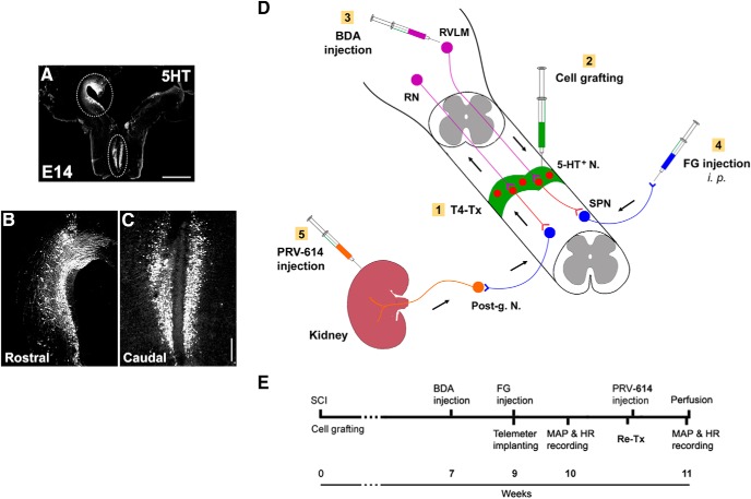
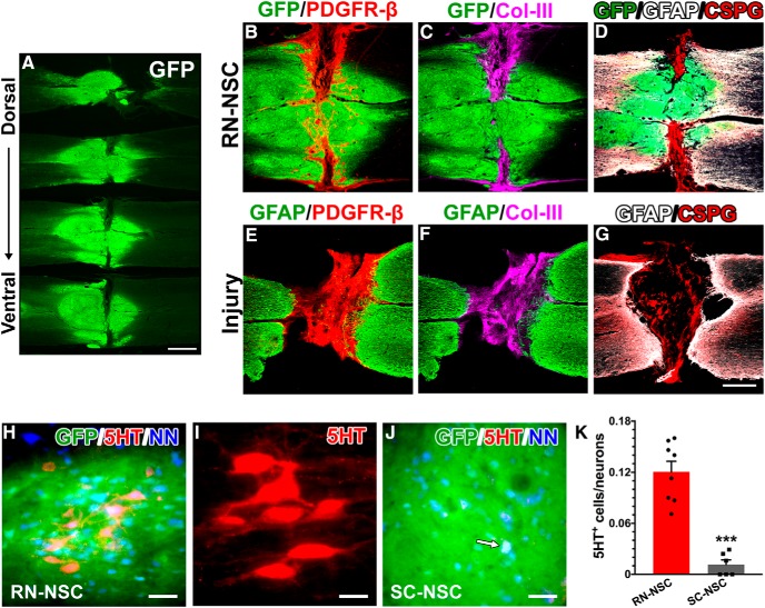
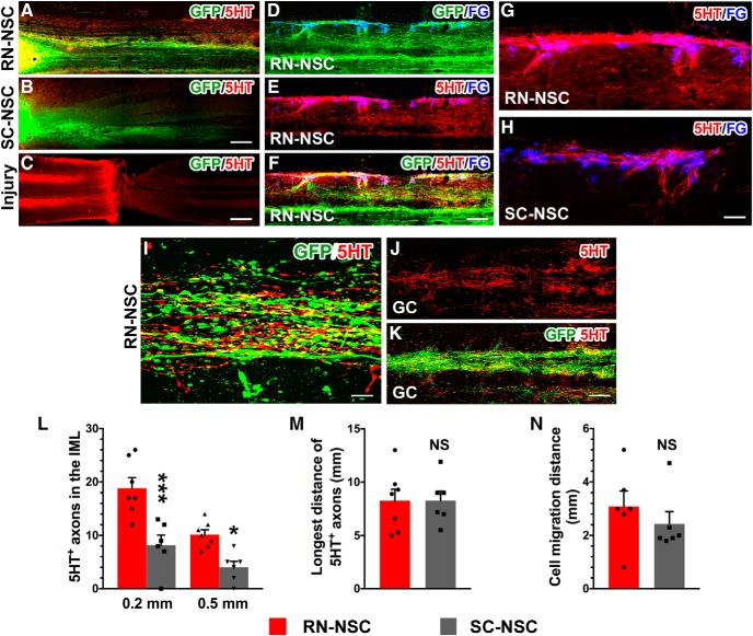
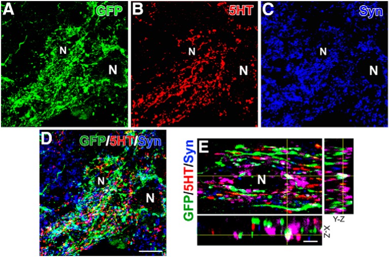
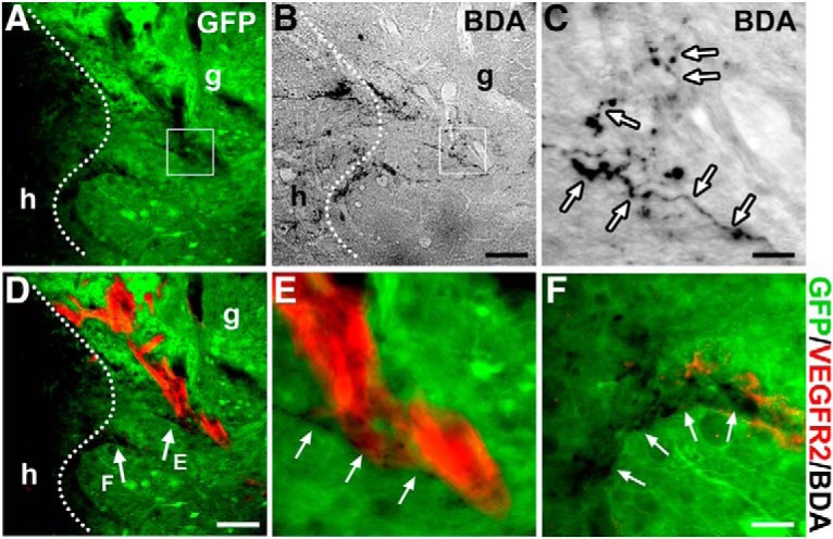
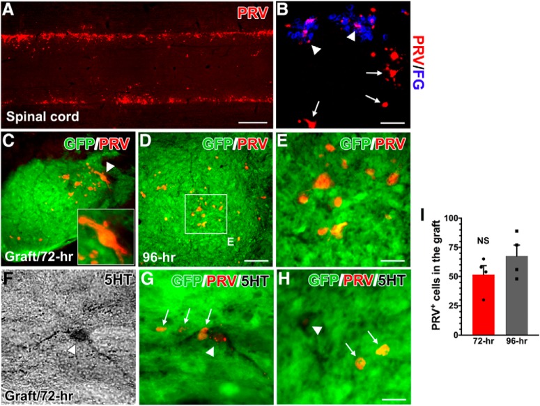
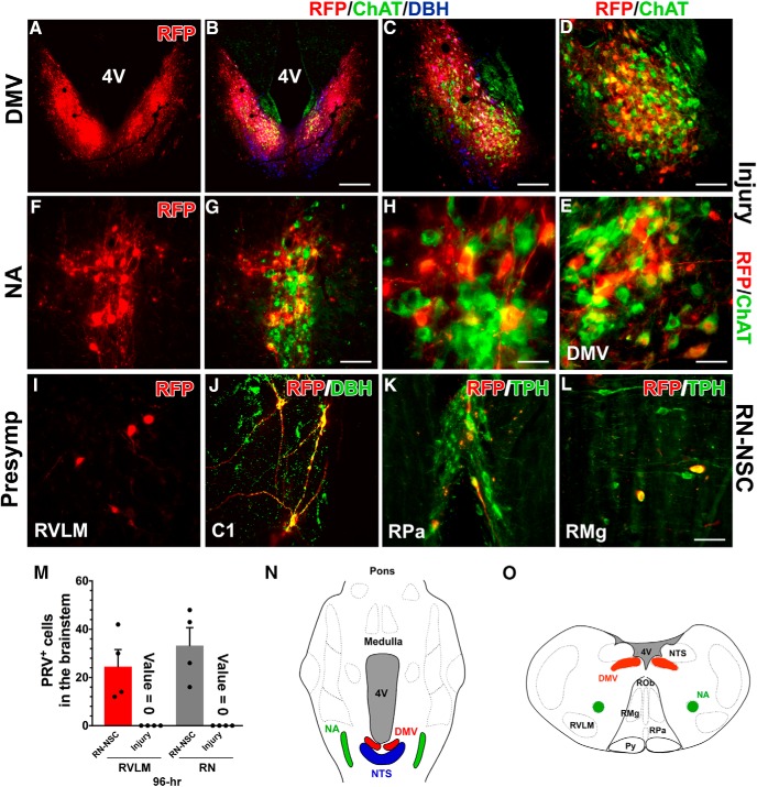
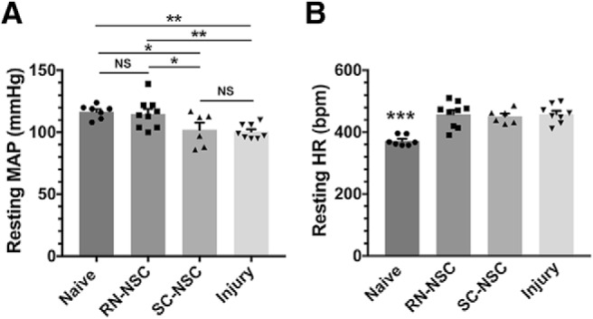
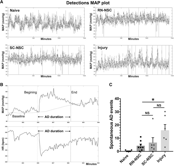
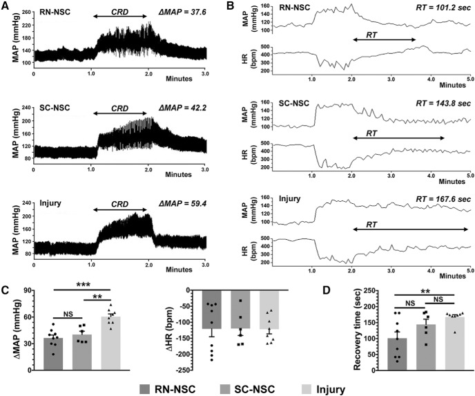
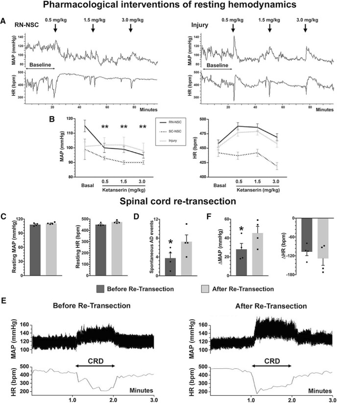
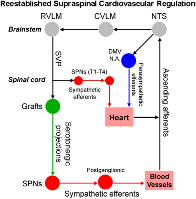
Cardiovascular dysfunction often occurs after high-level spinal cord injury. Disrupting supraspinal vasomotor pathways affects basal hemodynamics and contributes to the development of autonomic dysreflexia (AD). Transplantation of early-stage neurons to the injured cord may reconstruct the descending projections to enhance cardiovascular performance. To determine the specific role of reestablishing serotonergic regulation of hemodynamics, we implanted serotonergic (5-HT+) neuron-enriched embryonic raphe nucleus-derived neural stem cells/progenitors (RN-NSCs) into a complete spinal cord transection lesion site in adult female rats. Grafting embryonic spinal cord-derived NSCs or injury alone served as 2 controls. Ten weeks after injury/grafting, histological analysis revealed well-survived grafts and partial integration with host tissues in the lesion site. Numerous graft-derived serotonergic axons topographically projected to the caudal autonomic regions. Neuronal tracing showed that host supraspinal vasomotor pathways regenerated into the graft, and 5-HT+ neurons within graft and host brainstem neurons were transsynaptically labeled by injecting pseudorabies virus (PRV-614) into the kidney, indicating reconnected serotonergic circuits regulating autonomic activity. Using an implanted telemeter to record cardiovascular parameters, grafting RN-NSCs restored resting mean arterial pressure to normal levels and remarkably alleviated naturally occurring and colorectal distension-induced AD. Subsequent pharmacological blockade of 5-HT2A receptors with ketanserin in RN-NSC-grafted rats reduced resting mean arterial pressure and increased heart rate in all but 2 controls. Furthermore, spinal cord retransection below RN-NSC grafts partially eliminated the recovery in AD. Collectively, these data indicate that RN-NSCs grafted into a spinal cord injury site relay supraspinal control of serotonergic regulation for sympathetic activity to improve cardiovascular function.SIGNIFICANCE STATEMENT Disruption of supraspinal vasomotor pathways results in cardiovascular dysfunction following high-level spinal cord injury. To reestablish the descending regulation of autonomic function, we transplanted serotonergic neuron enriched embryonic raphe nucleus-derived neural stem cells/progenitors into the lesion site of completely transected rat spinal cord. Consequently, grafted raphe nucleus-derived neural stem cells/progenitors acted as a neuronal relay to reconnect supraspinal center and spinal sympathetic neurons below the injury. The reconstituted serotonergic regulation of sympathetic activity led to the improvement of hemodynamic parameters and mitigated autonomic dysreflexia. Based on morphological and physiological results, this study validates the effectiveness of transplanting early-stage serotonergic neurons into the spinal cord for cardiovascular functional recovery after spinal cord injury.

Keywords: cell transplantation; hemodynamics; spinal cord transection; transsynaptic tracing.


Figures

Figure 1.
Figure 1.
Schematics illustrate the experimental procedures. A–C, Donor cells are dissected from the RN in the GFP transgenic rat embryonic brainstem. Immunostaining for 5-HT indicates (A) the distribution of numerous early-stage serotonergic neurons in the rostral (B) and caudal (C) RN in an E14 brainstem. Dotted lined areas are approximately the regions dissected for cell grafting. D, A diagram represents cell transplantation and neuronal tract tracing. Immediately after spinal cord transection at T4 level (T4-Tx), E14 NSCs are implanted into the lesion site of the spinal cord. Animals survive for 10 weeks. Three weeks before death, BDA is injected into the RVLM of brainstem to anterogradely trace descending vasomotor pathways that regenerated into the graft. FG is injected intraperitoneally to retrogradely label SPNs in the spinal cord. RN-NSC-grafted rats received injections of PRV-614 into bilateral kidneys to retrogradely and transsysnaptically label neurons in the graft and host brainstem. E, A timeline shows the experimental arrangement. Post-g. N, Sympathetic postganglionic neurons. Scale bars: A, 2 mm; C, 200 μm.
Figure 2.
Figure 2.
Grafted E14 BS-NSCs partially integrate into the spinal cord. A, Surviving GFP+ BS-NSC implants are shown in a series of longitudinal spinal cord sections from the dorsal to ventral. Grafts form a bridge in the middle part and connect the rostral and caudal cords. B–G, Immunostaining revealed that (B) numerous PDGFR-β+ fibroblasts are present in the portions of the lesion devoid of GFP+ tissue. Col-III (C) and CSPG (D) are heavily deposited in this region. There is a little expression of GFAP at the interface of host/graft. In injury controls (E–G), however, a dense GFAP+ glial scarring is often present at the stumps of spinal cord. Intense Col-III and CSPG are stained into the lesion site of spinal cord. H–K, Abundant serotonergic neurons grow in RN-NSC grafts (H,I), whereas very few are detected (arrow) in SC-NSC grafts (J). These neurons express NeuN, indicating that they have developed mature. K, Cell quantification demonstrated that the percentage of serotonergic neurons in RN-NSC grafts is significantly higher (unpaired t test, ***p < 0.001) than those in SC-NSC grafts (n = 6 or 8 per group). Scale bars: A, 2 mm; G, 0.5 mm; H, J, 50 μm; I, 20 μm.
Figure 3.
Figure 3.
RN-NSC grafts derive abundant serotonergic fibers and project long distance to the caudal autonomic regions. A–C, Both grafted (A) RN-NSCs and (B) SC-NSCs initiate numerous GFP+ extensions toward the distal spinal cord while the lesion interrupts neuronal pathways in (C) injury controls, indicated by disrupted 5-HT+ serotonergic fibers (n = 6 or 8 per group). D–H, In the spinal cord grafted with RN-NSCs (D–G), dense 5-HT+ fibers are overlapped with GFP and spanned several segments to the caudal IML, innervating to FG-labeled SPNs. In contrast, much less 5-HT+ fibers are observed in SC-NSC grafted cords (H). I, Colocalization of 5-HT+/GFP+ axons in the IML caudal to the lesion is shown under a higher magnification. J, K, Ample GFP+/5-HT+ fibers distribute in the caudal gray commission (GC) around the central canal. L–N, Axonal quantification demonstrated that (L) a greater number of serotonergic fibers emerged from RN-NSC grafts than those from SC-NSC grafts (two-way ANOVA followed by Tukey's adjustment: *p < 0.05; ***p < 0.001 between groups), although the longest distance of 5-HT+ fibers (M) had no difference between the two groups (unpaired t test, p > 0.05), (N) nor did the cell migration from the edge of spinal cord stumps (unpaired t test, p > 0.05). NS, not significant. Scale bars: B, 200 μm; C, 400 μm; F, 150 μm; H, 50 μm; I, 12 μm; K, 100 μm.
Figure 4.
Figure 4.
RN-NSC graft-derived serotonergic fibers form synapses in the caudal autonomic region. A–D, Confocal analysis demonstrates that numerous GFP+/5-HT+ axon terminals innervate to the IML, where the bouton-like structures form plentiful synapses onto adjacent neurons (N), identified by expressing the presynaptic marker synaptophysin. E, A higher magnification in 3 dimensions confirms established synapses (white). Scale bars: D, 20 μm; E, 5 μm.
Figure 5.
Figure 5.
Descending vasomotor pathways regenerate into NSC grafts. A–C, BDA injection into the RVLM labels regenerated supraspinal axon terminals within the GFP+ RN-NSC-grafted region in the spinal cord (n = 5). Dotted lines indicate the interface of host (h)/graft (g). Labeled descending axon terminals (arrows) mainly emerge in the rostral part of GFP+ grafts. C, Higher magnification of the boxed region in A and B. D–F, Costaining of BDA and immunofluorescent labeling of VEGFR-2 disclose that penetrated axon terminals often apposite along nascent blood vessels in GFP+ implants. Scale bars: B, D, 100 μm; C, 12.5 μm; F, 30 μm.
Figure 6.
Figure 6.
PRV transsynaptic tracing labels neurons in the graft. Ten weeks after cell transplantation, PRV-614 was injected into the bilateral kidneys to transsynaptically trace reestablished supraspinal sympathetic pathways in RN-NSC-grafted rats (n = 8). A, B, At 72 h after inoculation (n = 4), immunolabeling shows that (A) numerous PRV-infected SPNs emerge in the IML of caudal spinal cord, which are (B) colabeled by FG (arrowheads). Some viral-infected neurons, but not FG-labeled (arrows), are observed in the middle area of gray matter, indicating the possibility of interneuronal identity linked to the SPNs. C–E, PRV injection transfects RN-NSCs in the grafts after 72 (C) or 96 h (D, E) (n = 4) proliferation and transportation times. C, Inset, A PRV-labeled neuron (arrowhead) in the graft with high magnification. F–H, Importantly, 5-HT antibody costaining verifies that a portion of PRV-infected neurons in the graft are serotonergic. I, Statistical analysis shows that the number of PRV-infected neurons in the graft is not significantly (NS) (unpaired t test, p > 0.05) different from 72 to 96 h after injection. This suggests that grafted RN-NSCs rebuild descending autonomic regulation pathways following SCI by forming synaptic connectivity with spinal SPNs. Scale bars: A, 1.7 mm; B, 75 μm; D, 80 μm; E, H, 20 μm.
Figure 7.
Figure 7.
PRV transsynaptic tracing labels neurons in the host brainstem. In either RN-NSC-grafted or injury control animals, PRV-infected neurons were identified within parasympathetic nuclei in the brainstem at both 72 and 96 h after inoculation (n = 3 or 4 per group). A–H, Representative photomicrograph shows RFP-immunolabeled PRV-infected neurons (A–E), the DMV, and (F–H) the NA, which are colabeled with ChAT indicative of cholinergic (green), at 96 h transportation time. In the DMV, RFP-labeled neurons (red) colocalized with (D, E) ChAT+ parasympathetic neurons (green) adjacent to the NTS that contains DBH+ noradrenergic neurons (blue). I–L, However, we observed PRV-infected neurons in sympathetic premotor brainstem regions (e.g., RVLM and caudal RN) only in RN-NSC-grafted rats at 96 h. I, Labeled neurons are detected in the RVLM. J, Infected C1 neurons within this region are DBH+. K, L, Infected neurons in the caudal RN, such as the nucleus raphe pallidus (RPa) and raphe magnus (RMg), are colabeled with TPH antibody, indicating serotonergic identity. No infected cell is detected within the presympathetic regions at the brainstem level in animals with injury control. M, Quantification of PRV-infected neurons in the RVLM or RN in RN-NSC-grafted or injury control rats at 96 h after infection. N, O, Parasympathetic and presympathetic structures in the brainstem are outlined in a 2D, (N) horizontal longitudinal or (O) transverse plane; albeit they may be located at different levels. Scale bars: B, 400 μm; C, 200 μm; D, G, 100 μm; E, 35 μm; H, 30 μm; L, 75 μm.
Figure 8.
Figure 8.
Grafting RN-BSCs improves resting hemodynamic performance following SCI. Telemetric recordings show that, 10 weeks after complete T4 spinal cord transection (n = 6 or 8 per group), rats with injury only exhibit significantly (A) lower resting MAP (one-way ANOVA, p = 0.005; Fisher's PLSD: *p < 0.05, **p < 0.01); and (B) higher HR (one-way ANOVA, p = 0.001; Fisher's PLSD: ***p < 0.001). However, the resting MAP in rats receiving RN-NSCs is elevated to the level of naive controls (p > 0.05), while the resting HR is still higher than the normal level (***p < 0.001). Transplantation of E14 SC-NSCs does not affect decreased MAP and increased HR following SCI (both p > 0.05 vs injury controls; both p < 0.05 vs naive rats). NS, not significant.
Figure 9.
Figure 9.
The incidence of naturally occurring AD reduces after grafting embryonic NSCs in the injured spinal cord. A, Representative MAP tracer is shown over a 24 h period (n = 6 or 8 per group). Vertical line indicates an episode of AD, detected by using the algorithm. B, A typical spontaneous AD is shown in time-stretched tracers of MAP and HR in a RN-NSC-grafted rat, which is manifested by suddenly increased MAP and simultaneously decreased HR. Vertical dotted lines indicate the beginning and end of AD. Horizontal black line indicates the baseline of MAP. Horizontal gray line indicates a smoothed curve. C, Statistical analysis reveals significantly less events of AD in rats grafted with RN-NSCs than in those with injury alone (one-way ANOVA p < 0.05, Fisher's PLSD: both *p < 0.05). Statistically, animals receiving RN-NSCs have no different occurrence of AD compared with those receiving SC-NSC graft (p = 0.91). NS, not significant.
Figure 10.
Figure 10.
Grafting RN-NSCs mitigates the severity of CRD-induced AD. A, C, An episode of AD develops in response to CRD, which mimics a visceral pain, in all tested rats (n = 6 or 8 per group). Compared with animals with injury alone, a lower extent of increased MAP is detected in animals grafted with either RN-NSCs or SC-NSCs (one-way ANOVA, p = 0.006; Fisher's PLSD, RN-NSCs: ***p < 0.001; SC-NSCs: **p < 0.01). The change of MAP is nonsignificantly smaller (p > 0.05) in RN-NSC-grafted animals than SC-NSC-grafted animals. In contrast, there is no difference in HR change during CRD between NSC-grafted animals and those with injury alone (ANOVA, p > 0.05), although HR decreases in all three groups corresponding to the stimulus. B, D, After 1 min CRD duration, the MAP and HR gradually recover to baseline level. Quantitative analysis demonstrates distinct recovery time among the three groups, in which the shortest time is displayed in rats grafted with RN-NSCs and the longest time in rats with injury alone. Statistics indicates significantly shorter recovery time in RN-NSC-grafted rats compared with those with injury alone (ANOVA, p = 0.018; Fisher's PLSD, **p < 0.01). However, the recovery time in SC-NSC-grafted rats, longer than RN-NSC-grafted rats but shorter than rats with injury alone, is nonsignificantly different versus both groups (Fisher's PLSD, both p > 0.05). NS, not significant.
Figure 11.
Figure 11.
Pharmacological intervention or spinal cord retransection compromises cardiovascular improvements. A, B, A serial dose of ketanserin is administered subcutaneously to block 5-HT2A receptors during hemodynamic recordings (n = 5 or 6 per group). A, A tracer of continuous recording shows, in response to the drug delivery, the resting MAP reduces in RN-NSC-grafted animals, whereas it does not display obvious change in those either grafted with SC-NSCs or injury controls. B, Statistical analysis confirmed that the resting MAP is significantly decreased (repeated-measures one-way ANOVA with Bonferroni corrections, all **p < 0.01) and HR is nonsignificantly increased (all p > 0.05) in RN-NSC-grafted rats. In contrast, blocking 5-HT2A receptors does not induce meaningful change in either rats grafted with SC-NSCs or injury controls (all p > 0.05). C–F, After spinal cord retransection below the level of RN-NSC grafts, (C) the resting MAP and HR do not change dramatically (paired t test, both p > 0.05). D, During 24 h recordings, retransection causes significantly more events of naturally occurring AD (paired t test, *p < 0.05). E, Representative examples of CRD-induced AD before and after spinal cord retransection (n = 4). A dramatic rise in MAP and decrease in HR can be seen when a balloon-tipped catheter is inflated in the colon. Markedly, the extent of MAP increase and HR decrease is exacerbated after retransection. F, Statistical analysis shows that CRD stimulation induces a greater increase in MAP (paired t test, *p < 0.05) and nonsignificant decrease in HR (p > 0.05) compared with those before retransection.
Figure 12.
Figure 12.
A schematic diagram illustrates the reestablished vasomotor pathways. Primary afferent fibers initiating from arterial chemoreceptors and baroreceptors project excitatory inputs to the NTS in the brainstem. Parasympathetic baroreceptor afferent fibers ascend in the vagus and glossopharyngeal cranial nerves, providing inhibitory inputs to the NTS. As a major recipient of primary sensory cardiovascular information, the NTS precisely regulates hemodynamic performance through the caudal ventrolateral medulla (CVLM) and the RVLM. At the brainstem level, neurons in the RVLM and other presympathetic nuclei originate supraspinal vasomotor fibers projecting to the SPNs in the IML of spinal cord, regulating cardiovascular function. High-level SCI often interrupts this descending control and results in disordered hemodynamics. Transplanting embryonic RN-NSCs into the lesion extends projections onto the SPNs in the caudal spinal cord. On the other hand, the cellular graft facilitates the regeneration of disrupted host supraspinal vasomotor pathways and forms synapses with grafted neurons. Thus, supraspinal autonomic pathways are rebuilt via a relay-based indirect circuit to regulate cardiovascular function following SCI.

Similar articles

Cited by

References

    1. Ahn J, Saltos TM, Tom VJ, Hou S (2018) Transsynaptic tracing to dissect supraspinal serotonergic input regulating the bladder reflex in rats. Neurourol Urodyn 37:2487–2494. 10.1002/nau.23762 - DOI - PMC - PubMed
    1. Akhavan M, Hoang TX, Havton LA (2006) Improved detection of fluorogold-labeled neurons in long-term studies. J Neurosci Methods 152:156–162. 10.1016/j.jneumeth.2005.09.010 - DOI - PubMed
    1. Assinck P, Duncan GJ, Hilton BJ, Plemel JR, Tetzlaff W (2017) Cell transplantation therapy for spinal cord injury. Nat Neurosci 20:637–647. 10.1038/nn.4541 - DOI - PubMed
    1. Bacon SJ, Smith AD (1988) Preganglionic sympathetic neurones innervating the rat adrenal medulla: immunocytochemical evidence of synaptic input from nerve terminals containing substance P, GABA or 5-hydroxytryptamine. J Auton Nerv Syst 24:97–122. 10.1016/0165-1838(88)90140-3 - DOI - PubMed
    1. Bonner JF, Connors TM, Silverman WF, Kowalski DP, Lemay MA, Fischer I (2011) Grafted neural progenitors integrate and restore synaptic connectivity across the injured spinal cord. J Neurosci 31:4675–4686. 10.1523/jneurosci.4130-10.2011 - DOI - PMC - PubMed

Publication types

MeSH terms

LinkOut - more resources