. 2020 Jan 2;11(1):37.
doi: 10.1038/s41467-019-13911-x.

Gut stem cell aging is driven by mTORC1 via a p38 MAPK-p53 pathway

Affiliations

Gut stem cell aging is driven by mTORC1 via a p38 MAPK-p53 pathway

Dan He et al. Nat Commun. .

Abstract

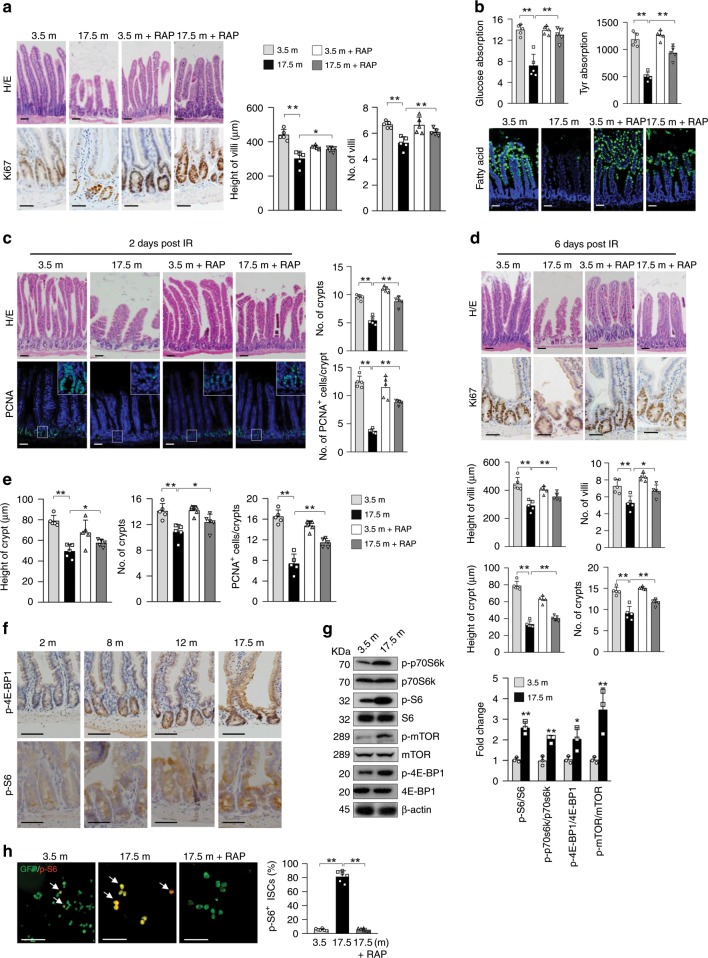
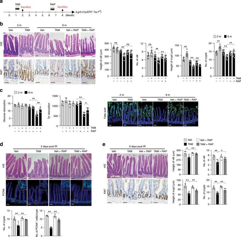
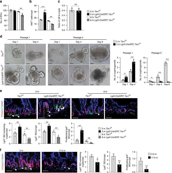
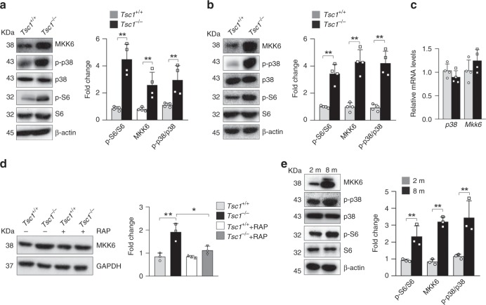
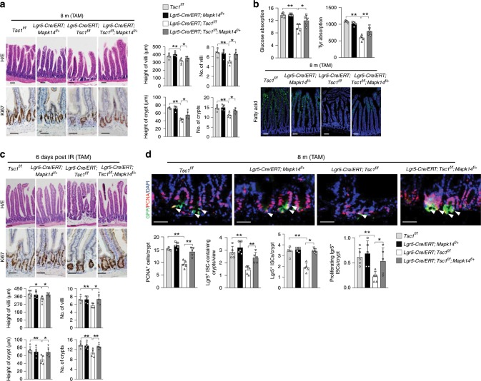
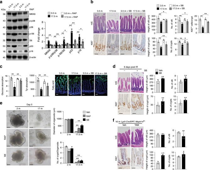
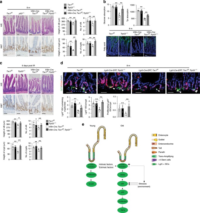
Nutrients are absorbed solely by the intestinal villi. Aging of this organ causes malabsorption and associated illnesses, yet its aging mechanisms remain unclear. Here, we show that aging-caused intestinal villus structural and functional decline is regulated by mTORC1, a sensor of nutrients and growth factors, which is highly activated in intestinal stem and progenitor cells in geriatric mice. These aging phenotypes are recapitulated in intestinal stem cell-specific Tsc1 knockout mice. Mechanistically, mTORC1 activation increases protein synthesis of MKK6 and augments activation of the p38 MAPK-p53 pathway, leading to decreases in the number and activity of intestinal stem cells as well as villus size and density. Targeting p38 MAPK or p53 prevents or rescues ISC and villus aging and nutrient absorption defects. These findings reveal that mTORC1 drives aging by augmenting a prominent stress response pathway in gut stem cells and identify p38 MAPK as an anti-aging target downstream of mTORC1.


Conflict of interest statement

The authors declare no competing interests.

Figures

Fig. 1
Fig. 1. Enhanced mTORC1 activation in ISCs/TA cells of geriatric mice drives villus natural aging.
ad Seventeen and half-month-old mice showed deterioration in villus and crypt structures (right panels: quantitation data) (a), decreased nutrient absorption activities (b), increased sensitivity to IR-induced decreases in the numbers of crypts and proliferating cells at day 2 post IR (c), and compromised regeneration (decreases in the height and number of villi and crypts) at day 6 (d) compared with young mice, which were partially rescued by 1.5 months of RAP treatment (3 mg/kg body weight) starting at 16 months of age. Data are expressed as mean ± SEM. N = 5 mice per group. *P < 0.05, **P < 0.01 (determined using Student’s t test). e Seventeen and half-month-old mice showed decreases in the height and number of crypts and the number of proliferating TA cells (based on (a)), which were rescued by RAP. Data are expressed as mean ± SEM. N = 5 mice per group. *P < 0.05, **P < 0.01 (determined using Student’s t test). f Representative images (proximal jejunum midline sections) showed that mTORC1 activation was increased with age in crypt cells. g Western blot results showed that mTORC1 activation was increased in the crypt samples of 17.5-month-old mice compared with 3.5-month-old mice. Isolated crypts were directly lysed and used for WB analysis. Data are expressed as mean ± SEM. N = 3 mice per group. *P < 0.05, **P < 0.01 (determined using Student’s t test). h More Lgr5+ ISCs isolated from 17.5-month-old mice showed mTORC1 activation than those from 3.5-month-old mice, which was suppressed by RAP treatment. Lgr5+ ISCs were isolated from the small intestines of Lgr5-GFP-CreERT mice with FACS sorting and stained for p-S6. Right panel: quantification data (mean ± SEM). N = 6 mice per group. **P < 0.01 (determined using Student’s t test).
Fig. 2
Fig. 2. Deletion of Tsc1 in Lgr5+ ISCs leads to villus premature aging.
a A schematic for conditional ablation of Tsc1 and RAP treatment in Lgr5-CreERT;Tsc1f/f mice (be). b H/E and Ki67 staining showed that ablation of Tsc1 in Lgr5+ ISCs led to crypt overgrowth at 2 months of age and deterioration of villus structure (the midline section of the proximal jejunum) at 8 months of age, which were partially rescued by RAP. Right panels: quantification data (mean ± SEM). N = 8 mice per group. **P < 0.01 (determined using Student’s t test). c Eight-month-old Lgr5-CreERT;Tsc1f/f mice showed decreased nutrient absorption activities for L-glucose (mmol/l), amino acids (ng/ml), and fatty acids, which were partially rescued by RAP treatment. Data are expressed as mean ± SEM. N = 5 mice per group. *P < 0.05, **P < 0.01 (determined using Student’s t test). d, e Eight-month-old Lgr5-CreERT;Tsc1f/f mice showed increased sensitivity to IR-induced decreases in the numbers of crypts and proliferating cells at day 2 post IR (d), and compromised regeneration (decreases in the height and number of villi and crypts) at day 6 post IR (e), which were partially rescued by RAP treatment. Data are expressed as mean ± SEM. N = 5 mice per group. *P < 0.05, **P < 0.01 (determined using Student’s t test).
Fig. 3
Fig. 3. Tsc1 deletion leads to defects in propagation of organoids and ISC/progenitor cells.
a, b Eight-month-old Lgr5-GFP-CreERT;Tsc1f/f mice showed decreases in the number of IECs (a) and the number of proliferating TA cells (b) (based on Fig. 2b). Data are expressed as mean ± SEM. N = 4 mice per group. **P < 0.01 (determined using Student’s t test). c Eight-month-old Lgr5-GFP-CreERT;Tsc1f/f mice showed no change in the villus-to-crypt ratios. N = 4. d Representative images showing that Tsc1−/− crypts (isolated from Villin-Cre;Tsc1f/f mice, since Lgr5-GFP-CreERT can label ~65% of the crypts) displayed increases in the size of the minigut organoids and the number of crypts, whereas serial passaging of the organoids revealed that Tsc1-deficient organoids quickly lost their ability to propagate. Right panels: quantification of the data (mean ± SEM). N = 3 mice per group. *P < 0.05, **P < 0.01 (determined using Student’s t test). e Eight-month-old Lgr5-GFP-CreERT;Tsc1f/f mice showed decreases in the numbers of ISCs and proliferating ISCs. Intestine sections were immunostained for PCNA, and cells positive for PCNA and GFP were counted and normalized to the total numbers of Lgr5+ ISCs. Bottom panels: quantitation data (mean ± SEM). N = 5 mice per group. **P < 0.01 (determined using Student’s t test). f The numbers of ISCs and proliferating ISCs were decreased in naturally aged 17.5-month-old mice. Right panels: quantitation data (mean ± SEM). N = 3 mice per group. *P < 0.05, **P < 0.01 (determined using Student’s t test).
Fig. 4
Fig. 4. Tsc1 deficiency increases MKK6 protein levels in an mTORC1-dependent manner.
a, b Western blot results showed that the levels of MKK6 and activation of p38 MAPKs were increased in villus samples of Villin-Cre; Tsc1f/f mice (a) and Tsc1-deficient organoids (b) compared with those in controls. Right panels: quantification data (mean ± SEM). N = 4 mice for a and 3 for b. **P < 0.01 (determined using Student’s t test). c Tsc1 deficiency did not significantly affect the mRNA levels of Mkk6 in crypt samples. The total RNA was isolated from the crypt samples, converted into cDNA, and then used for quantitative PCR analysis. N = 4 mice per group. d Tsc1 deficiency led to an increase in radio-labeled methionine incorporation into MKK6, which were suppressed by RAP. Primary enteroblast cultures were pulse-labeled with 35S Met and Cys for 1 h. The cells were collected, and MKK6 was immunoprecipitated and fractionated onto SDS-PAGE gels, which were dried; the signals were detected with a PhosphorImager. Right panels: quantification data (mean ± SEM). N = 3 mice per group. *P < 0.05 (determined using Student’s t test). e Western blot analysis revealed that crypt samples from 8-month-old Lgr5-GFP-CreERT;Tsc1f/f mice showed increases in MKK6 expression, p38 MAPK activation, and mTORC1 activation compared with those from 2-month-old mice. Right panels: quantification data (mean ± SEM). N = 3 per group. *P < 0.05 (determined using Student’s t test).
Fig. 5
Fig. 5. MKK6-p38α MAPK signaling mediates villus premature aging caused by Tsc1 deficiency.
ac Mapk14 haplodeficiency rescued the deterioration in villus structures (a), decreased nutrient absorption activities (b), and compromised regeneration (c) at 8 month of age. Data are expressed as mean ± SEM. N = 5 mice per group. *P < 0.05, **P < 0.01 (determined using Student’s t test). d Mapk14 haplodeficiency rescued the decreases in the numbers of ISCs and proliferating ISCs in 8-month-old Lgr5-GFP-CreERT;Tsc1f/f mice. The intestinal sections were immunostained for PCNA, and cells positive for PCNA and GFP were counted and normalized to the number of Lgr5+ ISCs. Bottom panels: quantitation data (mean ± SEM). N = 5 mice per group. *P < 0.05, **P < 0.01 (determined using Student’s t test).
Fig. 6
Fig. 6. Inhibition of p38 MAPK rescues natural aging of mouse villi.
a Western blot results showed that 17.5-month-old mouse crypt samples displayed elevated MKK6 expression, increased p38 MAPK activation, and increased p53 and p16 levels, which were suppressed by RAP treatment for 1.5 months starting at 16 months of age. Right panel: quantification data (mean ± SEM). N = 3 mice per group. *P < 0.05, **P < 0.01 (determined using Student’s t test). bd Inhibition of p38 MAPK largely rescued the deterioration in villus and crypt structures (b), decreased nutrient absorption activities (c), and compromised regeneration (d) in 17.5-month-old normal mice. The mice were treated with SB203580 for 1.5 months. Data are expressed as mean ± SEM. N = 5 mice per group. *P < 0.05, **P < 0.01 (determined using Student’s t test). e Representative images showing that SB203580 (2 μM) or RAP (0.5 μM) partially rescued the defects in growth and crypt formation of minigut organoids isolated from 17.5-month-old mice. The inhibitors were added to the culture at day 1 of the organoid cultures. Bottom panels: quantification data (mean ± SEM). N = 3 mice per group. **P < 0.01 (determined using Student’s t test). f Lgr5-CreERT;Mapk14f/+ mice (TAM injected at 2 months of age) did not show aging-like villus structural defects at 16 months of age. The intestines were sectioned and stained with H/E or Ki67 antibodies. Right panels: quantification data (mean ± SEM). N = 5 mice per group. *P < 0.05, **P < 0.01 (determined using Student’s t test).
Fig. 7
Fig. 7. Ablation of Trp53 prevents mTORC1-driven villus aging.
ac Deletion of Trp53 rescued the deterioration in villus and crypt structures (a), decreased nutrient absorption activities (b), and compromised regeneration (c) in 8-month-old Villin-Cre;Tsc1f/f mice. Data are expressed as mean ± SEM. N = 5 mice per group. *P < 0.05, **P < 0.0 (determined using Student’s t test). d Trp53 ablation rescued the decrease in the numbers of ISCs and proliferating ISCs in 8-month-old Lgr5-GFP-CreERT;Tsc1f/f mice. The intestinal sections were immunostained for PCNA, and cells positive for PCNA and GFP were counted and normalized to the total numbers of Lgr5+ ISCs. Bottom panels: quantification data (mean ± SEM). N = 5 mice per group. *P  < 0.05, **P < 0.0 (determined using Student’s t test). e A model for controlling gut stem and progenitor cell aging by mTORC1.

References

    1. Sharpless NE, Sherr CJ. Forging a signature of in vivo senescence. Nat. Rev. Cancer. 2015;15:397–408. doi: 10.1038/nrc3960. - DOI - PubMed
    1. Soenen S, Rayner CK, Jones KL, Horowitz M. The ageing gastrointestinal tract. Curr. Opin. Clin. Nutr. Metab. Care. 2016;19:12–18. doi: 10.1097/MCO.0000000000000238. - DOI - PubMed
    1. Clevers H, Batlle E. SnapShot: the intestinal crypt. Cell. 2013;152:1198–1198. doi: 10.1016/j.cell.2013.02.030. - DOI - PubMed
    1. Barker N, et al. Identification of stem cells in small intestine and colon by marker gene Lgr5. Nature. 2007;449:1003–1007. doi: 10.1038/nature06196. - DOI - PubMed
    1. Snippert HJ, et al. Intestinal crypt homeostasis results from neutral competition between symmetrically dividing Lgr5 stem cells. Cell. 2010;143:134–144. doi: 10.1016/j.cell.2010.09.016. - DOI - PubMed

Publication types

MeSH terms