

Comparison of four different internal fixation methods in the treatment of distal clavicle fractures
- PMID: 31897095
- PMCID: PMC6923748
- DOI: 10.3892/etm.2019.8233
Comparison of four different internal fixation methods in the treatment of distal clavicle fractures
Abstract
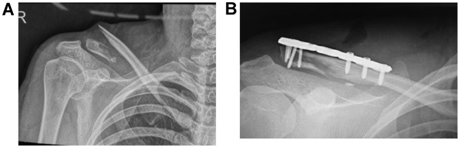
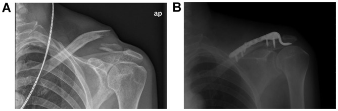
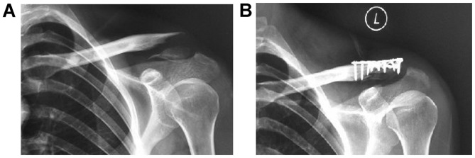
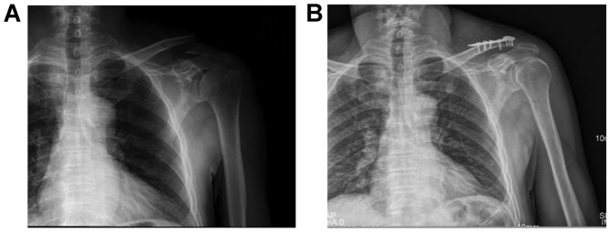
This study compared the clinical efficacy of four internal fixation methods in the treatment of distal clavicle fractures, in an effort to guide appropriate selection and application in the clinic. Eighty-four patients with distal clavicle-comminuted fractures were treated with a distal clavicle anatomic plate (group A), clavicular hook plate (group B), double-plate vertical fixation (group C), or T-shaped steel plate internal fixation (group D). The Constant-Murley scoring system was used to evaluate the shoulder joint function. The fracture healing time, VAS, and postoperative complications were compared and analyzed among the four groups. According to the Constant-Murley evaluation standard, the excellent and good rates of the four groups were 94.4, 73.1, 95 and 80% in groups A-D, respectively. The excellent and good rates of Constant-Murley evaluation standard in groups A and C were significantly better than those in groups B and D (P<0.05). VAS in the distal clavicle anatomic plate group (group A), double-plate vertical fixation group (group C), and T-shaped steel plate internal fixation group (group D) were significantly better than the clavicular hook plate group (group B) (P<0.05). The incidence of postoperative complications in the clavicular hook plate group (group B) was 15.4% and in the T-shaped steel plate internal fixation group (group D) was 15%, which were significantly higher than those of the distal clavicle anatomic plate group (group A) and double-plate vertical internal fixation group (group C) (P<0.05). The treatment of distal clavicle fractures using either one of the four internal fixation techniques can obtain better clinical results. The distal clavicle anatomic plate and double-plate vertical internal fixation techniques are associated with a decreased incidence of shoulder pain, an increase in the range of motion of the shoulder, and a reduction in complications, and thus, are preferable for the early functional recovery of limbs.
Keywords: bone plates; clavicle; dual plate; fracture fixation.
Copyright: © Li et al.
Figures
References
-
- Martetschläger F, Kraus TM, Schiele CS, Sandmann G, Siebenlist S, Braun KF, Stöckle U, Freude T, Neumaier M. Treatment for unstable distal clavicle fractures (Neer 2) with locking T-plate and additional PDS cerclage. Knee Surg Sports Traumatol Arthrosc. 2013;21:1189–1194. doi: 10.1007/s00167-012-2089-0. - DOI - PubMed
LinkOut - more resources
Full Text Sources