. 2019 Dec 16;5(4):2055217319894604.
doi: 10.1177/2055217319894604. eCollection 2019 Oct-Dec.

PARP-1 deregulation in multiple sclerosis

Affiliations

PARP-1 deregulation in multiple sclerosis

Maria Meira et al. Mult Scler J Exp Transl Clin. .

Abstract

Background: Poly (ADP-ribose) polymerase 1 (PARP-1) plays pivotal roles in immune and inflammatory responses. Accumulating evidence suggests PARP-1 as a promising target for immunomodulation in multiple sclerosis and natalizumab-associated progressive multifocal leukoencephalopathy.

Objective: This study explores expression of PARP-1 and downstream effectors in multiple sclerosis and during natalizumab treatment.

Methods: Transcriptional expressions were studied by real-time reverse transcriptase polymerase chain reaction on CD4+T/CD8+T/CD14+/B cells and peripheral blood mononuclear cells from healthy volunteers, untreated and natalizumab-treated non-progressive multifocal leukoencephalopathy and progressive multifocal leukoencephalopathy multiple sclerosis patients.

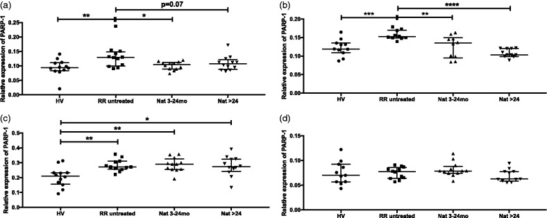
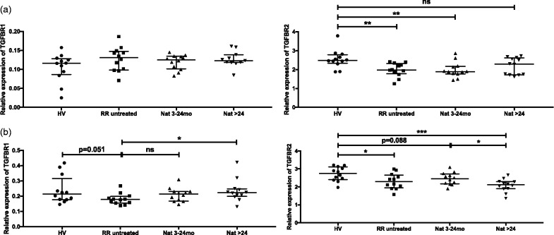
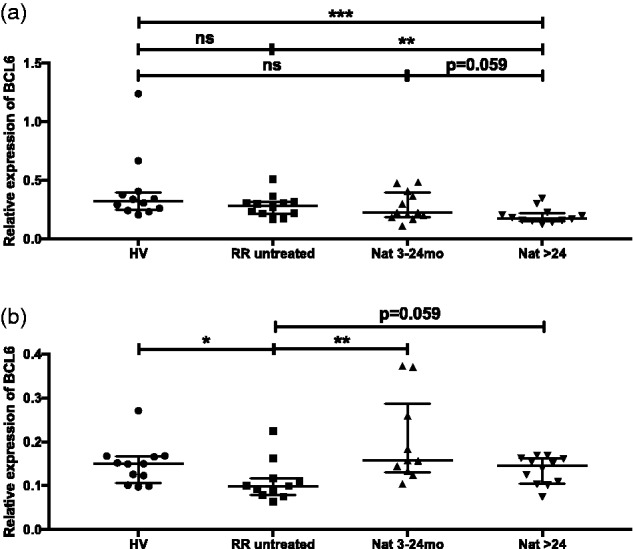
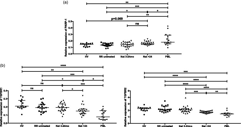
Results: PARP-1 expression was higher in CD4+T, CD8+T and B cells from untreated patients compared to healthy volunteers. Natalizumab treatment restored deregulated PARP-1 expression in T cells but not in B cells. Sustained upregulation of PARP-1 was associated with decreased expression of downstream PARP-1 factors such as TGFBR1/TGFBR2/BCL6 in B cells. Notably, a higher expression of PARP-1 was detected in progressive multifocal leukoencephalopathy patients.

Conclusions: Given the importance of PARP-1 in inflammatory processes, its upregulation in multiple sclerosis lymphocyte populations suggests a potential role in the immune pathogenesis of multiple sclerosis. Strikingly higher PARP-1 expression in progressive multifocal leukoencephalopathy cases suggests its involvement in progressive multifocal leukoencephalopathy disease pathomechanisms. These results further support the value of PARP-1 inhibitors as a potential novel therapeutic strategy for multiple sclerosis and natalizumab-associated progressive multifocal leukoencephalopathy.

Keywords: JCV; PARP-1; multiple sclerosis; natalizumab; progressive multifocal leukoencephalopathy (PML).


Figures

Figure 1.
Figure 1.
Expression of poly (ADP-ribose) polymerase 1 (PARP-1) mRNA in CD4+T, CD8+T, B cells and monocytes. Transcriptional expression of PARP-1 was analysed with real-time reverse transcriptase polymerase chain reaction in CD4+T (a), CD8+T cells (b), B cells (c) and monocytes (d) from healthy volunteer, untreated patients and natalizumab-treated patients (3–24 months or >24 months). Relative expression levels (median with interquartile range) are depicted. ****P < 0.0001; ***P < 0.001; **P < 0.01; *P < 0.05.
Figure 2.
Figure 2.
Expression of TGFBR1 and TGFBR2 mRNA in CD4+T and B cells. Transcriptional expressions of TGFBR1 (left panels) and TGFBR2 (right panels) were analysed with real-time reverse transcriptase polymerase chain reaction in CD4+T (a) and B cells (b) from healthy volunteers, untreated and natalizumab-treated patients (3–24 months; >24 months). Relative expression levels (median with interquartile range) are depicted. ***P < 0.001; **P < 0.01, *P < 0.05; ns: not significant.
Figure 3.
Figure 3.
Expression of BCL6 mRNA in B cells and CD8+T cells. Transcriptional expression of BCL6 was analysed with real-time reverse transcriptase polymerase chain reaction in B cells (a) and CD8+T cells (b) from healthy volunteers, untreated and natalizumab-treated patients (3–24 months; >24 months). Relative expression levels (median with interquartile range) are depicted. ***P < 0.001; **P < 0.01; *P < 0.05; ns: not significant.
Figure 4.
Figure 4.
Expression of poly (ADP-ribose) polymerase 1 (PARP-1), TGFBR1 and TGFBR2 mRNA in peripheral blood mononuclear cells (PBMCs). Transcriptional expression of PARP-1 (a), TGFBR1 ((b), left panel) and TGFBR2 ((b), right panel) were analysed with real-time reverse transcriptase polymerase chain reaction in PBMCs from healthy volunteers, untreated, natalizumab-treated patients (3–24 months; >24 months) and natalizumab-associated progressive multifocal leukoencephalopathy (PML) patients. Relative expression levels (median with interquartile range) are depicted. ****P < 0.0001; ***P < 0.001; **P < 0.01; *P < 0.05; ns: not significant.

References

    1. Reich DS, Lucchinetti CF, Calabresi PA. Multiple sclerosis. N Engl J Med 2018; 378: 169–180. - PMC - PubMed
    1. Kappos L, O’Connor PW, Polman CH, et al. Clinical effects of natalizumab on multiple sclerosis appear early in treatment course. J Neurol 2013; 260: 1388–1395. - PMC - PubMed
    1. Mills EA, Mao-Draayer Y. Understanding progressive multifocal leukoencephalopathy risk in multiple sclerosis patients treated with immunomodulatory therapies: a bird's eye view. Front Immunol 2018; 9: 138. - PMC - PubMed
    1. Berger JR. Classifying PML risk with disease modifying therapies. Mult Scler Relat Disord 2017; 12: 59–63. - PubMed
    1. Gupte R, Liu Z, Kraus WL. PARPs and ADP-ribosylation: recent advances linking molecular functions to biological outcomes. Genes Dev 2017; 31: 101–126. - PMC - PubMed