

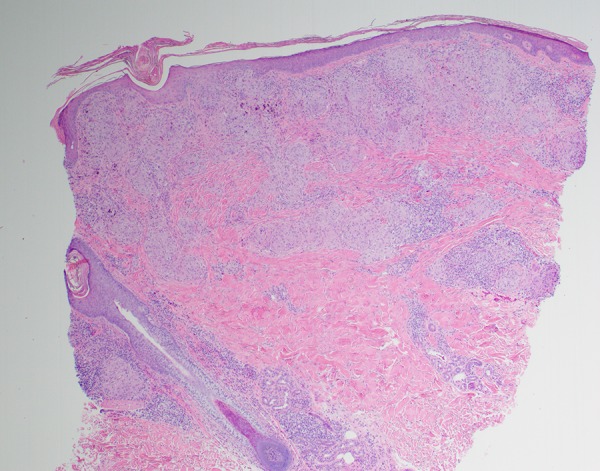
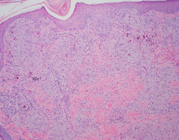
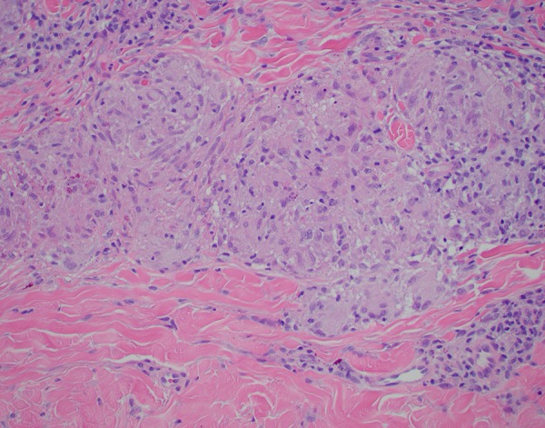
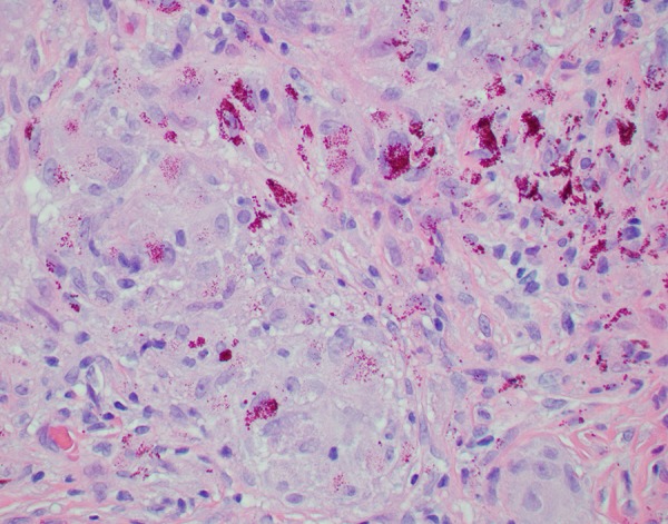
Educational Case: Granulomatous Dermatitis
- PMID: 31897421
- PMCID: PMC6920582
- DOI: 10.1177/2374289519892559
Educational Case: Granulomatous Dermatitis
Abstract
The following fictional case is intended as a learning tool within the Pathology Competencies for Medical Education (PCME), a set of national standards for teaching pathology. These are divided into three basic competencies: Disease Mechanisms and Processes, Organ System Pathology, and Diagnostic Medicine and Therapeutic Pathology. For additional information, and a full list of learning objectives for all three competencies, see http://journals.sagepub.com/doi/10.1177/2374289517715040.1.
Keywords: exogenous antigens; granulomatous dermatitis; noncaseating granulomatous dermatitis; organ system pathology; pathology competencies; skin; tattoo-related disorder.
© The Author(s) 2019.
Conflict of interest statement
Declaration of Conflicting Interests: The author(s) declared no potential conflicts of interest with respect to the research, authorship, and/or publication of this article.
Figures
References
-
- Noe MH, Rosenbach M. Cutaneous sarcoidosis. Curr Opin Pulm Med. 2017;23:482–486. - PubMed
-
- Simunovic C, Shinohara MM. Complications of decorative tattoos: recognition and management. Am J Clin Dermatol. 2014;15:525–536. - PubMed
-
- Mohan H, Bal A, Dhami GP. Non-infectious granulomatous dermatitis: a clinicopathological study. J Cutan Pathol. 2006;33:767–771. - PubMed
-
- Wick MR. Granulomatous and histiocytic dermatitides. Sem Diag Pathol. 2017;34:301–311. - PubMed
LinkOut - more resources
Full Text Sources
