. 2019 Dec 18;5(12):eaav2720.
doi: 10.1126/sciadv.aav2720. eCollection 2019 Dec.

Direct single-molecule quantification reveals unexpectedly high mechanical stability of vinculin-talin/α-catenin linkages

Affiliations

Direct single-molecule quantification reveals unexpectedly high mechanical stability of vinculin-talin/α-catenin linkages

Shimin Le et al. Sci Adv. .

Abstract

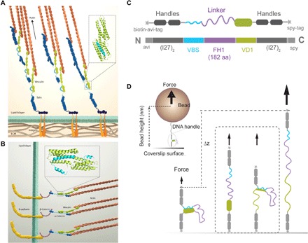
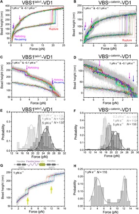
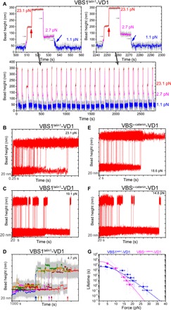
The vinculin-mediated mechanosensing requires establishment of stable mechanical linkages between vinculin to integrin at focal adhesions and to cadherins at adherens junctions through associations with the respective adaptor proteins talin and α-catenin. However, the mechanical stability of these critical vinculin linkages has yet to be determined. Here, we developed a single-molecule detector assay to provide direct quantification of the mechanical lifetime of vinculin association with the vinculin binding sites in both talin and α-catenin, which reveals a surprisingly high mechanical stability of the vinculin-talin and vinculin-α-catenin interfaces that have a lifetime of >1000 s at forces up to 10 pN and can last for seconds to tens of seconds at 15 to 25 pN. Our results suggest that these force-bearing intermolecular interfaces provide sufficient mechanical stability to support the vinculin-mediated mechanotransduction at cell-matrix and cell-cell adhesions.


Figures

Fig. 1
Fig. 1. Schematics of experimental design.
(A and B) Vinculin-mediated mechanotransduction pathways at FAs (A) and AJs (B). The key proteins involved in the mechanical linkages are illustrated. The example VBS—VD1 structures [Protein Data Bank: 1xwj (24) and 5y04 (23)] are highlighted in the zoomed-in box on each panel. (C) Design of the single-molecule VBS—VD1 detector (N to C terminus): avi-tag, two repeats of titin I27 domain, vinculin D1 domain, formin FH1 domain, talin/α-catenin VBS, two repeats of titin I27 domain, and spy-tag. Between two adjacent domains, there is a short flexible linker (GGGSG). (D) Single-molecule detection of the VBS—VD1 interaction: VBS—VD1 complex forms at low forces (left) and ruptures at higher forces (right) with higher extension (i.e., the end-to-end distance of the detector along force direction). The illustration in the dashed box in the middle describes two additional possible rupture transitions during force increase: (i) VD1 and VBS disengaged while the VD1 remain folded and (ii) the VD1 partially unfolds and then leads to the rupture of the linked VBS—VD1 complex.
Fig. 2
Fig. 2. Force-dependent rupture of the VBS—VD1 complex.
(A and B) Force bead height curves of VBS—VD1 detectors during force cycles with force increase at a loading rate of 1 pN s−1 and force decrease at a loading rate of −0.1 pN s−1 for the interfaces formed between the VD1 and the talin VBS1 (A) or α-catenin VBS (B), respectively. The red dashed boxes indicate the region of VBS—VD1 rupture, magenta arrows indicate the two-step refolding of the VD1 domain, and the blue arrows indicate repairing of VD1 and VBS. Each colored line indicates one independent force-increase and force-decrease cycle, smoothed (10-point fast Fourier transform) from raw data (gray). (C and D) The zoom-in of refolding and repairing events during force-decrease scans of the VBS—VD1 detectors. (E and F) The normalized histograms of VBS—VD1 rupture forces at force loading rates of 0.2, 1, and 5 pN s−1. The gray lines are corresponding to a Gaussian fitting of the histograms (the goodness of fit, R2 > 0.9). (G and H) Force bead height curves of VD1 during force-increase scans at a loading rate of 1 pN s−1 (G) and the resulting normalized histograms of VD1 unfolding forces (H). The yellow-green arrows indicate the unfolding events of VD1. The gray line is the double-Gaussian fitting of the histogram (the goodness of fit, R2 > 0.9). The corresponding numbers of data points (N) for the histograms are labeled on the figure panels. The sketch in the top panel shows the domains involved in the construct for Fig. 2 (G and H).
Fig. 3
Fig. 3. Force-dependent lifetime of the VBS—VD1 complex.
(A) Experimental procedures of force-jump assay. Bottom: Typical example of 29 cycles of the force-jump experiments. Top: Zoomed-in view of two force-jump cycles. The red arrows indicate the rupture of the complex, and the blue arrows indicate the re-formation of the complex. The light gray horizontal arrows indicate the bead height change mainly caused by the rotation of the bead due to torque rebalance during force jump. (B to F) Examples of the extension change time traces of the VBS—VD1 complex rupture at various forces. Each large extension jump step indicates an event of VBS—VD1 complex rupture. In (F), the ~10-nm extension increase/decrease steps are the force-induced unfolding/refolding of the I27 domains in the detectors (fig. S3). The colored arrows indicate the VBS—VD1 rupture events at these forces. (G) The force-dependent lifetime of the VBS—VD1 complex. The blue/magenta hollow cycles represent the individual lifetimes measured at the corresponding forces. The blue/magenta solid squares represent the characteristic lifetime at the corresponding forces. The light blue/magenta line is the fitting curve based on Bell’s model, and the blue/magenta line is the fitting curve based on Arrhenius law. The horizontal error bars represent the 10 to 15% uncertainty in force calibration due to the heterogeneity of the beads (18). The vertical error bars represent the SE.

Similar articles

Cited by

References

    1. Bays J. L., DeMali K. A., Vinculin in cell–cell and cell–matrix adhesions. Cell. Mol. Life Sci. 74, 2999–3009 (2017). - PMC - PubMed
    1. Iskratsch T., Wolfenson H., Sheetz M. P., Appreciating force and shape — The rise of mechanotransduction in cell biology. Nat. Rev. Mol. Cell Biol. 15, 825–833 (2014). - PMC - PubMed
    1. Vogel V., Sheetz M., Local force and geometry sensing regulate cell functions. Nat. Rev. Mol. Cell Biol. 7, 265–275 (2006). - PubMed
    1. Borgon R. A., Vonrhein C., Bricogne G., Bois P. R. J., Izard T., Crystal structure of human vinculin. Structure 12, 1189–1197 (2004). - PubMed
    1. Xu W., Baribault H., Adamson E. D., Vinculin knockout results in heart and brain defects during embryonic development. Development 125, 327–337 (1998). - PubMed

Publication types

LinkOut - more resources