Review
. 2020 Mar;77(5):789-805.
doi: 10.1007/s00018-019-03420-x. Epub 2020 Jan 2.

Senescence in the pathogenesis of age-related macular degeneration

Affiliations
Review

Senescence in the pathogenesis of age-related macular degeneration

Janusz Blasiak. Cell Mol Life Sci. 2020 Mar.

Abstract

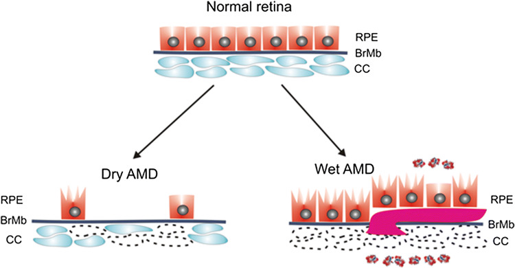
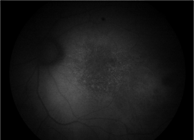
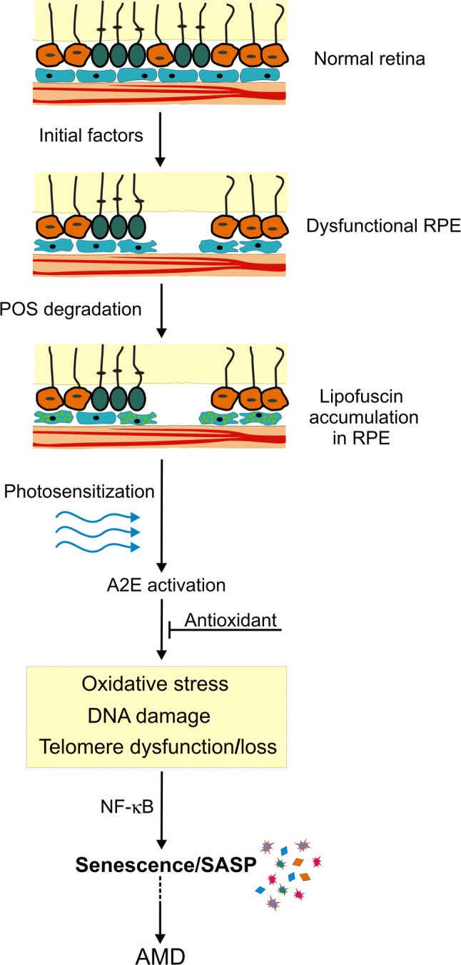
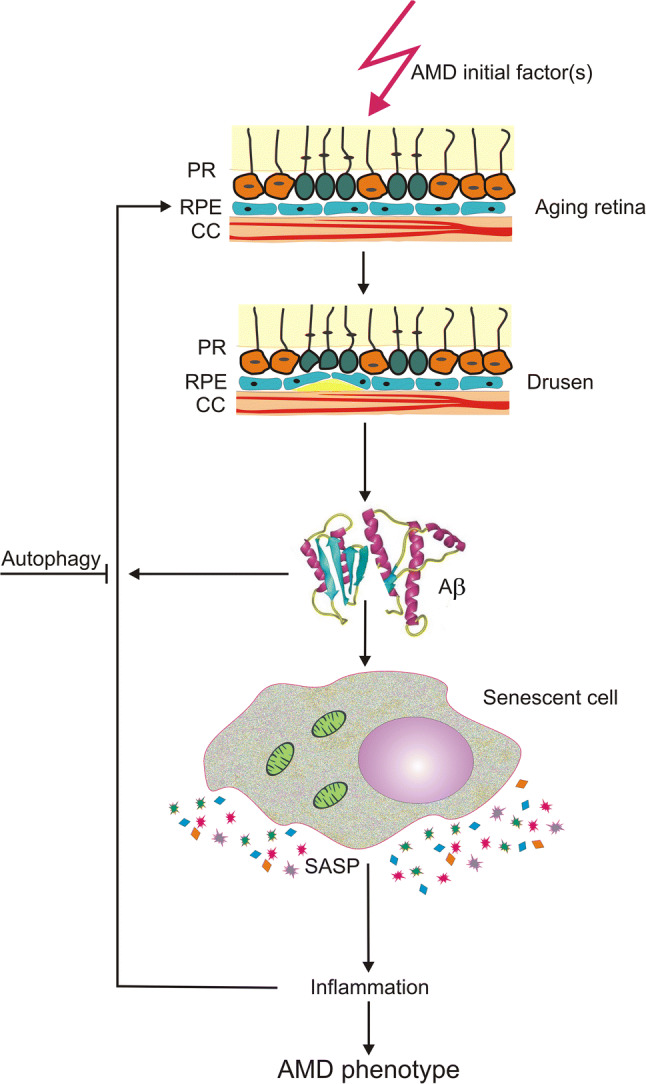
Age-related macular degeneration (AMD) is a complex eye disease underlined by the death of photoreceptors and degeneration of retinal pigment epithelium (RPE) and choriocapillaris (CC). The mechanism(s) responsible for massive and progressive retinal degeneration is not completely known. Senescence, a state of permanent inhibition of cell growth, may be induced by many factors important for AMD pathogenesis and results in senescence-associated secretory phenotype (SASP) that releases growth factors, cytokines, chemokines, proteases and other molecules inducing inflammation and other AMD-related effects. These effects can be induced in the affected cell and neighboring cells, leading to progression of AMD phenotype. Senescent cells also release reactive oxygen species that increase SASP propagation. Many other pathways of senescence-related AMD pathogenesis, including autophagy, the cGAS-STING signaling, degeneration of CC by membrane attack complex, can be considered. A2E, a fluorophore present in lipofuscin, amyloid-beta peptide and humanin, a mitochondria-derived peptide, may link AMD with senescence. Further studies on senescence in AMD pathogenesis to check the possibility of opening a perspective of the use of drugs killing senescent cells (senolytics) and terminating SASP bystander effects (senostatics) might be beneficial for AMD that at present is an incurable disease.

Keywords: A2E; Age-related macular degeneration (AMD); Amyloid-beta; DNA damage; Inflammaging; Oxidative stress; Senescence; Stress-induced premature senescence.


Conflict of interest statement

The author does not declare any conflict of interest associated with this paper.

Figures

Fig. 1
Fig. 1
Dry and wet AMD. The normal retina contains photoreceptors (not presented here) that are in contact with the retinal pigment epithelium (RPE)/Bruch’s membrane (BrMb)/choriocapillaris (CC) complex, which is underlined by large choroidal blood vessels (not shown). In dry age-related macular degeneration (AMD), RPE cells are progressively lost with disease progression. Some of the RPE cells in dry AMD may be damaged and some CC are lost (broken line). In wet AMD, CC are almost completely lost, become hypoxic and produce hypoxia-inducible growth factors, including vascular endothelial growth factor (VEGF, multi-color small objects) that induce formation of choroidal neovascularization (CNV) presented as a large purple vessel, penetrating BrMb and RPE. In advanced stage of wet AMD, almost all RPE cells are damaged, but they still reside on the top of CNV vessels [5]
Fig. 2
Fig. 2
Regeneration of macular cells. Retinal pigment epithelium (RPE) cells in the normal macula are dormant and do not proliferate due to spatial constraints. Degenerated or dead RPE cells from the macula can be replaced by RPE cells from the periphery of the retina where they are more relaxed and can proliferate. RPE stem cells (RPESCs) can self-renew in vitro upon activation and it is hypothesized that they could do so in vivo. If the cells are exposed to stress, they may not be able to proliferate as they may be senescent. SIPS - stress-induced premature senescence
Fig. 3
Fig. 3
Three general phases of cellular senescence. Senescence can be initiated by various factors that lead to the activation of the p53/p21 and p16INK4a/Rb pathways, resulting in an irreversible cell cycle arrest. Early stages of senescence are associated with chromatin remodeling, forming DNA segments with chromatin alterations reinforcing senescence (DNA-SCARS) and telomere-induced dysfunctional foci (TIF) as well as senescence-associated heterochromatin foci (SAHF). Senescent cells start to release various molecules, including chemokines, cytokines, growth factors and others, determining senescence-associated secretory phenotype (SASP). Senescent cells increase mitochondrial metabolism and become resistant to apoptosis. Further changes associated with aging and long-lasting cellular damage result in variation in senescent phenotype and chronic inflammation
Fig. 4
Fig. 4
Senescence-associated secretory phenotype (SASP) may contribute to age-related macular degeneration (AMD) in several pathways. SASP results in release of cytokines, chemokines, growth factor and other molecules that may change the phenotype of the source cells and neighboring cells. These changes may be in line with pathways of AMD pathogenesis. BRB blood–retina barrier, BrMb Bruch’s membrane
Fig. 5
Fig. 5
Lipofuscin accumulation in age-related macular degeneration (AMD) retina. Fundus autofluorescence image from an AMD macula shows increased accumulation of lipofuscin granules with increased autofluorescence signal (courtesy of Professor Kai Kaarniranta of University of Eastern Finland, Finland)
Fig. 6
Fig. 6
N-Retinylidene-N-retinylethanolamine (A2E) photosensitization-mediated senescence in the pathogenesis of age-related macular degeneration (AMD). A retinal pigment epithelium (RPE) damaging factor may impair the ability of RPE cells to phagocytose photoreceptor outer segments (POS) resulting in lipofuscin accumulation (light green spots) and AMD-like phenotype. Lipofuscin contains A2E that, upon photosensitization, may induce oxidative stress, DNA damage and telomere dysfunction/loss that leads to senescence and nuclear factor kappa B subunit 1 (NF-κB)-mediated senescence-associated secretory phenotype (SASP) that may result in damaging neighboring normal RPE cells and expansion of the AMD phenotype. Activated A2E may be neutralized by antioxidants
Fig. 7
Fig. 7
Amyloid-beta (Aβ) peptide in senescence-mediated pathogenesis of age-related macular degeneration (AMD). A factor of AMD pathogenesis may initiate drusen (yellow) formation between choriocapilaris (CC) and retinal pigment epithelium (RPE) cells. Amyloid-beta (Aβ) peptide is a component of drusen and can induce senescence in the affected cell, which acquires senescence-associated secretory phenotype (SASP), releasing inflammatory cytokines and inducing local inflammation that contribute to the expansion of AMD phenotype in the same cell and neighboring cells. Autophagy may be a protective mechanism against these detrimental changes in the retina. PR photoreceptors, Bruch’s membrane is not presented

References

    1. Ratnayaka JA, Serpell LC, Lotery AJ. Dementia of the eye: the role of amyloid beta in retinal degeneration. Eye (Lond) 2015;29(8):1013–1026. doi: 10.1038/eye.2015.100. - DOI - PMC - PubMed
    1. Hogan MJ. Role of the retinal pigment epithelium in macular disease. Trans Am Acad Ophthalmol Otolaryngol. 1972;76(1):64–80. - PubMed
    1. Saint-Geniez M, Kurihara T, Sekiyama E, Maldonado AE, Amore PA. An essential role for RPE-derived soluble VEGF in the maintenance of the choriocapillaris. Proc Natl Acad Sci. 2009;106(44):18751. doi: 10.1073/pnas.0905010106. - DOI - PMC - PubMed
    1. Strauss O. The retinal pigment epithelium in visual function. Physiol Rev. 2005;85(3):845–881. doi: 10.1152/physrev.00021.2004. - DOI - PubMed
    1. Bhutto I, Lutty G. Understanding age-related macular degeneration (AMD): relationships between the photoreceptor/retinal pigment epithelium/Bruch's membrane/choriocapillaris complex. Mol Aspects Med. 2012;33(4):295–317. doi: 10.1016/j.mam.2012.04.005. - DOI - PMC - PubMed

MeSH terms

Substances