. 2020 May;23(2):231-247.
doi: 10.1007/s10456-019-09703-y. Epub 2020 Jan 3.

Continuous endoglin (CD105) overexpression disrupts angiogenesis and facilitates tumor cell metastasis

Affiliations

Continuous endoglin (CD105) overexpression disrupts angiogenesis and facilitates tumor cell metastasis

Claudia Ollauri-Ibáñez et al. Angiogenesis. 2020 May.

Abstract

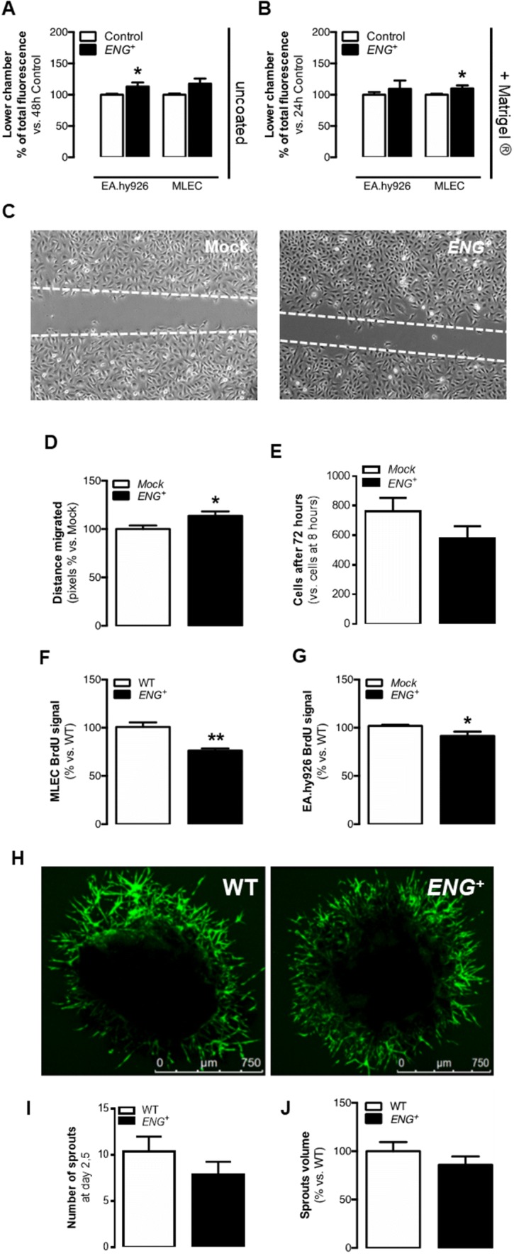
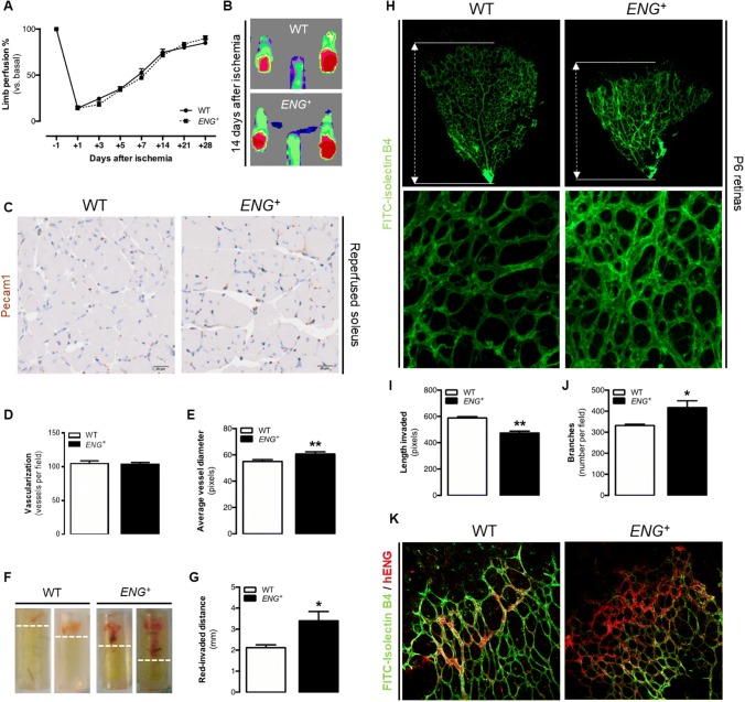
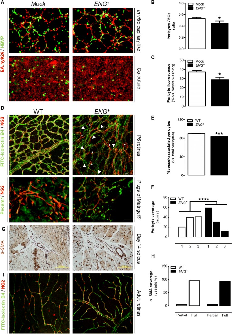
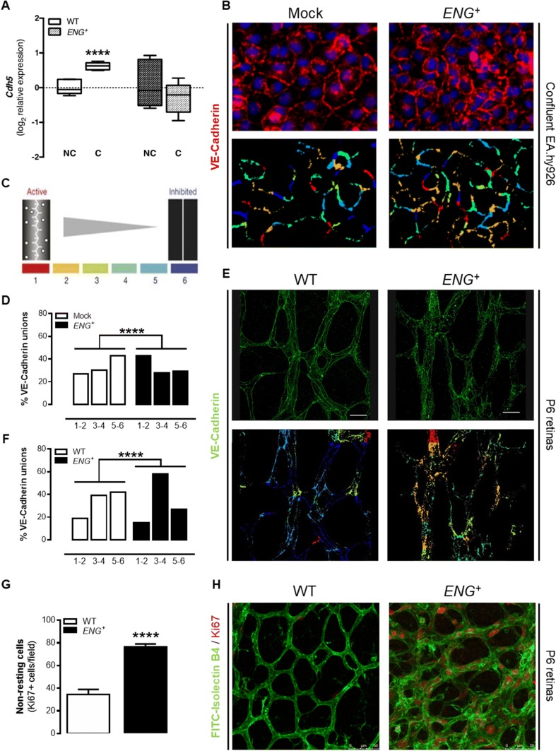
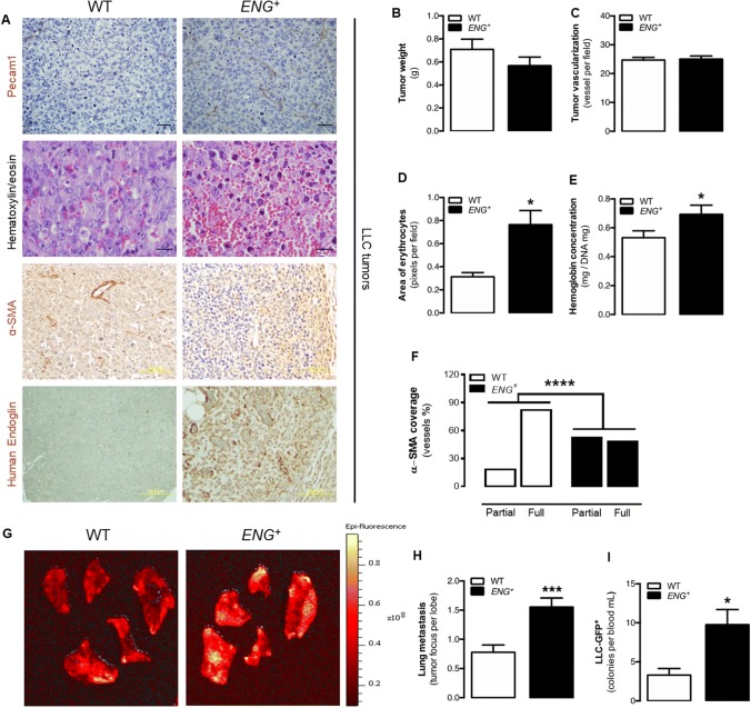
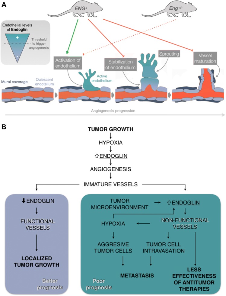
Endoglin (CD105) is an auxiliary receptor for members of the TFG-β superfamily. Whereas it has been demonstrated that the deficiency of endoglin leads to minor and defective angiogenesis, little is known about the effect of its increased expression, characteristic of several types of cancer. Angiogenesis is essential for tumor growth, so high levels of proangiogenic molecules, such as endoglin, are supposed to be related to greater tumor growth leading to a poor cancer prognosis. However, we demonstrate here that endoglin overexpression do not stimulate sprouting or vascularization in several in vitro and in vivo models. Instead, steady endoglin overexpression keep endothelial cells in an active phenotype that results in an impairment of the correct stabilization of the endothelium and the recruitment of mural cells. In a context of continuous enhanced angiogenesis, such as in tumors, endoglin overexpression gives rise to altered vessels with an incomplete mural coverage that permit the extravasation of blood. Moreover, these alterations allow the intravasation of tumor cells, the subsequent development of metastases and, thus, a worse cancer prognosis.

Keywords: Angiogenesis; CD105; Cancer; Endoglin; Metastasis.


Conflict of interest statement

The authors declare that they have no conflict of interest.

Figures

Fig. 1
Fig. 1
Permanent endoglin overexpression modifies EC physiology in vitro but does not alter sprouting. a Quantification of EC migration through the uncoated transwell with a VEGF gradient after 48 h for EA.hy926 and MLEC cells [n(Mock) = 3, n(ENG+) = 3; n(WT) = 3, n(ENG+) = 3; p (EA.hy926) = 0.0297, p (MLEC) = 0.0642]. b Quantification of EC migration through the Matrigel®-coated transwell with a VEGF gradient after 24 h for EA.hy926 and MLEC cells [n(Mock) = 3, n(ENG+) = 3; n(WT) = 3, n(ENG+) = 3; p (EA.hy926) = 0.4453, p (MLEC) = 0.0344]. c EA.hy926 scratch closure after 14.5 h in culture. d Quantification of the distance migrated by EA.hy926 cells though the scratch after 14.5 h [n(Mock) = 3, n(ENG+) = 3; p = 0.0250]. e Ratio of EA.hy926 cell counts after 72 h vs. after 8 h in culture [n(Mock) = 3, n(ENG+) = 3; p = 0.2030]. f BrdU incorporation after 24 h in MLECs [n(WT) = 3, n(ENG+) = 3; p = 0.0277]. g BrdU incorporation after 4 h in EA.hy926 cells [n(Mock) = 3, n(ENG+) = 3; p = 0.0272]. h FITC-lectin-labeled sprout growth from aortic rings isolated from WT and ENG+ mice. i Number of sprouts grown from aortic rings 2.5 days after the seedtime [n(WT) = 42, n(ENG+) = 42;p = 0.2920]. j Quantification of the volume occupied by sprouts from aortic rings [n(WT) = 25, n(ENG+) = 20; p = 0.2875]
Fig. 2
Fig. 2
Continuous endoglin overexpression impairs in vivo angiogenesis. a Ratio of ischemic to non-ischemic limb perfusion following femoral artery ligation as measured by laser Doppler flow analysis in mice, represented as the percentage of the basal value (before artery ligation) at 1, 3, 5, 7, 14, 21 and 28 days post-ischemia [n(WT) = 9, n(ENG+) = 9; p = 0.4989]. b Laser Doppler images showing mice hindlimb perfusion 14 days after ischemia. c Pecam1 immunostaining in the ischemic soleus muscle 14 days post-ischemia. d Quantification of the number of Pecam1-positive vessels in the ischemic soleus muscle [n(WT) = 4, n(ENG+) = 6; p = 0.8204]. e Average Pecam1-positive vessel diameter in the ischemic soleus muscle [n(WT) = 3, n(ENG+) = 3; p = 0.0098]. f DIVAA 9 days after implantation in mice, showing blood invasion. g Quantification of DIVAA red-invaded distance from the tube end 9 days after implantation [n(WT) = 16, n(ENG+) = 28; p = 0.0383]. h Upper panel: FITC-lectin staining of ECs in the retinal vasculature of p6 pups and representation of the plexus progression. Lower panel: Structure of the vessel plexus of the retina in P6 pups. i Quantification of plexus progression in the retinas of P6 pups [n(WT) = 7, n(ENG+) = 3; p < 0.0001]. j Quantification of the ramification of the retinas of P6 pups [n(WT) = 7, n(ENG+) = 3; p = 0.0227]. k Human endoglin (red) and FITC-lectin (green) staining in the angiogenic front of the retinal vasculature of P6 pups. The angiogenic front is oriented towards the upper left of the image in both cases
Fig. 3
Fig. 3
Continuous endoglin overexpression impairs pericyte recruitment in vitro and delays vessel maturation in vivo. a Upper panel: Pseudocapillary-like structures formed by EA.hy926 cells cocultured with HBVPs in Matrigel®. Lower panel: HBVP attachment to EA.hy926 monolayers in culture. b Quantification of the ratio between HBVPs and ECs in pseudocapillary-like structures [n(Mock) = 3, n(ENG+) = 3; p = 0.0433]. c Quantification of the HBVP fluorescent signal over EA.hy926 monolayers [n(Mock) = 3, n(ENG+) = 3; p = 0.0128]. d Upper panel: NG2 (red) and FITC-lectin (green) staining in the retinal vasculature of P6 pups, showing merged signals (yellow) in WT retinas and noncovered endothelium (arrowheads) and mural cells not bound to vessels (asterisk) in ENG+ retinas. Lower panel: NG2 (red) and CD31 (green) staining in plugs of Matrigel®. e Quantification of the ratio of pericytes that are bound to the endothelium with respect to the total number of pericytes in the retinal vasculature of P6 pups [n(WT) = 4, n(ENG+) = 6; p < 0.0001]. f Quantification of pericyte recovery in plugs of Matrigel® vessel, where 1 represents low coverage and 3 represents high coverage [n(WT) = 4, n(ENG+) = 3; p < 0.0001]. g α-SMA immunostaining in the ischemic soleus muscle 14 days post-ischemia. h Quantification of the ratio of vessels in ischemic soleus muscle that are partially or fully covered by α-SMA immunostaining [n(WT) = 3, n(ENG+) = 3; p = 1]. i NG2 (red) and FITC-lectin (green) staining in the retinal vasculature of adult mice
Fig. 4
Fig. 4
Continuous endoglin overexpression alters endothelium stability in vivo. a qPCR analysis of Cdh5 expression in confluent (C) and nonconfluent (NC) MLECs [n(WT) = 6, n(ENG+) = 6; p = 0.0237]. b Upper panel: VE-cadherin staining in confluent EA.hy926 ECs. Lower panel: pattern of VE-cadherin junctions in confluent EA.hy926 ECs, using the “patch algorithm” in MATLAB™. c Schematic illustration of patch classification numbers and colors used to quantify the pattern of VE-cadherin junctions. d Quantification of each type of junction in confluent EA.hy926 ECs [n(WT) = 10, n(ENG+) = 10; p < 0.0001]. e Upper panel: VE-cadherin staining in the retinal vasculature of P6 pups. Lower panel: Pattern of VE-cadherin junctions in the retinal vasculature of P6 pups, using the “patch algorithm” in MATLAB™. f Quantification of each type of junction in the retinal vasculature of P6 pups [n(WT) = 13, n(ENG+) = 12; p < 0.0001]. g Quantification of non-resting (Ki67+) cells in the central area of the retinal vasculature of P6 pups [n(WT) = 4, n(ENG+) = 5; p < 0.0001]. h Ki67 (red) and FITC-lectin (green) staining in the central area of the retinal vasculature of P6 pups
Fig. 5
Fig. 5
Permanent endoglin overexpression does not increase growth and vascularization in tumors but prevent vessel maturation and facilitates tumor cell metastasis. a First panel: Pecam1 immunostaining in the tumor tissue showing tumor vessels. Second panel: Hematoxylin–eosin staining of tumor tissue showing characteristic blood extravasation, blood lakes and edema. Third panel: α-SMA immunostaining in the tumor tissue showing vessel maturation. Fourth panel: human endoglin immunostaining in the tumor tissue. b Weight of tumors implanted in mice after 10 days [n(WT) = 35, n(ENG+) = 26; p = 0.3329]. c Quantification of the number of Pecam1-positive vessels in tumor tissue [n(WT) = 4, n(ENG+) = 6; p = 0.8758]. d Quantification of the area filled by erythrocytes in the tumor revealed by hematoxylin–eosin staining [n(WT) = 4, n(ENG+) = 6; p = 0.0484]. e Quantification of hemoglobin concentration in the tumor tissue [n(WT) = 30, n(ENG+) = 21; p = 0.0427]. f Quantification of the ratio of vessels in the tumor tissue that is partially or totally covered by α-SMA immunostained tissue [n(WT) = 8, n(ENG+) = 13; p < 0.0001]. g Epi-fluorescence images of mouse lung lobe metastases of LLC-GFP+ tumor cells. h Quantification of tumor metastatic foci per mouse lung lobe [n(WT) = 8, n(ENG+) = 9; p = 0.0004]. i Quantification of circulating LLC-GFP+ tumor cells in mice [n(WT) = 8, n(ENG+) = 9; p = 0.0107]
Fig. 6
Fig. 6
Models of endothelial endoglin regulation during physiological and tumor angiogenesis. a Endoglin levels in the endothelium need to be carefully regulated for the correct activation (upregulation) and subsequent stabilization (downregulation to basal levels) of the newly formed vessel. Consistent endoglin overexpression (ENG+) promotes endothelium activation but impedes vessel stabilization and maturation. A lack of endoglin (Eng+/−) may prevent proper activation of the endothelium and triggering of angiogenesis. b Tumoral continuous endoglin overexpression could difficult vessel normalization which reduces the effectiveness of anti-tumor therapies, enhances the aggressiveness of tumor cells by further increasing hypoxia and promotes the appearance of metastases. On the contrary, downregulation of endoglin after tumor angiogenesis promotes vessel stabilization and thus tumor growth control, which could explain the positive effect of anti-endoglin therapies in cancer control

Similar articles

Cited by

References

    1. Manetti M, Guiducci S, Ibba-Manneschi L, Matucci-Cerinic M. Mechanisms in the loss of capillaries in systemic sclerosis: angiogenesis versus vasculogenesis. J Cell Mol Med. 2010;14:1241–1254. doi: 10.1111/j.1582-4934.2010.01027.x. - DOI - PMC - PubMed
    1. Núñez-Gómez E, Pericacho M, Ollauri-Ibáñez C, et al. The role of endoglin in post-ischemic revascularization. Angiogenesis. 2017;20:1–24. doi: 10.1007/s10456-016-9535-4. - DOI - PubMed
    1. Eilken HM, Adams RH. Dynamics of endothelial cell behavior in sprouting angiogenesis. Curr Opin Cell Biol. 2010;22:617–625. doi: 10.1016/j.ceb.2010.08.010. - DOI - PubMed
    1. Geudens I, Gerhardt H. Coordinating cell behaviour during blood vessel formation. Development. 2011;138:4569–4583. doi: 10.1242/dev.062323. - DOI - PubMed
    1. Bernabeu C, Lopez-Novoa JM, Quintanilla M. The emerging role of TGF-β superfamily coreceptors in cancer. Biochim Biophys Acta. 2009;1792:954–973. doi: 10.1016/j.bbadis.2009.07.003. - DOI - PubMed

Publication types