

A novel procedure of endobronchial ultrasound-guided transbronchial needle aspiration for pulmonary parenchymal lesions: The ZUTAM technique
- PMID: 31898623
- PMCID: PMC6961091
- DOI: 10.4103/lungindia.lungindia_187_19
A novel procedure of endobronchial ultrasound-guided transbronchial needle aspiration for pulmonary parenchymal lesions: The ZUTAM technique
Abstract
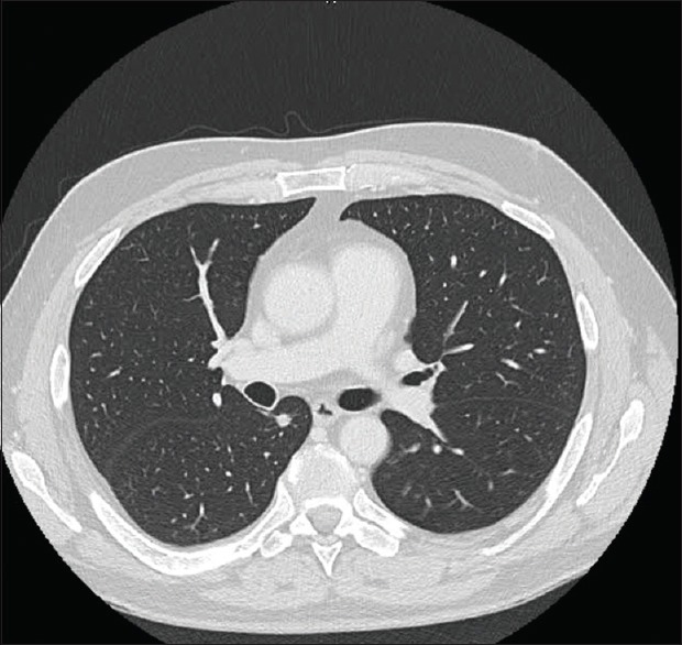
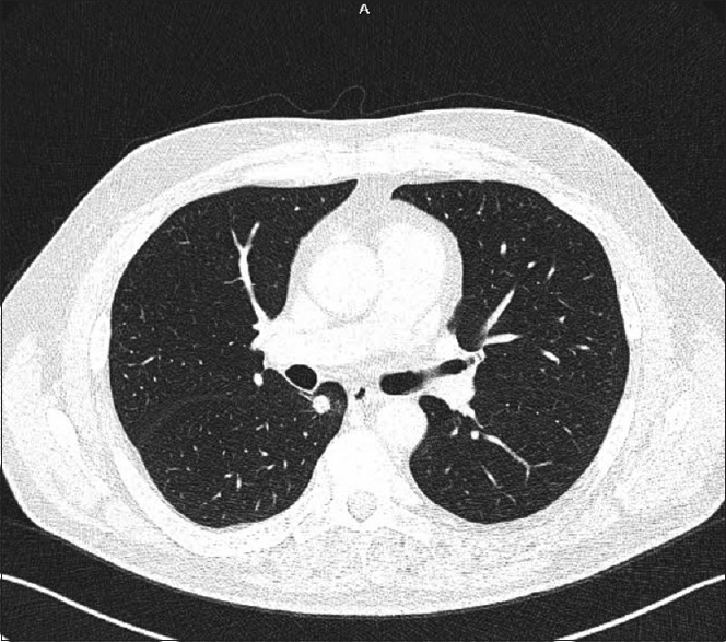
Convex probe-endobronchial ultrasound (CP-EBUS) has been proven to be safe and accurate for identifying malignancy and granulomatous disease affecting the mediastinum and hilum. CP-EBUS can be used for intraparenchymal lesions also and has been shown to be efficacious. A subset of lesions particularly suited for CP-EBUS are those completely surrounded by lung parenchyma, centrally located, and typically close to but without an airway leading directly to them. We report a case of transbronchial needle aspiration (TBNA) done from a nodule of size 11 mm in the superior segment of the right lower lobe. EBUS-TBNA was done from this lesion, which was 5 mm away from the bronchus in the lung parenchyma with intervening normal lung tissue in between. TBNA was performed by compressing the abutting normal lung tissue, thus causing compression collapse of the intervening normal lung. We labeled this Zealous Unique Trans Arterial Maneuver as the "ZUTAM" technique.
Keywords: Bronchoscopy; Zealous Unique Trans Arterial Maneuver; cancer (lung); endobronchial ultrasound-guided transbronchial needle aspiration; parenchymal lesions.
Conflict of interest statement
None
Figures


References
-
- Chao TY, Chien MT, Lie CH, Chung YH, Wang JL, Lin MC. Endobronchial ultrasonography-guided transbronchial needle aspiration increases the diagnostic yield of peripheral pulmonary lesions: A randomized trial. Chest. 2009;136:229–36. - PubMed
-
- Warren WA, Sobieszczyk MJ, Sarkar Sy, Krimsky WS. Endobronchial ultrasound bronchoscopy: Current uses, innovations and future directions. AME Med J. 2018;3:70.
-
- Du Rand IA, Barber PV, Goldring J, Lewis RA, Mandal S, Munavvar M, et al. British Thoracic Society guideline for advanced diagnostic and therapeutic flexible bronchoscopy in adults. Thorax. 2011;66(Suppl 3):iii1–21. - PubMed
-
- Dhooria S, Sehgal IS, Aggarwal AN, Agarwal R. Convex-probe endobronchial ultrasound: A Decade of progress. Indian J Chest Dis Allied Sci. 2016;58:21–35. - PubMed
-
- Paone G, Nicastri E, Lucantoni G, Dello Iacono R, Battistoni P, D'Angeli AL, et al. Endobronchial ultrasound-driven biopsy in the diagnosis of peripheral lung lesions. Chest. 2005;128:3551–7. - PubMed
Publication types
LinkOut - more resources
Full Text Sources
Miscellaneous
