. 2020 May;72(5):946-959.
doi: 10.1016/j.jhep.2019.12.016. Epub 2019 Dec 31.

IL-17 signaling in steatotic hepatocytes and macrophages promotes hepatocellular carcinoma in alcohol-related liver disease

Affiliations

IL-17 signaling in steatotic hepatocytes and macrophages promotes hepatocellular carcinoma in alcohol-related liver disease

Hsiao-Yen Ma et al. J Hepatol. 2020 May.

Abstract

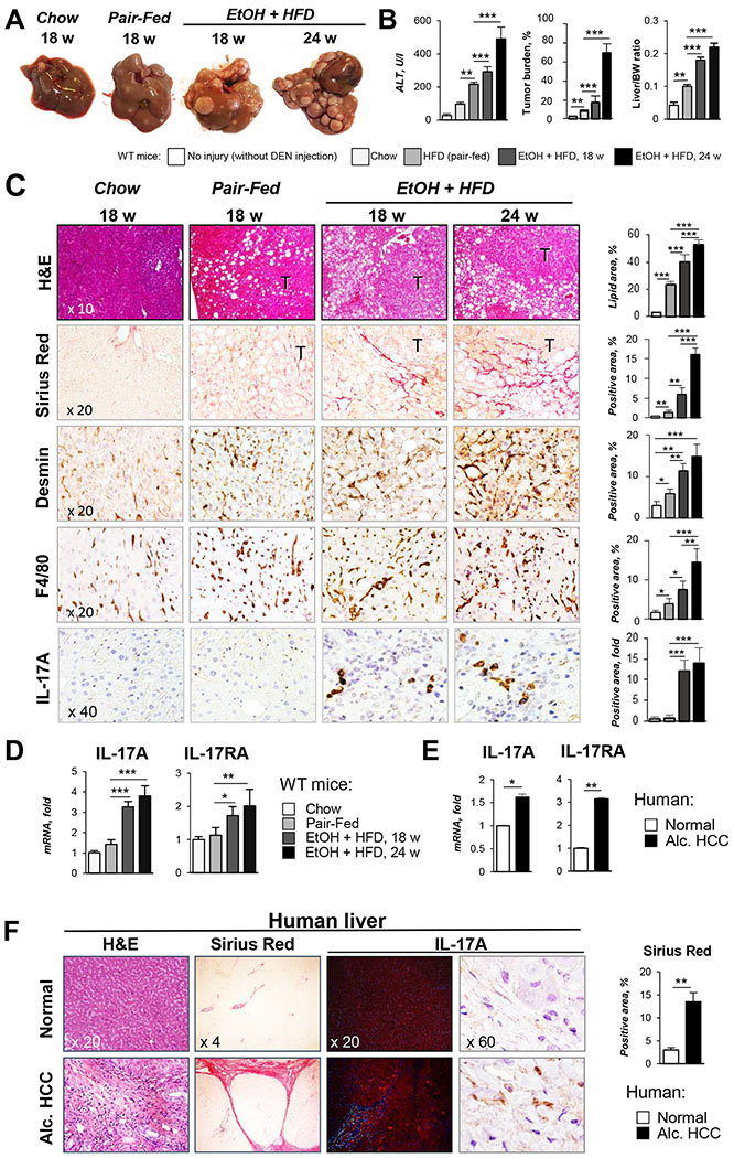
Background & aims: Chronic alcohol consumption is a leading risk factor for the development of hepatocellular carcinoma (HCC), which is associated with a marked increase in hepatic expression of pro-inflammatory IL-17A and its receptor IL-17RA.

Methods: Genetic deletion and pharmacological blocking were used to characterize the role of IL-17A/IL-17RA signaling in the pathogenesis of HCC in mouse models and human specimens.

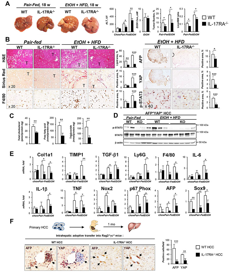
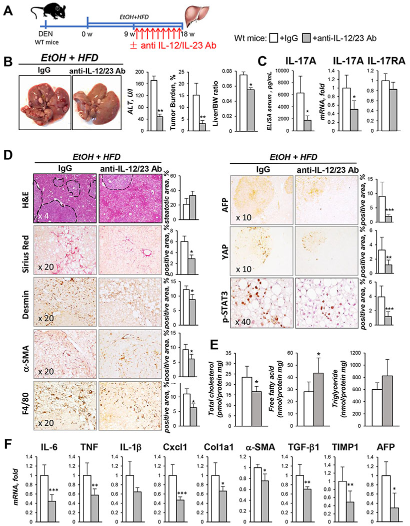
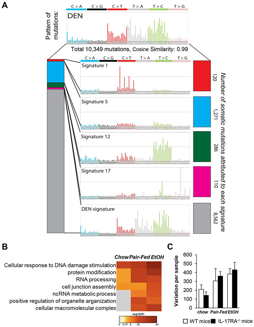
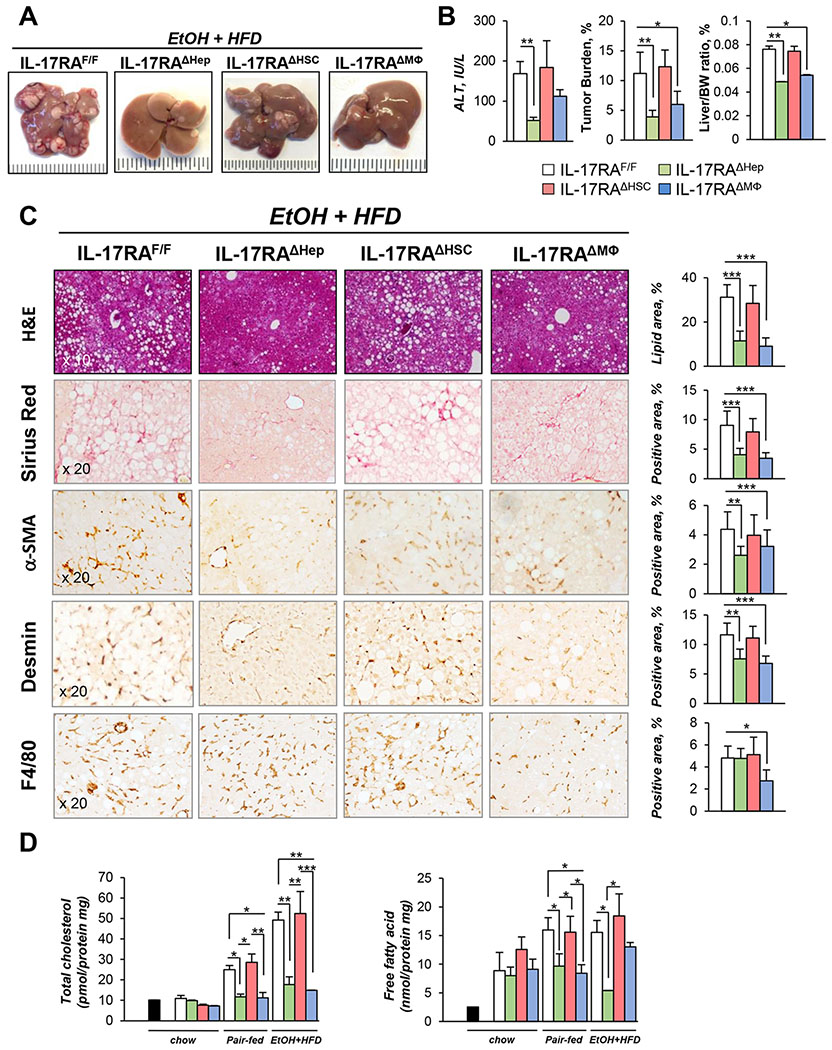
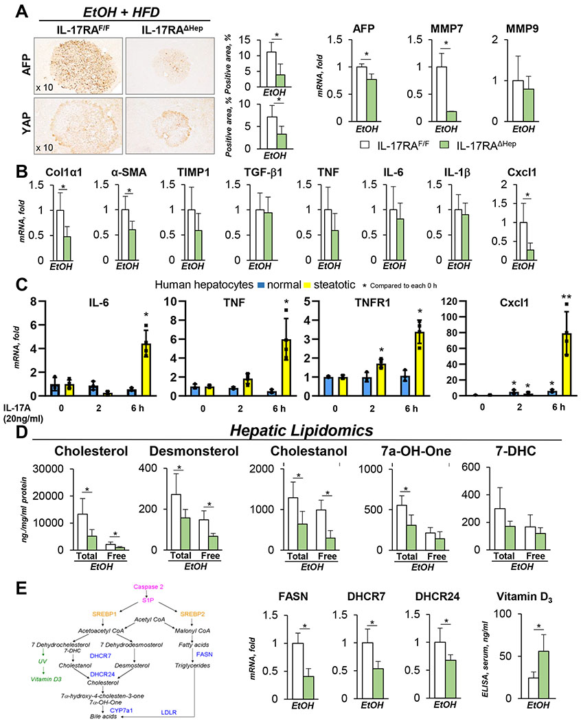
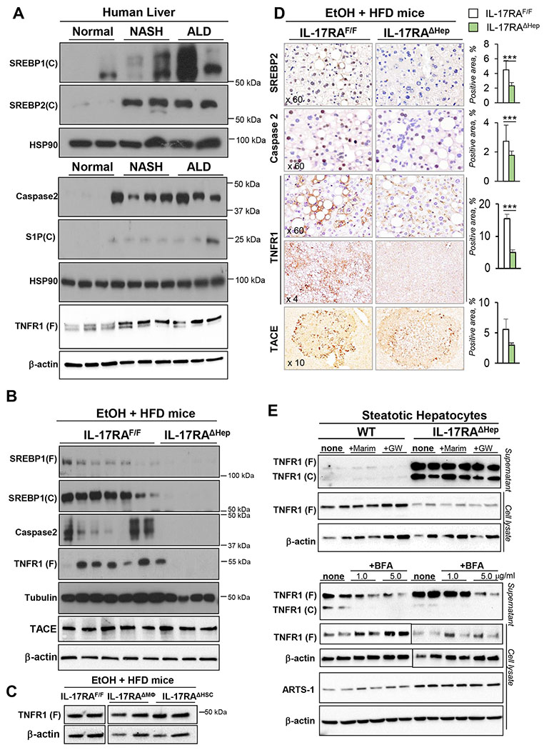
Results: We demonstrate that the global deletion of the Il-17ra gene suppressed HCC in alcohol-fed diethylnitrosamine-challenged Il-17ra-/- and major urinary protein-urokinase-type plasminogen activator/Il-17ra-/- mice compared with wild-type mice. When the cell-specific role of IL-17RA signaling was examined, the development of HCC was decreased in both alcohol-fed Il-17raΔMΦ and Il-17raΔHep mice devoid of IL-17RA in myeloid cells and hepatocytes, but not in Il-17raΔHSC mice (deficient in IL-17RA in hepatic stellate cells). Deletion of Il-17ra in myeloid cells ameliorated tumorigenesis via suppression of pro-tumorigenic/inflammatory and pro-fibrogenic responses in alcohol-fed Il-17raΔMΦ mice. Remarkably, despite a normal inflammatory response, alcohol-fed Il-17raΔHep mice developed the fewest tumors (compared with Il-17raΔMΦ mice), with reduced steatosis and fibrosis. Steatotic IL-17RA-deficient hepatocytes downregulated the expression of Cxcl1 and other chemokines, exhibited a striking defect in tumor necrosis factor (TNF)/TNF receptor 1-dependent caspase-2-SREBP1/2-DHCR7-mediated cholesterol synthesis, and upregulated the production of antioxidant vitamin D3. The pharmacological blocking of IL-17A/Th-17 cells using anti-IL-12/IL-23 antibodies suppressed the progression of HCC (by 70%) in alcohol-fed mice, indicating that targeting IL-17 signaling might provide novel strategies for the treatment of alcohol-induced HCC.

Conclusions: Overall, IL-17A is a tumor-promoting cytokine, which critically regulates alcohol-induced hepatic steatosis, inflammation, fibrosis, and HCC.

Lay summary: IL-17A is a tumor-promoting cytokine, which critically regulates inflammatory responses in macrophages (Kupffer cells and bone-marrow-derived monocytes) and cholesterol synthesis in steatotic hepatocytes in an experimental model of alcohol-induced HCC. Therefore, IL-17A may be a potential therapeutic target for patients with alcohol-induced HCC.

Keywords: ALD; Alcoholic liver disease; Cholesterol synthesis; Fibrosis; HCC; Hepatocellular carcinoma; IL-17 signaling; Inflammation; Mutational signatures.


Conflict of interest statement

Conflict of interest The authors declare they have no conflicts of interest. Please refer to the accompanying ICMJE disclosure forms for further details.

Figures

Figure 1.
Figure 1.. IL-17 signaling is activated in experimental model of EtOH/DEN-induced HCC in mice and patients with ALD.
(A) Gross liver images from DEN-challenged WT mice: chow-(n=3, 9 months), and pair-fed (HFD n=4) for 18 weeks, and EtOH-fed (EtOH+HFD) for 18 weeks (n= 8) and 24 weeks (n= 7). (B) Serum ALT, tumor burden, and liver/body weight (BW) ratio were calculated. (C) Livers were stained with H&E, Sirius Red, anti-Desmin, anti-F4/80, and anti-IL-17A Abs. Positive area was calculated as percent (micrographs were taken using x10, x20 and x40 objectives). (D) Hepatic expression of IL-17A and IL-17RA mRNA was analyzed by qRT-PCR (E-F) Livers from HCC patients with ALD (Alc. HCC, n=3, stage 3-4), or patients with no fibrosis (Normal, n=3) were (E) analyzed for expression of IL-17A and IL-17RA mRNA or (F) stained with H&E, Sirius Red, and anti-IL-17A Abs (x4, x20, and x60 objectives). Positive Sirius Red staining was calculated as percent. Statistical analysis was performed using 2-tailed Student’s t test or ANOVA for comparisons between 2 groups or more than 2 groups. Data are means ± SD, *p<0.05, **p<0.01, ***p<0.001.
Figure 2.
Figure 2.. Global deletion of IL-17RA protects EtOH+HFD-fed DEN-challenged IL-17RA−/− mice from HCC.
(A). Gross liver images from DEN-challenged IL-17RA−/− and WT mice: chow-(n=4/group, 9 months, Figure S2A), pair-fed (HFD, n≥4/group) and EtOH-fed (EtOH+HFD, n≥8/group) for 18 weeks. Serum ALT, EtOH level, tumor burden, and liver/body weight (BW) ratio are shown. (B) Livers were stained with H&E, Sirius Red, and anti-F4/80, anti-AFP, anti-YAP, anti-phospho-STAT3 Abs, positive area was calculated as percent (x10, x20, and x40 objectives). (C) Cholesterol, fatty acids, and triglycerides were measured in livers of EtOH-fed IL-17RA−/− and wt mice. (D) Expression of phospho-STAT3 in tumors was assessed by Western blot. (E) Hepatic expression of fibrogenic, inflammatory genes and HCC markers was analyzed using qRT-PCR. (F) Primary WT or IL-17RA-deficient AFP+YAP+ HCC from EtOH-fed mice were injected (1 x 10⁶ cells) into the livers of Rag2−/−γc−/− pups (3 days old, n≥6/group). Mice were sacrificed at 1 month of age, livers were stained for AFP and YAP, representative macrographs are taken x60 objective; the number of engrafted AFP+ and YAP+ cells/field was calculated. Statistical analysis was performed using 2-tailed Student’s t test. Data are means ± SD, *p<0.05, **p<0.01, ***p<0.001. (see Figures S1, S3).
Figure 3.
Figure 3.. Therapeutic blocking of IL-12/IL-23/IL-17 ameliorates development of HCC in EtOH+HFD-fed DEN-challenged mice.
(A) After 9 weeks of EtOH+HFD-feeding, DEN-injured WT male mice were administered with anti-IL-12/IL-23 Ab (40mg/kg, once a week, for 9 weeks) or IgG (n≥8/group). (B) Gross liver images from EtOH+HFD-fed DEN-injured wt mice ± anti-IL-12/IL-23 Ab. Serum ALT, tumor burden, liver/body weight (BW) ratio are shown. (C) Serum and hepatic levels of IL-17A and IL-17RA were measured by ELISA and qRT-PCR. (D) Livers were stained with H&E, Sirius Red, anti-Desmin, anti-αSMA, and anti-F4/80, anti-AFP, anti-YAP, anti-phospho-STAT3 Abs, positive area was calculated as percent (x10, x20, and x40 objectives). (E) Cholesterol, fatty acids, and triglycerides were measured in livers of EtOH-fed wt mice ± anti-IL-12/IL-23 Ab. (F) Expression of inflammatory and fibrogenic genes was analyzed using qRT-PCR. Statistical analysis was performed using 2-tailed Student’s t test. Data are means ± SD, *p<0.05, **p<0.01, ***p<0.001.
Figure 4.
Figure 4.. Mutational landscapes of HCC from EtOH+HFD/DEN-injured WT and IL-17RA−/− mice are similar to that in human HCC.
(A-C) Paired-end RNA-Seq analysis was used to identify coding somatic gene mutations and gene expression profile of AFP+YAP+ HCC (4 mm, 1 tumor/mouse) and non-tumor tissue from DEN-challenged WT and IL-17RA−/− mice: chow- (n=3/group), pair-fed (n=3/group), and EtOH-fed (n=8/group). The data are normalized vs recurrent somatic mutations identified in livers of non-injured C57BL/6 mice. (A) HCC mutational signature deciphered from EtOH+HFD-fed WT and IL-17RA−/− mice, and COSMIC signatures 1, 5, 12, and 17, characteristic for HCC patients with NASH and ALD, and are displayed according to the 96 substitution classification defined by the substitution class and the sequence context immediately 3′ and 5′ to the mutated base, (see Figure S4). (B) Gene set enrichment analysis (GSEA) identified the gene/pathway-specific mutations enriched in WT and IL-17RA-deficient HCC. (C) Mutational frequency in HCC from WT and IL-17RA−/− mice is shown. Enrichment of variant counts per sample was observed between feeding groups, but not between different genotypes of mice. Associations were assessed using Chi-square tests for categorical variables and ANOVA for quantitative features. The strength of association among gene mutation events was modeled using a binomial logistic regression. Statistical analysis was performed using 2-tailed Student’s t test. Data are means ± SD, *p<0.05, **p<0.01, ***p<0.001. (see Figure S5)
Figure 5.
Figure 5.. IL-17 signaling in myeloid cells and steatotic hepatocytes is critical for HCC growth in EtOH+HFD-fed DEN-challenged mice.
(A) Gross liver images from EtOH+HFD-fed (18 weeks) DEN-injured IL-17RAΔMΦ mice (n=4, IL-17RAF/F mice x LyzMCre mice), IL-17RAΔHSC mice (n=5, IL-17RAF/F mice x LratCre mice), IL-17RAΔHep mice (n=7, generated by crossing IL-17RAF/F mice x AlbCre mice), and WT IL-17RAF/Flittermates (n=7). The efficiency of Cre-LoxP recombination for each Cre-mouse line has been tested. Mice were chow-fed (n=3/group, 9 months), pair-fed (HFD, n≥4/group) and EtOH-fed (EtOH+HFD) for 18 weeks. (B) Serum ALT, tumor burden, liver body weight (BW) ratio is shown. (C) Livers were stained with H&E, Sirius Red, anti-αSMA, anti-Desmin, and anti-F4/80 Abs, positive area was calculated as percent (x20 objectives). (D) Cholesterol, fatty acids were measured in livers of uninjured (black bar); chow-, pair-, EtOH+HFD-fed mice. Statistical analysis was performed using 2-tailed Student’s t test. Data are means ± SD, *p<0.05, **p<0.01, ***p<0.001. (see Figure S2C–E, S6).
Figure 6.
Figure 6.. EtOH+HFD-fed DEN-challenged IL-17RAΔHep mice exhibit a defect in cholesterol synthesis.
(A) Expression of HCC markers was analyzed in tumors of EtOH+HFD-fed DEN-challenged IL-17RAΔHep mice using immunostaining (x10 objective) and qRT-PCR. (B) Expression of inflammatory and fibrogenic genes was assessed using qRT-PCR (see Figure S6C). (C) Human primary steatotic (from patients with ALD, n=4) and normal hepatocytes (from patients with no liver injury, n=3) were in vitro stimulated with IL-17A (20 ng/ml, or vehicle). Gene expression was analyzed using qRT-PCR, the data are average of four independent experiments (see Figure S7). (D) Lipidomic analysis of free and bound lipids in the livers of EtOH+HFD-fed IL-17RAF/Fand IL-17RAΔHep mice. (E) Schematic representation of cholesterol and fatty acid synthesis pathways. mRNA expression of FASN, DHCR7 and DHCR24 was assessed using qRT-PCR (and RNA-Seq, see Figure S8). Serum levels of Vitamin D3 were measured using ELISA. Statistical analysis was performed using 2-tailed Student’s t test. Data are means ± SD, *p<0.05, **p<0.01, ***p<0.001.
Figure 7.
Figure 7.. EtOH+HFD-fed DEN-challenged IL-17RAΔHep mice exhibit a defect in Caspase 2-dependent activation of SREBP1/2.
(A) Expression of cleaved (C) or full length (F) SREBP-1, SREBP-2, Caspase2, S1P, and TNFR1 was analyzed by Western blotting in livers from patients with no liver injury (Normal), NASH, or ALD. (B) Expression of cleaved (C) or full length (F) SREBP-1, SREBP-2, Caspase2, TNFR1, and TACE in livers of EtOH+HFD-fed IL-17RAF/F mice and IL-17RAΔHep littermates, representative images of ≥3 independent experiments are shown. (C) Expression of full length (F) TNFR1 was analyzed by Western blot of livers of EtOH+HFD-fed IL-17RAF/F mice, IL-17RAΔMΦ and IL-17RAΔHSC littermates, representative images of ≥3 independent experiments are shown. (D) Expression of SREBP-2, Caspase2, TNFR1 (Ab1: Santa Cruz, upper panel; Ab2, Abcam, lower panel), and TACE was analyzed in livers of EtOH+HFD-fed IL-17RAF/F mice and IL-17RAΔHep mice using immunohistochemistry (x60, x4, x10 objectives), positive area was calculated as a percent. (E) Primary WT and IL-17RA-deficient steatotic hepatocytes from EtOH+HFD-fed IL-17RAF/F mice and IL-17RAΔHep mice were cultured ± Marimastat (10 μM), GW280264X (2 μM), or ± Brefeldin A (BFA, 1 or 5 μg/ml) for 6 h. Expression of full length (F) or cleaved (C) TNFR1 and ARTS-1 was analyzed using Western blotting of cell lysates and supernatants from WT mice and IL-17RAΔHep. BFA inhibited TNFR1 exocytosis in WT and IL-17RA-deficient hepatocytes in a dose-dependent manner. Statistical analysis was performed using 2-tailed Student’s t test. Data are means ± SD, *p<0.05, **p<0.01, ***p<0.001.

Comment in

References

    1. Morgan RL, Baack B, Smith BD, et al. Eradication of hepatitis C virus infection and the development of hepatocellular carcinoma: a meta-analysis of observational studies. Ann Intern Med 2013;158:329–37. - PubMed
    1. Gao B, Bataller R. Alcoholic liver disease: pathogenesis and new therapeutic targets. Gastroenterology 2011;141:1572–85. - PMC - PubMed
    1. Brandl K, Hartmann P, Jih LJ, et al. Dysregulation of serum bile acids and FGF19 in alcoholic hepatitis. J Hepatol 2018;69:396–405. - PMC - PubMed
    1. Goossens N, Hoshida Y. Is Hepatocellular Cancer the Same Disease in Alcoholic and Nonalcoholic Fatty Liver Diseases? Gastroenterology 2016;150:1710–7. - PMC - PubMed
    1. Alexandrov LB. Understanding the origins of human cancer. Science 2015;350:1175. - PubMed

Publication types

MeSH terms