Review
. 2020 Jul:77:100827.
doi: 10.1016/j.preteyeres.2019.100827. Epub 2019 Dec 30.

Progress in treating inherited retinal diseases: Early subretinal gene therapy clinical trials and candidates for future initiatives

Affiliations
Review

Progress in treating inherited retinal diseases: Early subretinal gene therapy clinical trials and candidates for future initiatives

Alexandra V Garafalo et al. Prog Retin Eye Res. 2020 Jul.

Abstract

Due to improved phenotyping and genetic characterization, the field of 'incurable' and 'blinding' inherited retinal diseases (IRDs) has moved substantially forward. Decades of ascertainment of IRD patient data from Philadelphia and Toronto centers illustrate the progress from Mendelian genetic types to molecular diagnoses. Molecular genetics have been used not only to clarify diagnoses and to direct counseling but also to enable the first clinical trials of gene-based treatment in these diseases. An overview of the recent reports of gene augmentation clinical trials by subretinal injections is used to reflect on the reasons why there has been limited success in this early venture into therapy. These first-in human experiences have taught that there is a need for advancing the techniques of delivery of the gene products - not only for refining further subretinal trials, but also for evaluating intravitreal delivery. Candidate IRDs for intravitreal gene delivery are then suggested to illustrate some of the disorders that may be amenable to improvement of remaining central vision with the least photoreceptor trauma. A more detailed understanding of the human IRDs to be considered for therapy and the calculated potential for efficacy should be among the routine prerequisites for initiating a clinical trial.

Keywords: ABCA4; BCM; CNGA3; CNGB3; Gene therapy; Genetic retinal degenerations; Leber congenital amaurosis; MERTK; MYO7A; Molecular mechanisms; NPHP5; OPN1LW; OPN1MW; PDE6B; REP1; RLBP1; RPE65; RPGR; RPGRIP1; Retinitis pigmentosa; TULP1.


Figures

Fig 1.
Fig 1.
Mendelian patterns of inheritance, resolution of genotype over time and comparison of genotypes in IRD centers. (A-C) Data from the Philadelphia IRD center. (A) Pie chart showing the Mendelian patterns of inheritance in clinically-diagnosed patients seen prior to 1990. (B) Cumulative number of unique genes identified as causal over time. (C) The proportion of patients with unresolved and resolved genotypes in the current patient population and the comparison of Mendelian patterns of inheritance for patients with resolved and unresolved molecular diagnoses. (D) Comparison of the genes identified in the Philadelphia and Toronto cohorts that each cause disease in ≥2% of patients. Common top genes are given in the center. Blue, Philadelphia cohort; Orange, Toronto cohort. (E) Comparison of the top 5 disease-causing genes in the Philadelphia and Toronto cohorts along with percentages of the cohorts that are positive for each disease-causing gene. Blue (solid and hashed), Philadelphia; Orange (solid and hashed), Toronto. Solid bars represent genes that are in the top 5 for respective center; hashed bars indicate genes that are not in the top 5 for that center but still contribute to the overall cohort.
Fig 2.
Fig 2.
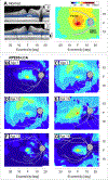
Photoreceptor layer thickness topography in young patients with RPE65-LCA (A). Left. OCT cross-sectional images through the horizontal (upper scan) and vertical (lower scan) meridians of a normal subject (icons at upper right of scans are the location of the image on a schematic fundus; ONL, highlighted in blue). Right. Normal ONL thickness topography with color scale above. Hatched area, location of the optic nerve head. (B-G) ONL thickness topography in 6 young patients with RPE65-LCA, illustrating that there can be abnormalities at these ages and differences between patients despite similar ages. All eyes are depicted as right eyes. F, fovea. Modified from Jacobson et al., 2008a, copyright by the Association for Research in Vision and Ophthalmology.
Fig 3.
Fig 3.
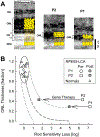
Early effects on rod-mediated vision of gene augmentation therapy in patients with RPE65-LCA. (A) OCT cross-sectional images (grayscale) from a normal subject and two patients (P2, P1) with RPE65-LCA. The sections are taken from ~5 mm superior retina in the normal subject and P2; section in P1 is from ~5 mm inferior retina. Schematics of ONL and RPE layers are overlaid (in yellow) on the OCT and illustrate the interpretation of reduced outer retinal structure in patients compared to normal. (B) Relationship of ONL thickness and rod-mediated sensitivity loss in normal subjects (triangles) and the two patients before therapy (unfilled large symbols) and after (up to 90 days; filled large symbols) gene therapy. The effect of the therapy led to rod sensitivity increase of ~5 log units (P2) and ~2 log units (P1). Ellipse, normal variability; broken lines, model of the expected relationship between structure and function based on quantum catch. Modified from Cideciyan et al., 2008, copyright by the National Academy of Sciences.
Fig 4.
Fig 4.
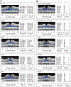
Foveal ONL thickness and central visual function, as measured with best-corrected visual acuity (VA, represented as decimal). (A) OCT scans of a normal subject (upper panel) and patients with IRDs that show normal or close to normal ONL thickness and minimally reduced or even normal VA. (B) Patients with IRD that show foveal ONL thickness comparable to ones in panel A, but severely impaired visual function. Representative OCT scans are along the vertical meridian from inferior retina (I) to superior (S) through the fovea. ONL is highlighted in blue. Foveal ONL thickness is depicted by white double arrow in normal scan. Dashed line in ONL thickness bar graph is lower limit of normal (n= 11; ages 26–62). Dashed line in VA bar graph is normal VA (20/20 or 1.0 in decimal representation).
Fig 5.
Fig 5.
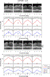
Predicted and measured sensitivities for CEP290-LCA patients with severe loss of visual acuity (VA) and fixation. The upper row shows OCT scans along the horizontal meridian across the fovea; VAs are given above the scans (LP=light perception, BLP=bare light perception, NLP=no light perception). Plots under scans are measured FST sensitivities with red and blue full-field stimuli in the dark-adapted state (symbols) and artificial intelligence predictions of localized retinal function based on residual retinal structure (lines). Gray regions depict normal cone-mediated sensitivity in dark-adapted conditions. Modified from Sumaroka et al., 2019, copyright by the Association for Research in Vision and Ophthalmology.

References

    1. Acland GM, Aguirre GD, Ray J, Zhang Q, Aleman TS, Cideciyan AV, et al. Gene therapy restores vision in a canine model of childhood blindness. Nat Genet. 2001;28(1):92–95. - PubMed
    1. Acland GM, Aguirre GD, Bennett J, Aleman TS, Cideciyan AV, Bennicelli J, et al. Long-term restoration of rod and cone vision by single dose rAAV-mediated gene transfer to the retina in a canine model of childhood blindness. Mol. Ther 2005;12(6):1072–1082. - PMC - PubMed
    1. Aguirre G Retinal degenerations in the dog. I. Rod dysplasia. Exp Eye Res. 1978;26(3):233–253. - PubMed
    1. Aleman TS, Han G, Serrano LW, Fuerst NM, Charlson ES, Pearson DJ, et al. Natural history of the central structural abnormalities in choroideremia: A prospective cross-sectional study. Ophthalmology. 2017;124(3):359–373. - PMC - PubMed
    1. Aleman TS, Huckfield RM, Serrano L, Vergillio G, Pearson DJ, Uyhazi KE, et al. AAV2-hCHM subretinal delivery to the macular in choroideremia: 2 year results of an ongoing phase I/II gene therapy trial. Invest Ophthalmol Vis Sci. 2019;60(9):5713.

Publication types

MeSH terms