. 2020 Jan 3;11(1):3.
doi: 10.1186/s13287-019-1508-2.

Small extracellular vesicles derived from embryonic stem cells restore ovarian function of premature ovarian failure through PI3K/AKT signaling pathway

Affiliations

Small extracellular vesicles derived from embryonic stem cells restore ovarian function of premature ovarian failure through PI3K/AKT signaling pathway

Mengyu Liu et al. Stem Cell Res Ther. .

Erratum in

Abstract

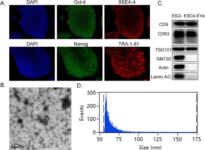
Background: Premature ovarian failure (POF) has a great impact on reproductive endocrine function in females, and it is an important cause of infertility. Previous studies have demonstrated that small extracellular vesicles (sEVs) derived from stem cells play an important role in tissue regeneration. This study aimed to investigate the therapeutic effect of sEVs derived from embryonic stem cells (ESCs-sEVs) on damaged ovaries and explore the underlying molecular mechanisms.

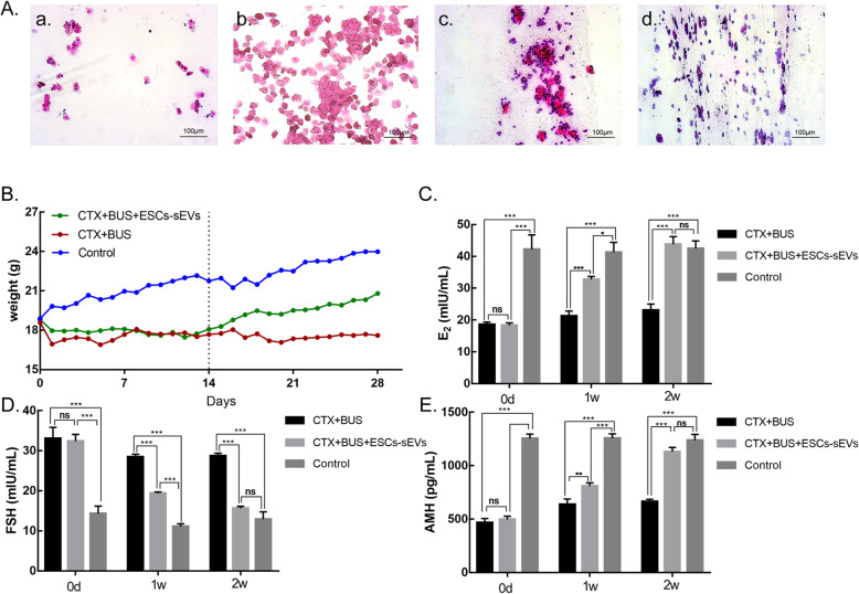
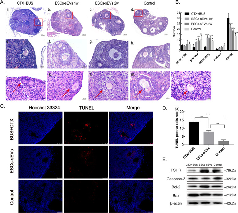
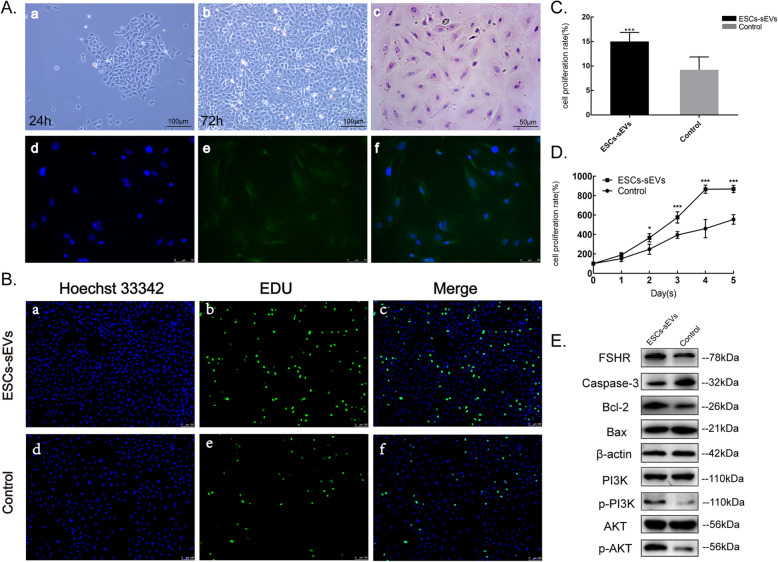
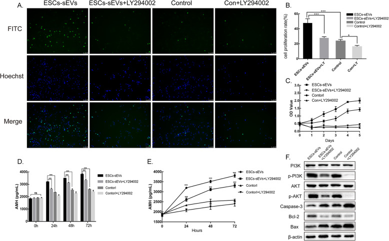
Methods: Mice POF models were established by injecting mice with cyclophosphamide and busulfan. Then, ESCs-sEVs were intravenously transplanted into POF mice. The plasma of mice was harvested at 1 and 2 weeks after treatment to analyze the levels of anti-Mullerian hormone (AMH), estradiol (E2), and follicle stimulating hormone (FSH) by ELISA. The morphology of ovaries and follicles was observed by H&E staining, and apoptosis of granulosa cells was detected by TUNEL. In vitro, EdU and CCK-8 tests were used to evaluate the proliferation of cultured granulosa cells stimulated by ESCs-sEVs. Western blotting was used to determine the expression of PI3K/AKT and apoptotic-related proteins.

Results: After transplantation of ESCs-sEVs, the levels of serum sex hormones recovered to normal levels. In addition, the number of follicles was significantly increased, and the number of apoptotic cells was decreased. The results in vitro revealed that ESCs-sEVs could significantly improve the proliferation rate of granulosa cells and increase the expression of phosphorylated PI3K and AKT. Meanwhile, the positive effect on proliferation and the negative effect on apoptosis observed in granulosa cells were obviously decreased when the PI3K/AKT signaling pathway was inhibited.

Conclusion: Our findings suggested that ESCs-sEVs could improve ovarian function by regulating the PI3K/AKT signaling pathway, which could provide a promising clinical therapy for POF.

Keywords: Ovarian function; PI3K/AKT signaling pathway; Premature ovarian failure (POF); Small extracellular vesicle derived from embryonic stem cells (ESCs-sEVs).


Conflict of interest statement

The authors declare that they have no competing interests.

Figures

Fig. 1
Fig. 1
Characterization of ESCs and ESCs-sEVs. a Immunofluorescence detected the pluripotency markers in ESCs, including Oct-4, SSEA-4, Nanog, and TRA-1-81. Scale bars = 50 μm. b The morphology of ESCs-sEVs by TEM. Scale bars = 200 μm. c ESCs-sEVs were positive for CD9, CD63, and TSG101 and negative for GM130, Actin, and Lamin A/C, as shown by Western-blotting analysis. d Particle size distribution of ESCs-sEVs was determined by Flow Nano Analyzer
Fig. 2
Fig. 2
ESCs-sEVs contributed to the estrous cycle, body weight, and hormone levels in mice. A Estrous cycle of mice: (a) proestrus, (b) estrus, (c) metestrus, and (d) diestrus. B The weight of mice with ESCs-sEVs gradually increased to normal levels, while the weight of the CTX + BUS group gradually decreased to stable levels. The dashed line indicates mice that received treatment for 14 days. C E2 was significantly increased compared to the CTX + BUS group. D FSH was significantly decreased compared to the CTX + BUS group. E AMH was significantly increased compared to the CTX + BUS group. Scale bars = 100 μm. (Data are presented as the mean ± SD, *P < 0.05, **P < 0.01, ***P < 0.001; ns, no significance)
Fig. 3
Fig. 3
ESCs-sEVs promoted the development of follicles. A H&E staining of ovaries. A (e–h) Magnifications (× 100) of the red squares in A (a–d) (× 200), respectively. A (j) Primordial follicle. A (k) Primary follicle. A (l) Secondary follicle. A (m) Mature follicle. A (n) Atretic follicle. B Summary of follicles in each group. C Apoptosis was measured by TUNEL staining. The cell nuclei were stained by DAPI (blue fluorescence), and the apoptotic cells were stained with Cy3 (red fluorescence). D The percentage of TUNEL-positive granulosa cells was measured in each group. Apoptotic cells were increased in the CTX + BUS group and decreased in the ESCs-sEVs group. Scale bar = 75 μm E Western blotting analysis detected the expression of FSHR and a protein related to apoptosis in ovarian tissue. Data are presented as the mean ± SD. *P < 0.05, **P < 0.01, ***P < 0.001
Fig. 4
Fig. 4
ESCs-sEVs promoted the proliferation and inhibited the apoptosis of ovary granulosa cells. A Morphology and identification of mouse ovarian granulosa cells. A (a) Granulosa cells adhered to the wall after 24 h (× 100). A (b) Granulosa cells proliferated significantly after 72 h (× 100). A (c) The phenotype of granulosa cells is shown by H&E staining (× 200), bars = 50 μm. A (d–f) Identification of ovarian granulosa cells by FSHR immunofluorescence (× 200). Bars = 100 μm. B ESCs-sEVs promoted the proliferation of granulosa cells, as detected by EdU tests; the cell nuclei are stained blue, and the cells with proliferation activity are stained green. C The proliferation ratio of granulosa cells was analyzed using EdU tests. D The cell proliferation curve of granulosa cells was measured by CCK-8 from day 1 to day 5. E The expression of FSHR, a protein related to apoptosis and the PI3K/AKT signaling pathway, was measured in granulosa cells by Western blotting analysis. Data are presented as the mean ± SD. *P < 0.05, **P < 0.01, ***P < 0.001
Fig. 5
Fig. 5
The effect of ESCs-sEVs on granulosa cells was inhibited by a PI3K/AKT inhibitor. a ESCs-sEVs promoted the proliferation of granulosa cells, while the PI3K/AKT pathway inhibitor LY294002 reversed this effect. b The proliferation ratio of granulosa cells with/without ESCs-sEVs or LY294002. c CCK-8 tests indicated that the proliferation activity of granulosa cells was obviously inhibited in the presence of LY294002 as the time passed, which was consistent with the EdU test. d ESCs-sEVs increased the level of AMH synthesized by granulosa cells, while it was suppressed by LY294002. e The AMH secretion curve is shown in different groups at 24 h, 48 h, and 72 h. f ESCs-sEVs increased the phosphorylation of PI3K and AKT, as detected by Western blotting, and they inhibited the downstream apoptosis pathway. The results are presented as the mean ± SD. *P < 0.05, **P < 0.01, ***P < 0.001, ESCs-sEVs versus control group. ###P < 0.001, ESCs-sEVs versus ESCs-sEVs + LY294002 group

References

    1. Kokcu A. Premature ovarian failure from current perspective. Gynecol Endocrinol. 2010;26(8):555–562. doi: 10.3109/09513590.2010.488773. - DOI - PubMed
    1. Bao R, Xu P, Wang Y, et al. Bone marrow derived mesenchymal stem cells transplantation rescues premature ovarian insufficiency induced by chemotherapy. Gynecol Endocrinol. 2018;34(4):320–326. doi: 10.1080/09513590.2017.1393661. - DOI - PubMed
    1. Li J, Yu Q, Huang H, et al. Human chorionic plate-derived mesenchymal stem cells transplantation restores ovarian function in a chemotherapy-induced mouse model of premature ovarian failure. Stem Cell Res Ther. 2018;9(1):81. doi: 10.1186/s13287-018-0819-z. - DOI - PMC - PubMed
    1. Sheikhansari G, Aghebati-Maleki L, Nouri M, et al. Current approaches for the treatment of premature ovarian failure with stem cell therapy. Biomed Pharmacother. 2018;102:254–262. doi: 10.1016/j.biopha.2018.03.056. - DOI - PubMed
    1. Yabut O, Bernstein HS. The promise of human embryonic stem cells in aging-associated diseases. Aging (Albany NY) 2011;3(5):494–508. doi: 10.18632/aging.100328. - DOI - PMC - PubMed

Publication types

Substances