. 2020 Jan 21;117(3):1312-1320.
doi: 10.1073/pnas.1915907117. Epub 2020 Jan 3.

The effector mechanism of siRNA spherical nucleic acids

Affiliations

The effector mechanism of siRNA spherical nucleic acids

Gokay Yamankurt et al. Proc Natl Acad Sci U S A. .

Abstract

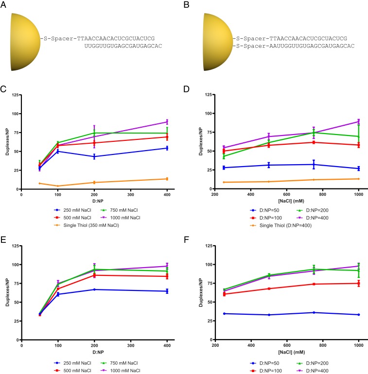
Spherical nucleic acids (SNAs) are nanostructures formed by chemically conjugating short linear strands of oligonucleotides to a nanoparticle template. When made with modified small interfering RNA (siRNA) duplexes, SNAs act as single-entity transfection and gene silencing agents and have been used as lead therapeutic constructs in several disease models. However, the manner in which modified siRNA duplex strands that comprise the SNA lead to gene silencing is not understood. Herein, a systematic analysis of siRNA biochemistry involving SNAs shows that Dicer cleaves the modified siRNA duplex from the surface of the nanoparticle, and the liberated siRNA subsequently functions in a way that is dependent on the canonical RNA interference mechanism. By leveraging this understanding, a class of SNAs was chemically designed which increases the siRNA content by an order of magnitude through covalent attachment of each strand of the duplex. As a consequence of increased nucleic acid content, this nanostructure architecture exhibits less cell cytotoxicity than conventional SNAs without a decrease in siRNA activity.

Keywords: gene regulation; siRNA processing; spherical nucleic acids.


Conflict of interest statement

The authors declare no competing interest.

Figures

Fig. 1.
Fig. 1.
The RNAi pathway in Drosophila and possible mechanisms for SNA processing. (A) The blue shade marks the canonical RNAi pathway in Drosophila: Dicer-2 and Loqs bind to long dsRNA and cleave it into a 22-nucleotide duplex siRNA. Dicer-2 and R2D2 bind to this siRNA and form the RDI complex. Then Ago2 is recruited, which cleaves the passenger strand, forming active RISC. Pathway 1 is characterized by the desorption of the siRNA from the surface of the SNA, which then behaves like free linear siRNA in its fate after desorption. Pathway 2 initiates with a Dicer-2/R2D2 complex binding to siRNA on the surface of the SNA to form a bound RDI complex. It then recruits Ago2 to the SNA, which forms RISC with the guide strand and is free to dissociate. Pathway 3 is characterized by Ago2 directly binding and processing the siRNA on the surface of the SNA and forms a “minimal RISC.” Finally, pathway 4 initiates with a Dicer-2/Loqs complex cleaving the SNA-bound siRNA, and the liberated siRNA then enters the canonical RNAi pathway. Common entry points into the pathway are shown in dashed boxes, while the catalytically active endpoints are shown in solid boxes. Solid and dashed arrows depict experimentally verified and hypothesized steps, respectively. Shading illustrates steps that involve linear (blue) or spherical (green) forms of RNA. (B) Injection of SNAs into Drosophila embryos effectively knocks down bicoid mRNA levels as measured by RT-qPCR. The targeting SNAs and siRNAs exhibit comparable knockdown efficiency with equimolar amounts of nucleic acid injected per embryo. A control SNA with heterologous sequence composition to bicoid does not significantly inhibit the mRNA level of bicoid (***P < 0.001). Each bar shows the mean of at least 3 independent experiments and 3 replicate RT-qPCR measurements. Error bars show aggregate SEM of biological and technical replicates. ns, not significant.
Fig. 2.
Fig. 2.
Release of siRNA from the SNA is Dcr-2 dependent. We incubated WT or mutant Drosophila embryo extracts with SNAs containing a radioactive guide strand. We analyzed the release of radioactive siRNA from the nanoparticle using an SDS/PAGE gel. (A) The quantification of the band intensities for 3 independent experiments for duplex RNA and guide strand RNA as a percentage of total radioactive signal. WT extract causes significant release of duplex siRNA from the SNA, indicating that a factor from the extract is required for siRNA processing. This release is abolished when extracts from Dcr-2 KO or CI embryos are used. Ago2 KO or CI extracts do not affect duplex siRNA release. Release of guide strand is not significant among any of the groups (***P < 0.001, ****P < 0.0001). Error bars show SEM of 3 independent experiments. (B) A sample SDS/PAGE gel used to quantify the bands for SNA, duplex, and guide strand. Left lane, no embryo extract control reaction. Right 2 lanes, single-stranded (ss) guide siRNA and duplex (ds) siRNA were run alone on the gel.
Fig. 3.
Fig. 3.
Complexes formed after the release of siRNA from SNAs are similar to those formed from linear siRNA duplexes. WT or mutant Drosophila embryo extracts were incubated with linear siRNA duplex (A) or SNAs (B) containing radioactive siRNA duplex. The SNAs were removed by centrifugation and the complexes formed were then separated on a native PAGE gel. Please note that both panels are from the same gel image with different brightness values, as SNAs complexes have weaker intensities. The asterisk indicates radioactivity in the well that did not enter the gel.
Fig. 4.
Fig. 4.
Dcr-2 is necessary for gene silencing in vivo by SNAs. Injection of linear siRNA duplexes or SNAs into Drosophila embryos with dcr-2 KO or CI genotypes, followed by quantification of bicoid gene knockdown. In both mutants, the targeting SNA shows no silencing activity, indicating that the SNA function is dependent on Dcr-2 catalytic activity (*P < 0.05). Each bar shows the mean of 3 independent experiments and 3 replicate qPCR measurements. Error bars show aggregate SEM of biological and technical replicates.
Fig. 5.
Fig. 5.
Doubly thiolated SNAs increase siRNA duplex loading on SNAs. Structure of (A) single-thiol and (B) doubly thiolated SNAs. Plots show duplex density as a function of D:NP ratio and NaCl concentration with disulfide duplex (C and D, respectively) and with thiol duplex (E and F, respectively). For both thiol and disulfide duplexes, increasing the D:NP ratio increases the duplex density until the electrostatic limit of oligonucleotide repulsion is reached for a given NaCl concentration. This limit can be overcome by addition of NaCl, which will increase duplex density until the oligonucleotides are depleted, or the absolute maximum density for a given sequence is reached. Even though both the thiol and disulfide forms of the duplex reach the absolute maximum loading density, using the thiol form of the duplex in the synthesis allows it to be reached with a lower D:NP ratio and NaCl concentration. Each data point shows the mean of 3 independent synthesis replicates and 3 independent duplex density measurements. The error bars show aggregated SEM of synthetic and technical replicates.
Fig. 6.
Fig. 6.
Doubly thiolated SNAs show gene knockdown activity and decreased cytotoxicity. (A) SKOV-3 cells were treated with SNAs targeting HER2 and matching linear siRNA controls, and target gene expression was assayed by qPCR. All HER2 targeting sequences show significant knockdown compared to nontargeting sequences (****P < 0.0001). However, none of the targeting sequences are statistically different. Each bar shows the mean of 3 independent experiments and 3 replicate qPCR measurements. Error bars show aggregate SEM of biological and technical replicates. (B) We quantified the relationship between gene knockdown efficiency of doubly thiolated SNAs and duplex density. We plotted the gene knockdown vs. duplex density between 40 and 160 duplexes per particle and fit each dataset (SI Appendix, Fig. S5). For both targeting and nontargeting SNAs, the average slope is zero, indicating that higher duplex densities do not hinder Dicer cleavage and hence SNA function. Each bar shows the mean of 4 independent experiments and 3 replicate qPCR measurements. Error bars show aggregate SEM of biological and technical replicates. (C) We treated cells with different concentrations of SNAs for 48 h and measured ATP in each well as a measure of cytotoxicity. Plots for the targeting sequence are shown here. The doubly thiolated SNA curve is shifted to the right, indicating that cytotoxicity is induced at higher siRNA concentrations. Each data point shows the mean of 4 biological replicates; error bars show the SEM of this mean. Dashed lines show the 95% confidence intervals for the fits. (D) We plotted the LD50 values from curves in C and SI Appendix, Fig. S8. For both the targeting and the nontargeting SNAs the LD50 value shifts by an order of magnitude, indicating that doubly thiolated SNAs are much less toxic to cells (****P < 0.0001). Error bars show SEM.

References

    1. Russ A. P., Lampel S., The druggable genome: An update. Drug Discov. Today 10, 1607–1610 (2005). - PubMed
    1. Conde J., Artzi N., Are RNAi and miRNA therapeutics truly dead? Trends Biotechnol. 33, 141–144 (2015). - PubMed
    1. Haussecker D., Kay M. A., RNA interference. Drugging RNAi. Science 347, 1069–1070 (2015). - PMC - PubMed
    1. Mirkin C. A., Letsinger R. L., Mucic R. C., Storhoff J. J., A DNA-based method for rationally assembling nanoparticles into macroscopic materials. Nature 382, 607–609 (1996). - PubMed
    1. Rosi N. L., et al. , Oligonucleotide-modified gold nanoparticles for intracellular gene regulation. Science 312, 1027–1030 (2006). - PubMed

Publication types