. 2020 Jan 3;4(1):2.
doi: 10.1186/s41747-019-0134-1.

Imaging of tumour response to immunotherapy

Affiliations

Imaging of tumour response to immunotherapy

Clarisse Dromain et al. Eur Radiol Exp. .

Abstract

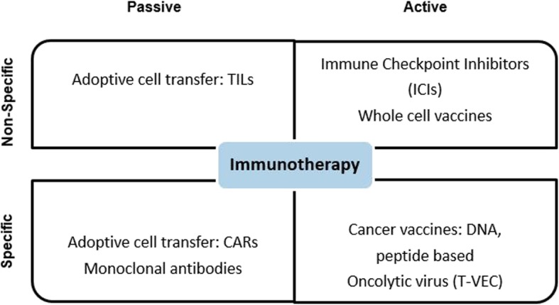
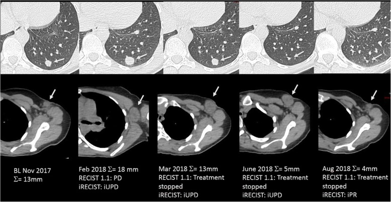
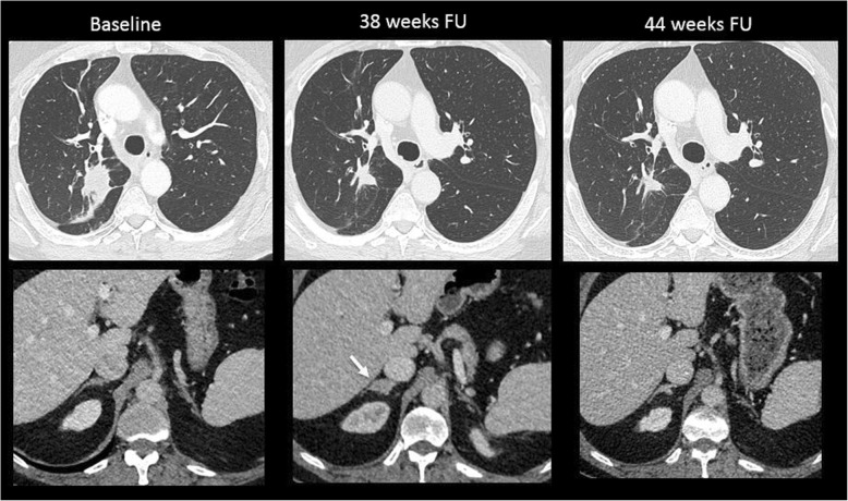
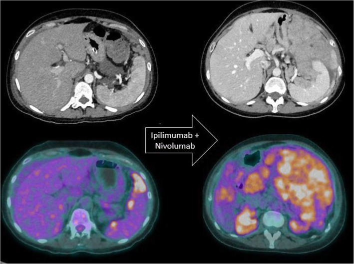
A wide range of cancer immunotherapy approaches has been developed including non-specific immune-stimulants such as cytokines, cancer vaccines, immune checkpoint inhibitors (ICIs), and adoptive T cell therapy. Among them, ICIs are the most commonly used and intensively studied. Since 2011, these drugs have received marketing authorisation for melanoma, lung, bladder, renal, and head and neck cancers, with remarkable and long-lasting treatment response in some patients. The novel mechanism of action of ICIs, with immune and T cell activation, leads to unusual patterns of response on imaging, with the advent of so-called pseudoprogression being more pronounced and frequently observed when compared to other anticancer therapies. Pseudoprogression, described in about 2-10% of patients treated with ICIs, corresponds to an increase of tumour burden and/or the appearance of new lesions due to infiltration by activated T cells before the disease responds to therapy. To overcome the limitation of response evaluation criteria in solid tumors (RECIST) to assess these specific changes, new imaging criteria-so-called immune-related response criteria and then immune-related RECIST (irRECIST)-were proposed. The major modification involved the inclusion of the measurements of new target lesions into disease assessments and the need for a 4-week re-assessment to confirm or not confirm progression. The RECIST working group introduced the new concept of "unconfirmed progression", into the irRECIST. This paper reviews current immunotherapeutic approaches and summarises radiologic criteria to evaluate new patterns of response to immunotherapy. Furthermore, imaging features of immunotherapy-related adverse events and available predictive biomarkers of response are presented.

Keywords: Cell- and tissue-based therapy; Immune checkpoint inhibitors; Immunotherapy; Pseudoprogression; Response evaluation criteria in solid tumors (RECIST).


Conflict of interest statement

The authors declare that they have no competing interests.

Figures

Fig. 1
Fig. 1
Different approaches of immunotherapy. CAR Chimeric antigen receptor, DNA Deoxyribonucleic acid, TILs Tumour-infiltrating lymphocytes, T-VEC Talimogene laherparepvec
Fig. 2
Fig. 2
Comparison of RECIST 1.1 and iRECIST criteria for evaluation of a 45-year-old woman with metastatic melanoma treated with ipilimumab (antiCTLA-4) and nivolumab (anti-PD-1). Baseline CT image (November 2017) shows a 13-mm lung metastasis (target lesion, upper panel, arrow) and a 10-mm short-axis axillary lymph node (non-target lesion, lower panel, arrow). On a 3-month follow-up, both lesions enlarged with an increase of 38% of the target lesion leading to a progressive disease (PD) and a stop of treatment according to RECIST 1.1 and an unconfirmed PD with maintained treatment according to iRECIST criteria. On the two following CT examinations (March and June 2018), the lung metastasis decreased in size, but the axillary lymph node was stable (unconfirmed PD) but still significantly enlarged compared to the baseline (still unconfirmed PD according to iRECIST criteria). Finally, on August 2018, CT images showed a decrease in size of both lesions, confirming the pseudoprogression with a response assessed to be -70%, leading to a partial response according to iRECIST criteria
Fig. 3
Fig. 3
Pseudoprogression in a 65-year-old patient with lung carcinoma treated with nivolumab (anti-PD-1). Baseline axial CT showed a lung mass in the upper right lobe with normal adrenal glands. At a 38-week follow-up (FU), there was a good reduction in the size of the lung mass, but a new lesion appeared in the right adrenal gland (arrow). The patient was maintained under the same treatment. At 44-week follow-up, the right adrenal mass disappeared, confirming the diagnosis of pseudoprogression
Fig. 4
Fig. 4
Paradoxical acceleration of tumour growth kinetics in a patient with metastatic melanoma treated with ipilimumab and nivolumab. Baseline axial CT image and corresponding 18F-FDG PET/CT image show few perisplenic peritoneal metastatic implants. Two months after the initiation of immunotherapy, both imaging modalities show a dramatic increase in peritoneal metastases.
Fig. 5
Fig. 5
18F-FDG PET/CT image of a stage IV enterocolitis during anti-CTLA-4 treatment in a patient with metastatic melanoma. Immunotherapy was interrupted, and a high-dose steroid therapy was started
Fig. 6
Fig. 6
Immune-related pneumonitis presenting as an organising pneumonia pattern in a patient with metastatic lung cancer that occurred after 13 cycles of anti-PD1 therapy. This axial CT image in lung windowing shows multifocal alveolar consolidations in a subpleural and peribronchovascular location, predominating at the level of the left upper lobe. Although suggestive of a diagnosis of organising pneumonia, infectious or tumoural lesions were excluded by means of a brochoalveolar lavage. Note that numerous round lucencies are visible within the alveolar consolidations, corresponding to associated centrilobular emphysema in this heavy smoker patient
Fig. 7
Fig. 7
Sarcoid-like reactions in a patient with metastatic melanoma treated with ipilimumab (antiCTLA-4) and nivolumab (anti-PD-1). Twelve-week follow-up (FU) 18F-FDG PET/CT images show typical hypermetabolic symmetric mediastinal and hilar lymph node enlargement, very suggestive of a sarcoid-like reaction. These features disappeared on the following 18F-FDG-PET/CT images at 18-week FU, confirming the diagnosis of sarcoid-like reaction under immunotherapy
Fig. 8
Fig. 8
A 75-year-old woman with a low-differentiated primary cardiac sarcoma with microsatellite instability, treated with pembrolizumab (anti-PD-1). Baseline contrast-enhanced MRI image shows a large retroatrial mass (arrow). Two months follow-up (FU) imaging shows a good reduction in the size of the mass assessed as -34% according to iRECIST criteria (partial response, arrow). One year FU imaging shows a complete response

References

    1. Kim R, Emi M, Tanabe K. Cancer immunoediting from immune surveillance to immune escape. Immunology. 2017;121:1–14. doi: 10.1111/j.1365-2567.2007.02587.x. - DOI - PMC - PubMed
    1. Eisenhauer EA, Therasse P, Bogaerts J, et al. New response evaluation criteria in solid tumours: revised RECIST guideline (version 1.1) Eur J Cancer. 2009;45:228–247. doi: 10.1016/j.ejca.2008.10.026. - DOI - PubMed
    1. Raja J, Ludwig JM, Gettinger SN, Schalper KA, Kim HS (2018) Oncolytic virus immunotherapy: future prospects for oncology. J Immunother Cancer 6:140. 10.1186/s40425-018-0458-z - PMC - PubMed
    1. Andtbacka RH, Kaufman HL, Collichio F, et al. Talimogene laherparepvec improves durable response rate in patients with advanced melanoma. J Clin Oncol. 2015;33:2780–2788. doi: 10.1200/JCO.2014.58.3377. - DOI - PubMed
    1. Raman SS, Hecht JR, Chan E. Talimogene laherparepvec: review of its mechanism of action and clinical efficacy and safety. Immunotherapy. 2019;11:705–723. doi: 10.2217/imt-2019-0033. - DOI - PubMed

Publication types

LinkOut - more resources