

Conventional Machine Learning and Deep Learning Approach for Multi-Classification of Breast Cancer Histopathology Images-a Comparative Insight
- PMID: 31900812
- PMCID: PMC7256154
- DOI: 10.1007/s10278-019-00307-y
Conventional Machine Learning and Deep Learning Approach for Multi-Classification of Breast Cancer Histopathology Images-a Comparative Insight
Abstract
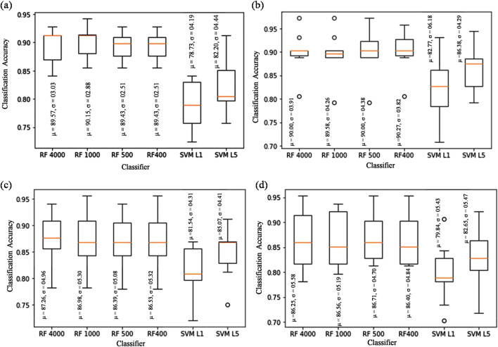
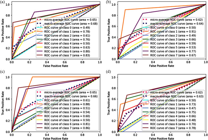
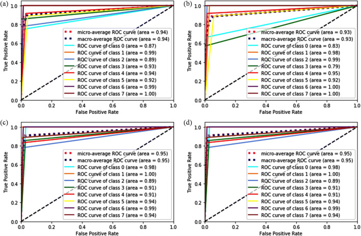
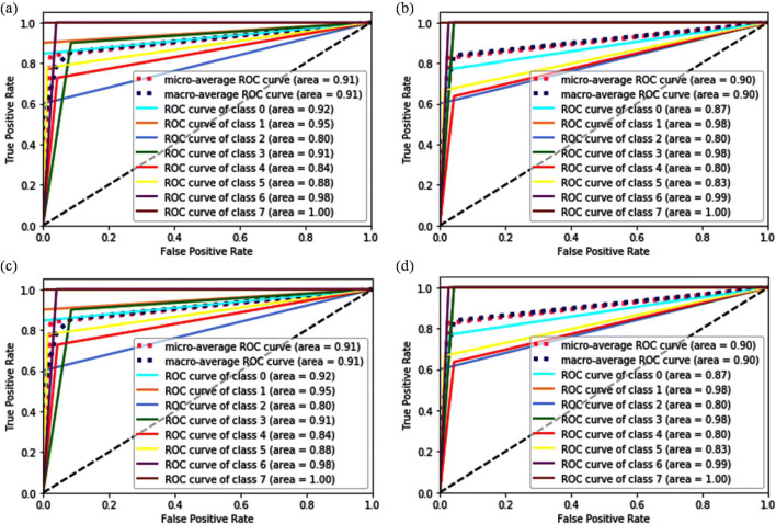
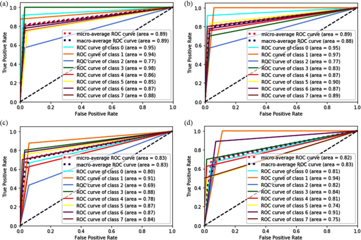
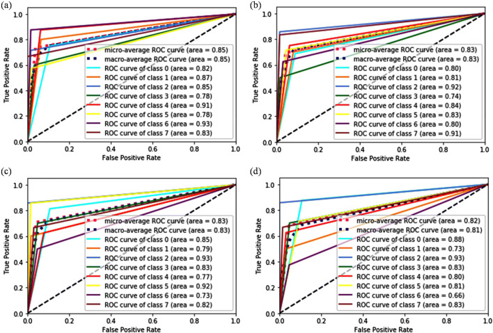
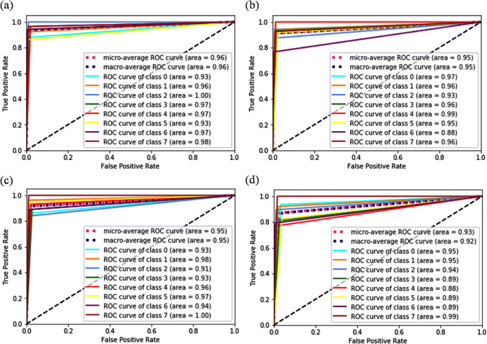
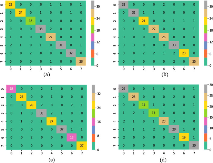
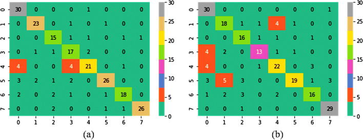
Automatic multi-classification of breast cancer histopathological images has remained one of the top-priority research areas in the field of biomedical informatics, due to the great clinical significance of multi-classification in providing diagnosis and prognosis of breast cancer. In this work, two machine learning approaches are thoroughly explored and compared for the task of automatic magnification-dependent multi-classification on a balanced BreakHis dataset for the detection of breast cancer. The first approach is based on handcrafted features which are extracted using Hu moment, color histogram, and Haralick textures. The extracted features are then utilized to train the conventional classifiers, while the second approach is based on transfer learning where the pre-existing networks (VGG16, VGG19, and ResNet50) are utilized as feature extractor and as a baseline model. The results reveal that the use of pre-trained networks as feature extractor exhibited superior performance in contrast to baseline approach and handcrafted approach for all the magnifications. Moreover, it has been observed that the augmentation plays a pivotal role in further enhancing the classification accuracy. In this context, the VGG16 network with linear SVM provides the highest accuracy that is computed in two forms, (a) patch-based accuracies (93.97% for 40×, 92.92% for 100×, 91.23% for 200×, and 91.79% for 400×); (b) patient-based accuracies (93.25% for 40×, 91.87% for 100×, 91.5% for 200×, and 92.31% for 400×) for the classification of magnification-dependent histopathological images. Additionally, "Fibro-adenoma" (benign) and "Mucous Carcinoma" (malignant) classes have been found to be the most complex classes for the entire magnification factors.
Keywords: Breast cancer; Handcrafted features; Histopathological images; Multi-classification; Transfer learning.
Conflict of interest statement
The authors declare that they have no competing interests.
Figures
Similar articles
-
Convolution Neural Network for Breast Cancer Detection and Classification Using Deep Learning.Asian Pac J Cancer Prev. 2023 Feb 1;24(2):531-544. doi: 10.31557/APJCP.2023.24.2.531. Asian Pac J Cancer Prev. 2023. PMID: 36853302 Free PMC article.
-
Feature Generalization for Breast Cancer Detection in Histopathological Images.Interdiscip Sci. 2022 Jun;14(2):566-581. doi: 10.1007/s12539-022-00515-1. Epub 2022 Apr 28. Interdiscip Sci. 2022. PMID: 35482216
-
Smart neural network and cognitive computing process for multi task nuclei detection segmentation and classification in breast cancer histopathology images.Sci Rep. 2025 May 26;15(1):18435. doi: 10.1038/s41598-025-02575-x. Sci Rep. 2025. PMID: 40419621 Free PMC article.
-
Classification of Histopathological Images from Breast Cancer Patients Using Deep Learning: A Comparative Analysis.Crit Rev Biomed Eng. 2023;51(4):41-62. doi: 10.1615/CritRevBiomedEng.2023047793. Crit Rev Biomed Eng. 2023. PMID: 37581350 Review.
-
Multi-features extraction based on deep learning for skin lesion classification.Tissue Cell. 2022 Feb;74:101701. doi: 10.1016/j.tice.2021.101701. Epub 2021 Nov 25. Tissue Cell. 2022. PMID: 34861582 Review.
Cited by
-
Investigating the effects of artificial intelligence on the personalization of breast cancer management: a systematic study.BMC Cancer. 2024 Jul 18;24(1):852. doi: 10.1186/s12885-024-12575-1. BMC Cancer. 2024. PMID: 39026174 Free PMC article.
-
Deep Learning With Radiomics for Disease Diagnosis and Treatment: Challenges and Potential.Front Oncol. 2022 Feb 17;12:773840. doi: 10.3389/fonc.2022.773840. eCollection 2022. Front Oncol. 2022. PMID: 35251962 Free PMC article. Review.
-
A review and comparative study of cancer detection using machine learning: SBERT and SimCSE application.BMC Bioinformatics. 2023 Mar 23;24(1):112. doi: 10.1186/s12859-023-05235-x. BMC Bioinformatics. 2023. PMID: 36959534 Free PMC article. Review.
-
Hybrid framework for automated generation of mammography radiology reports.Comput Struct Biotechnol J. 2025 Jul 16;27:3229-3239. doi: 10.1016/j.csbj.2025.07.018. eCollection 2025. Comput Struct Biotechnol J. 2025. PMID: 40741541 Free PMC article.
-
Artificial classification of cervical squamous lesions in ThinPrep cytologic tests using a deep convolutional neural network.Oncol Lett. 2020 Oct;20(4):113. doi: 10.3892/ol.2020.11974. Epub 2020 Aug 12. Oncol Lett. 2020. PMID: 32863926 Free PMC article.
References
-
- Breast Cancer. Available at http://www.who.int/cancer/prevention/diagnosis-screening/breast-cancer/en/.
-
- Breast Cancer Facts & Figures 2017-2018. Available at https://www.cancer.org/content/dam/cancer-org/research/cancer-facts-and-....
-
- Wilson ML, Fleming KA, Kuti MA, Looi LM, Lago N, Ru K: Access to pathology and laboratory medicine services: A crucial gap. The Lancet, 2018 - PubMed
-
- Robboy SJ, Weintraub S, Horvath AE, Jensen BW, Alexander CB, Fody EP, Crawford JM, Clark JR, Cantor-Weinberg J, Joshi MG, Cohen MB, Prystowsky MB, Bean SM, Gupta S, Powell SZ, Speights VO Jr, Gross DJ, Black-Schaffer WS. Pathologist workforce in the United States: I. Development of a predictive model to examine factors influencing supply. Archives of Pathology and Laboratory Medicine. 2013;137:1723–1732. doi: 10.5858/arpa.2013-0200-OA. - DOI - PubMed
MeSH terms
LinkOut - more resources
Full Text Sources
Medical
