. 2020 Jan 1;161(1):bqz016.
doi: 10.1210/endocr/bqz016.

Inhibition of FGFR Signaling Partially Rescues Osteoarthritis in Mice Overexpressing High Molecular Weight FGF2 Isoforms

Affiliations

Inhibition of FGFR Signaling Partially Rescues Osteoarthritis in Mice Overexpressing High Molecular Weight FGF2 Isoforms

Liping Xiao et al. Endocrinology. .

Abstract

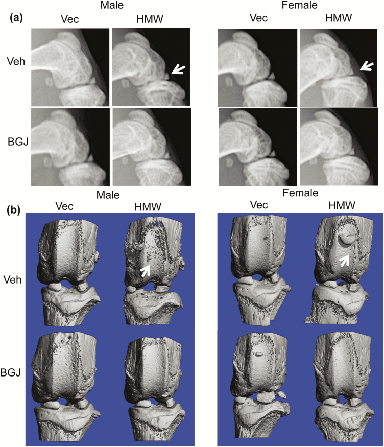
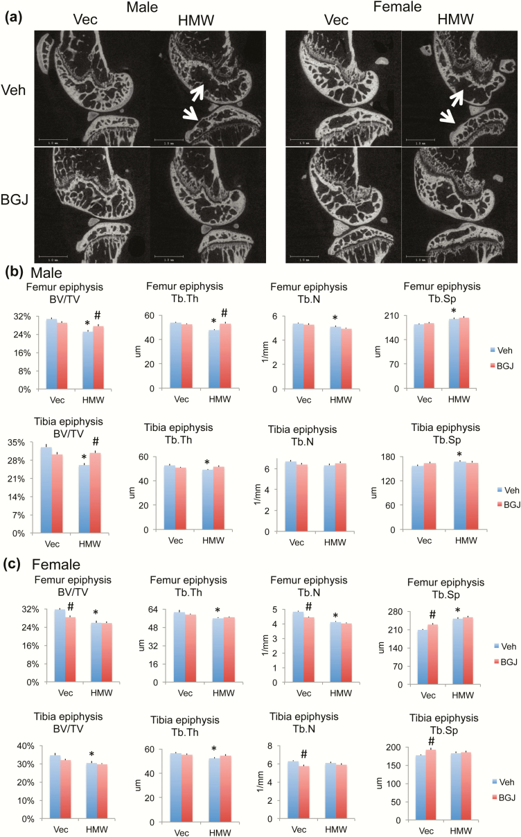
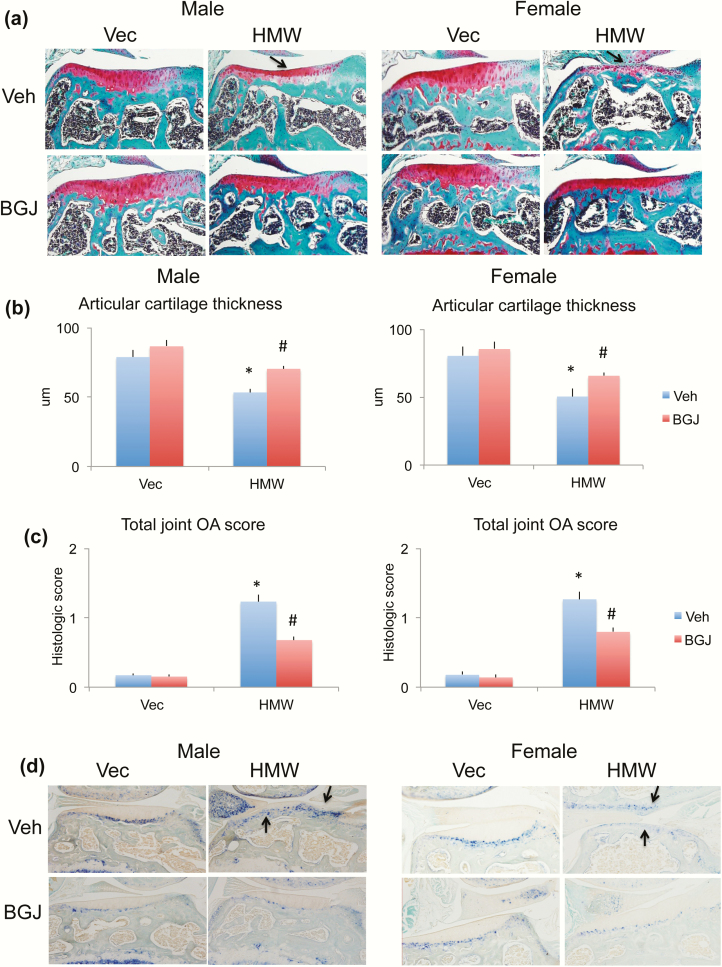
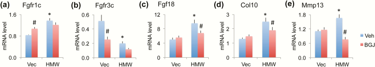
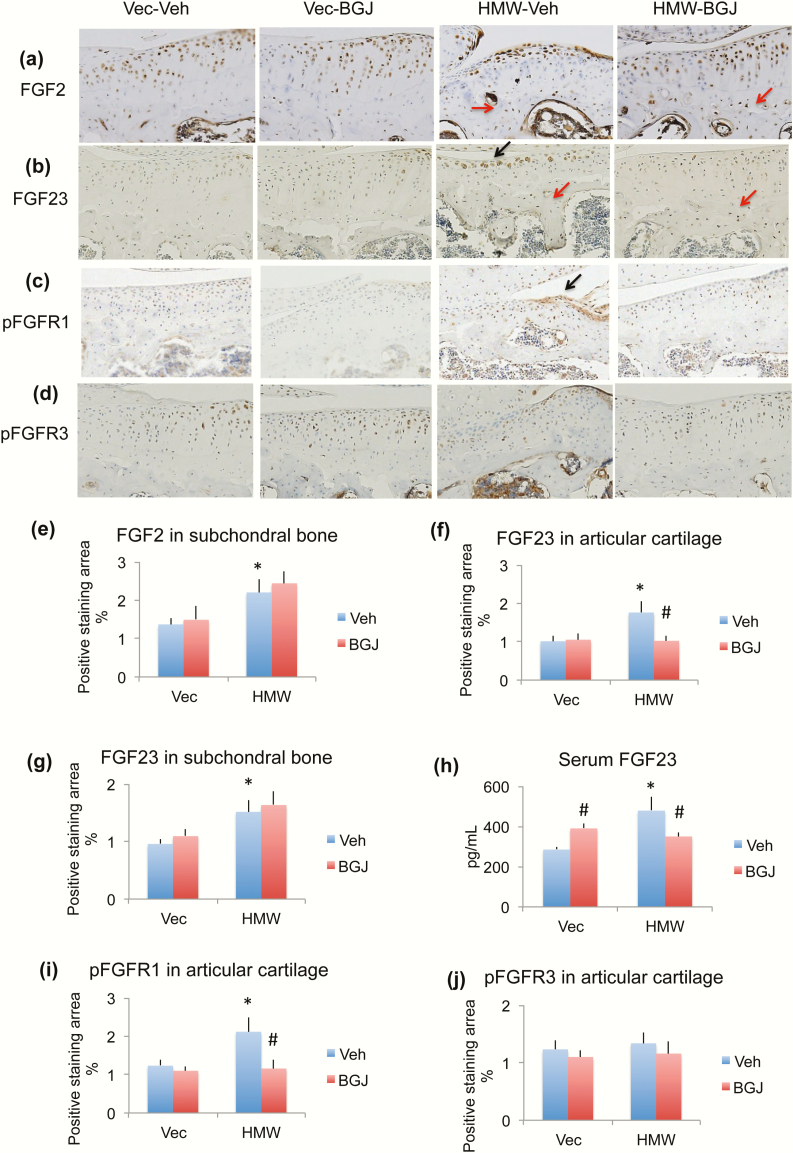
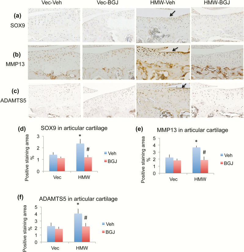
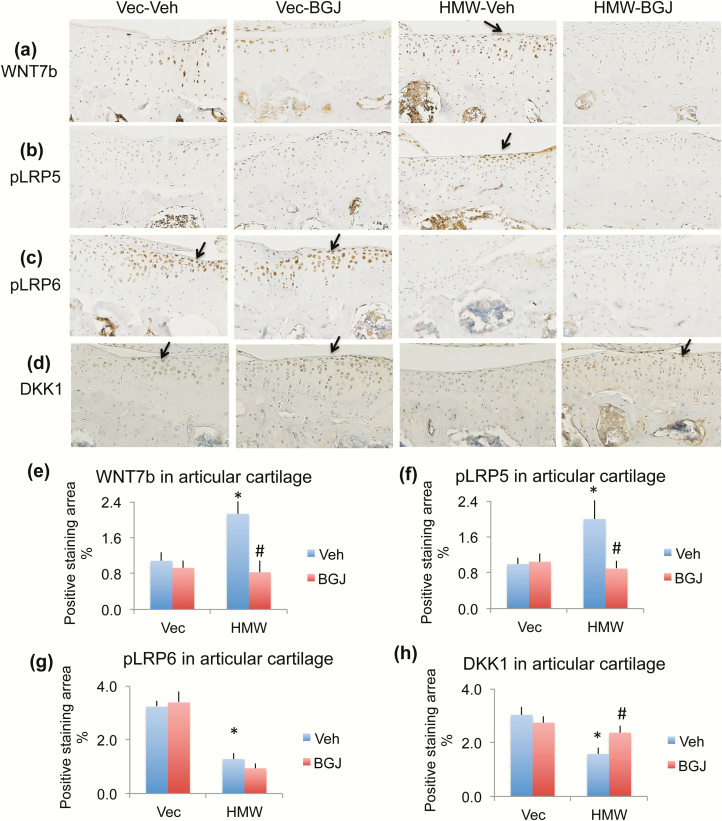
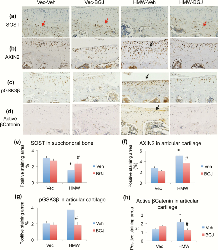
Fibroblast growth factor 2 (FGF2) and fibroblast growth factor receptors (FGFRs) are key regulatory factors in osteoarthritis (OA). HMWTg mice overexpress the high molecular weight FGF2 isoforms (HMWFGF2) in osteoblast lineage and phenocopy both Hyp mice (which overexpress the HMWFGF2 isoforms in osteoblasts and osteocytes) and humans with X-linked hypophosphatemia (XLH). We previously reported that, similar to Hyp mice and XLH subjects who develop OA, HMWTg mice also develop an OA phenotype associated with increased degradative enzymes and increased FGFR1 compared with VectorTg mice. Therefore, in this study, we examined whether in vivo treatment with the FGFR tyrosine kinase inhibitor NVP-BGJ398 (BGJ) would modulate development of the OA phenotype in knee joints of HMWTg mice. VectorTg and HMWTg mice (21 days of age) were treated with vehicle or BGJ for 13 weeks. Micro-computed tomography images revealed irregular shape and thinning of the subchondral bone with decreased trabecular number and thickness within the epiphyses of vehicle-treated HMWTg knees, which was partially rescued following BGJ treatment. Articular cartilage thickness was decreased in vehicle-treated HMWTg mice, and was restored to the cartilage thickness of VectorTg mice in the BGJ-treated HMWTg group. Increased OA degradative enzymes present in HMWTg vehicle-treated joints decreased after BGJ treatment. OA in HMWTg mice was associated with increased Wnt signaling that was rescued by BGJ treatment. This study demonstrates that overexpression of the HMWFGF2 isoforms in preosteoblasts results in osteoarthropathy that can be partially rescued by FGFR inhibitor via reduction in activated Wnt signaling.

Keywords: FGF receptor inhibitor; FGF2 HMWTg mice; cartilage; osteoarthritis.


Figures

Figure 1.
Figure 1.
Radiographic and microCT images of knee joint from VectorTg and HMWTg mice treated with vehicle or BGJ. (a) Sagittal digital x-ray images of HMWTg knees show the flattened tibia plateau, narrowing of the joint space, osteophytes, and uneven joint surfaces in the HMWTg vehicle-treated group in both male and female mice, which was rescued following BGJ treatment in both sexes. (b) Representative 3-dimensional microCT images show erosion of articular cartilage in the HMWTg vehicle-treated group in both male and female mice, whereas BGJ treatment resulted in a phenotype resembling that of the VectorTg groups in both sexes. Representative images from n = 7–9 males/group, n = 7–8 females/group.
Figure 2.
Figure 2.
MicroCT analysis of subchondral bone of knee joint from VectorTg and HMWTg mice treated with vehicle or BGJ. (a) Representative 2-dimensional sagittal microCT images reveal irregular shape and thinning of the subchondral bone in HMWTg vehicle-treated group in both male and female mice, which was rescued following BGJ treatment in both sexes. Morphometric parameters calculated from 3-dimensional microCT of femoral and tibiae epiphyses of (b) male and (c) female mice. There is significantly decreased bone volume per total volume (BV/TV), trabecular thickness (Tb.Th), trabecular number (Tb.N), and increased trabecular spacing (Tb.Sp) in femoral epiphysis of HMWTg vehicle-treated knees compared with VectorTg vehicle-treated epiphyses in both sexes. BGJ treatment significantly increased abnormal BV/TV and Tb.Th in male mice. There were significantly decreased BV/TV and Tb.Th in tibia epiphysis of HMWTg vehicle-treated knees compared with VectorTg vehicle-treated epiphyses in both genders. BGJ treatment significantly increased abnormal BV/TV in male mice. n = 7–9 males/group, n = 7–8 females/group. *compared with VectorTg-vehicle group P < 0.05; #compared with corresponding vehicle group P < 0.05 by 2-way ANOVA.
Figure 3.
Figure 3.
Comparison of proteoglycan content and alkaline phosphatase of knee joint from VectorTg and HMWTg mice treated with vehicle or BGJ. (a) Safranin-O stained representative images showed a dramatic decrease in proteoglycan content in HMWTg knees (arrows) in both male and female mice, which was partially rescued with BGJ treatment in both sexes. (b) Mean lateral tibial articular cartilage thickness analyzed by OsteoMeasure showed a decrease in cartilage thickness in both male and female HMWTg mice, which was partially rescued with BGJ treatment in both sexes. n = 7–9 males/group, n = 7–8 females/group. *compared with VectorTg-vehicle group P < 0.05; #compared with corresponding vehicle group P < 0.05 by 2-way ANOVA. (c) Mean total joint OA score was significantly increased HWTg-vehicle group compared with VectorTg-vehicle group in both male and female mice, which was partially rescued with BGJ treatment in both sexes. n = 7–9 males/group, n = 7–8 females/group. *compared with VectorTg-vehicle group P < 0.05; #compared with corresponding vehicle group P < 0.05 by 2-way ANOVA. (d) Alkaline phosphatase staining showed more alkaline phosphatase-positive articular chondrocytes in HMWTg vehicle-treated joints of both male and female mice, these were partially normalized with BGJ treatment in both sexes. Representative images from n = 7–9 males/group, n = 7–8 females/group.
Figure 4.
Figure 4.
Effect of BGJ on OA regulatory genes and OA marker genes in knees of VectorTg and HMWTg mice. RNA extracted from whole knee joint was used for gene expression. qPCR analysis of (a) Fgfr1c, (b) Fgfr3c, (c) Fgf18, (d) Col10, and (e) Mmp13. n = 7–9 males/group. *compared with VectorTg-vehicle group P < 0.05; #compared with corresponding vehicle group P < 0.05 by 2-way ANOVA.
Figure 5.
Figure 5.
Effect of BGJ on OA regulatory proteins in knees of VectorTg and HMWTg mice. Representative immunohistochemical staining images for (a) FGF2, (b) FGF23, (c) pFGFR1, and (d) pFGFR3. Quantitative analysis of percentage of positive staining area for (e) FGF2 in subchondral bone, (f) FGF23 in articular cartilage, (g) FGF23 in subchondral bone, (i) pFGFR1 in articular cartilage, and (j) pFGFR3 in articular cartilage. (h) Serum FGF23 level. (a and e) FGF2 immunohistochemical staining showed that FGF2 expression in cartilage was similar among 4 groups. However, FGF2 expression in the osteocytes of subchondral bone was markedly increased in the HMWTg vehicle-treated group; BGJ treatment did not alter FGF2 expression in either VectorTg mice or HMWTg mice, n = 4–5 males/group. (b, f, and g) FGF23 protein expression was significantly increased in chondrocyte of articular cartilage and osteocytes of subchondral bone in knees from HMWTg vehicle-treated mice compared with VectorTg vehicle-treated mice. BGJ treatment significantly rescued abnormal FGF23 expression in articular cartilage, but not in subchondral bone, n = 7–9 males/group. (h) Serum FGF23 level was significantly increased in HMWTg vehicle-treated mice compared with VectorTg vehicle-treated mice. BGJ treatment significantly increased FGF23 serum level in VectorTg mice, but significantly decreased serum FGF23 level in HMWTg mice, n = 7–9 males/group. (c and i) phosphorylated FGFR1 (pFGFR1) was significantly increased in HMWTg-vehicle group compared with VectorTg-vehicle group, which was rescued with BGJ treatment, n = 7–9 males/group. (d and j) pFGFR3 protein expression was similar among all 4 groups, n = 4–5 males/group. *compared with VectorTg-vehicle group P < 0.05; #compared with corresponding vehicle group P < 0.05 by 2-way ANOVA.
Figure 6.
Figure 6.
Effect of BGJ on OA marker proteins in knees of VectorTg and HMWTg mice. (a and d) Representative immunohistochemical staining images for (a) SOX9, (b) MMP13, and (c) ADAMTS5. Quantitative analysis of percentage of positive staining area for (d) SOX9, (e) MMP13, and (f) ADAMTS5 in articular cartilage. (a and d) SOX9 protein expression was significantly increased in chondrocytes of articular cartilage of knees from HMWTg-vehicle group compared with VectorTg-vehicle group, which was rescued with BGJ treatment, n = 7–9 males/group. (b and e) MMP13 and (c and f) ADAMTS5 expression were increased in knees of HMWTg vehicle-treated mice compared with VectorTg vehicle-treated mice; these were rescued with BGJ treatment, n = 7–9 males/group. *compared with VectorTg-vehicle group P < 0.05; #compared with corresponding vehicle group P < 0.05 by 2-way ANOVA.
Figure 7.
Figure 7.
Effect of BGJ on Wnt signaling component proteins in knees of VectorTg and HMWTg mice. Representative immunohistochemical staining images for (a) WNT7b, (b) phosphorylated LRP5 (pLRP5), (c) pLRP6, and (d) DKK1. Quantitative analysis of percentage of positive staining area for (e) WNT7b, (f) pLRP5, (g) pLRP6, and (h) DKK1 in articular cartilage. (a and e) WNT7b was greatly increased in HMWTg vehicle-treated cartilage compared with VectorTg vehicle-treated group, which was rescued with BGJ treatment, n = 7–9 males/group. (b and f) pLRP5 was strongly expressed in HMWTg vehicle-treated cartilage compared with VectorTg vehicle-treated group, which was rescued with BGJ treatment, n = 7–9 males/group. (c and g) pLRP6 protein was decreased in HMWTg vehicle-treated and HMWTg BGJ-treated articular cartilage, compared with VectorTg cartilage, n = 7–9 males/group. (d and h) DKK1 was significantly decreased in chondrocytes of articular cartilage of HMWTg-vehicle knees compared with VectorTg vehicle-treated mice, which was rescued with BGJ treatment, n = 4–5 males/group. *compared with VectorTg-vehicle group P < 0.05; #compared with corresponding vehicle group P < 0.05 by 2-way ANOVA.
Figure 8.
Figure 8.
Effect of BGJ on Wnt signaling component proteins in knees of VectorTg and HMWTg mice. Representative immunohistochemical staining images for (a) SOST, (b) AXIN2, (c) phosphorylated GSK3β (pGSK3β), and (d) active β-catenin. Quantitative analysis of percentage of positive staining area for (e) SOST in subchondral bone, (f) AXIN2 in articular cartilage, (g) pGSK3β in articular cartilage, and (h) active β-catenin in articular cartilage. (a and e) SOST was only expressed in osteocytes in the subchondral bone, but not expressed in articular cartilage. SOST expression in the subchondral bone of HMWTg vehicle-treated mice was much lower than VectorTg vehicle-treated mice, which was rescued with BGJ treatment, n = 4–5 males/group. (b and f) AXIN2 was increased in the cartilage of HMWTg vehicle-treated mice compared with VectorTg vehicle-treated mice. BGJ treatment markedly inhibited the increased AXIN2 in HMWTg mice, n = 4–5 males/group. (c and g) pGSK3β was strongly expressed in HMWTg vehicle-treated cartilage compared with VectorTg vehicle-treated and VectorTg BGJ-treated groups, and was rescued with BGJ treatment, n = 7–9 males/group. (d and h) Active β-Catenin protein expression was significantly increased in articular cartilage of knees from HMWTg-vehicle group compared with VectorTg-vehicle group, which was rescued with BGJ treatment, n = 7–9 males/group. *compared with VectorTg-vehicle group P < 0.05; #compared with corresponding vehicle group P < 0.05 by 2-way ANOVA.

Similar articles

Cited by

References

    1. Econs MJ, Francis F. Positional cloning of the PEX gene: new insights into the pathophysiology of X-linked hypophosphatemic rickets. Am J Physiol. 1997;273(4):F489–F498. - PubMed
    1. Hardy DC, Murphy WA, Siegel BA, Reid IR, Whyte MP. X-linked hypophosphatemia in adults: prevalence of skeletal radiographic and scintigraphic features. Radiology. 1989;171(2):403–414. - PubMed
    1. Reid IR, Hardy DC, Murphy WA, Teitelbaum SL, Bergfeld MA, Whyte MP. X-linked hypophosphatemia: a clinical, biochemical, and histopathologic assessment of morbidity in adults. Medicine (Baltimore). 1989;68(6):336–352. - PubMed
    1. Liang G, Katz LD, Insogna KL, Carpenter TO, Macica CM. Survey of the enthesopathy of X-linked hypophosphatemia and its characterization in Hyp mice. Calcif Tissue Int. 2009;85(3):235–246. - PMC - PubMed
    1. Liang G, Vanhouten J, Macica CM. An atypical degenerative osteoarthropathy in Hyp mice is characterized by a loss in the mineralized zone of articular cartilage. Calcif Tissue Int. 2011;89(2):151–162. - PubMed

Publication types

MeSH terms