. 2020 Feb 3;30(3):408-420.e5.
doi: 10.1016/j.cub.2019.11.064. Epub 2020 Jan 2.

The Initiation of Meiotic Sex Chromosome Inactivation Sequesters DNA Damage Signaling from Autosomes in Mouse Spermatogenesis

Affiliations

The Initiation of Meiotic Sex Chromosome Inactivation Sequesters DNA Damage Signaling from Autosomes in Mouse Spermatogenesis

Hironori Abe et al. Curr Biol. .

Abstract

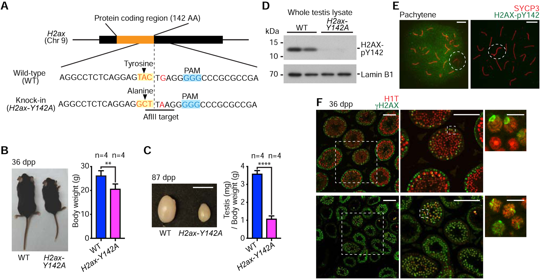
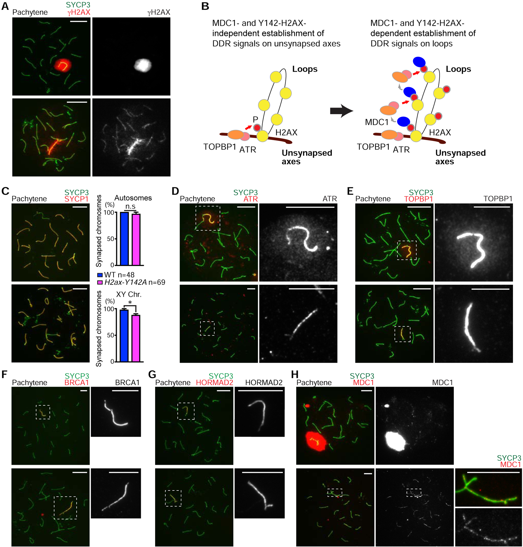
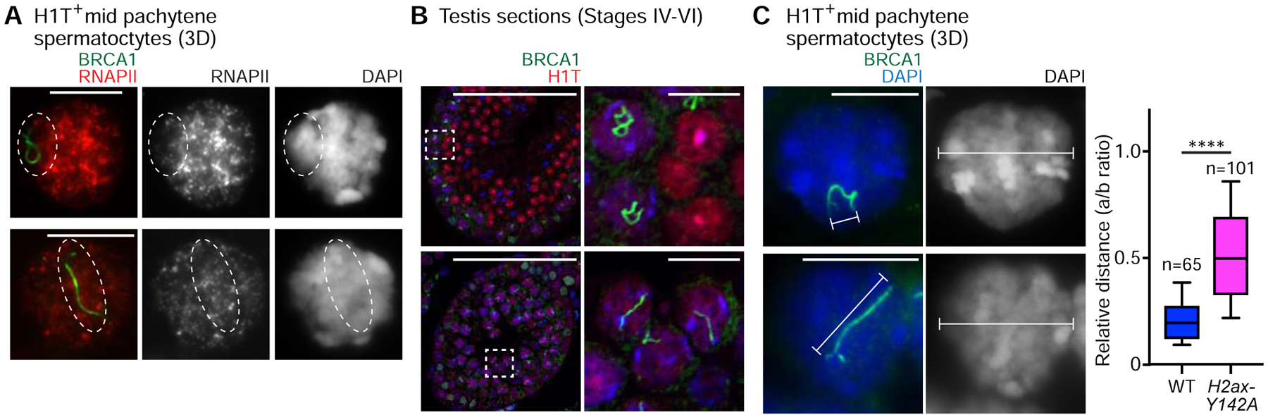
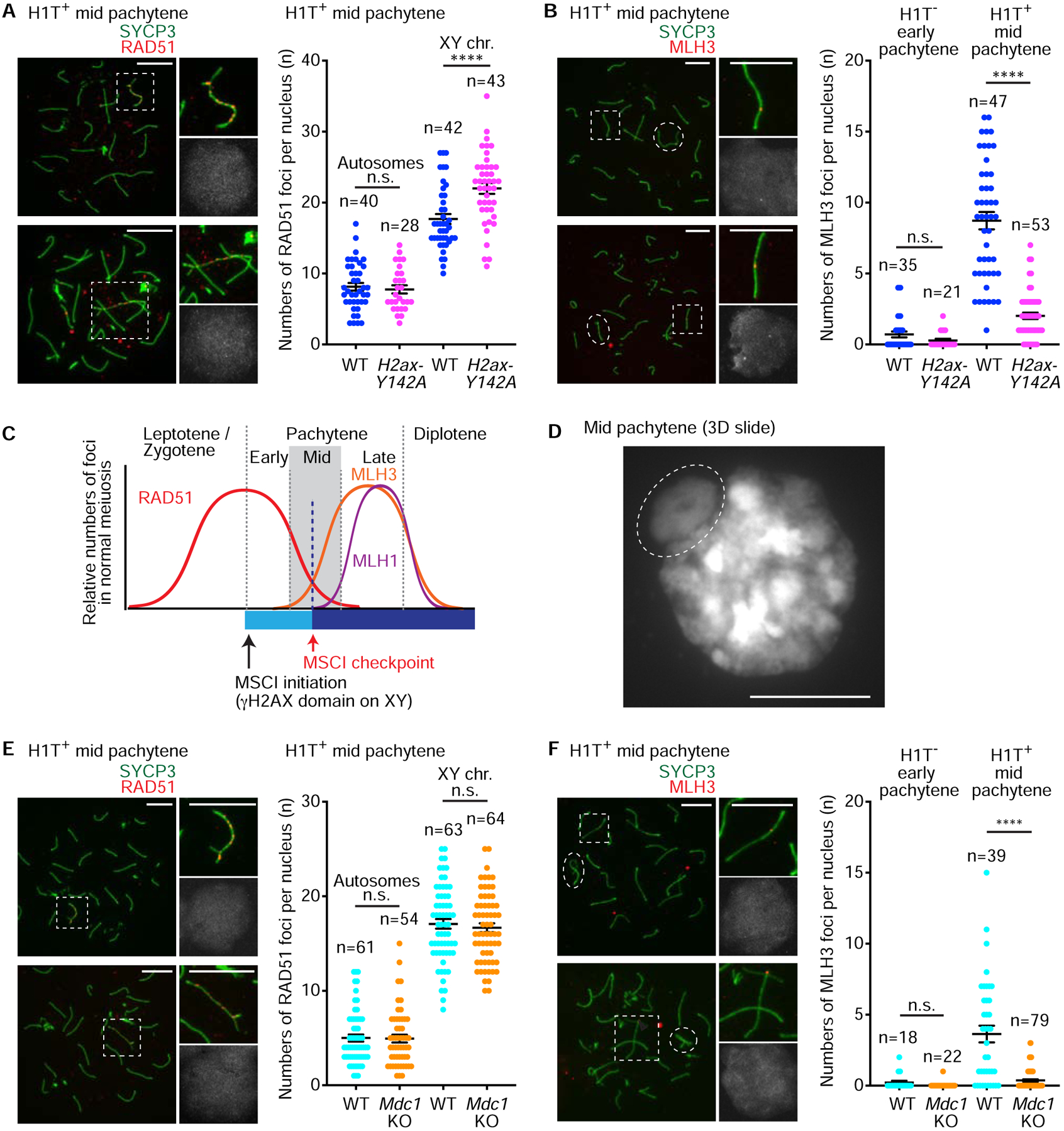
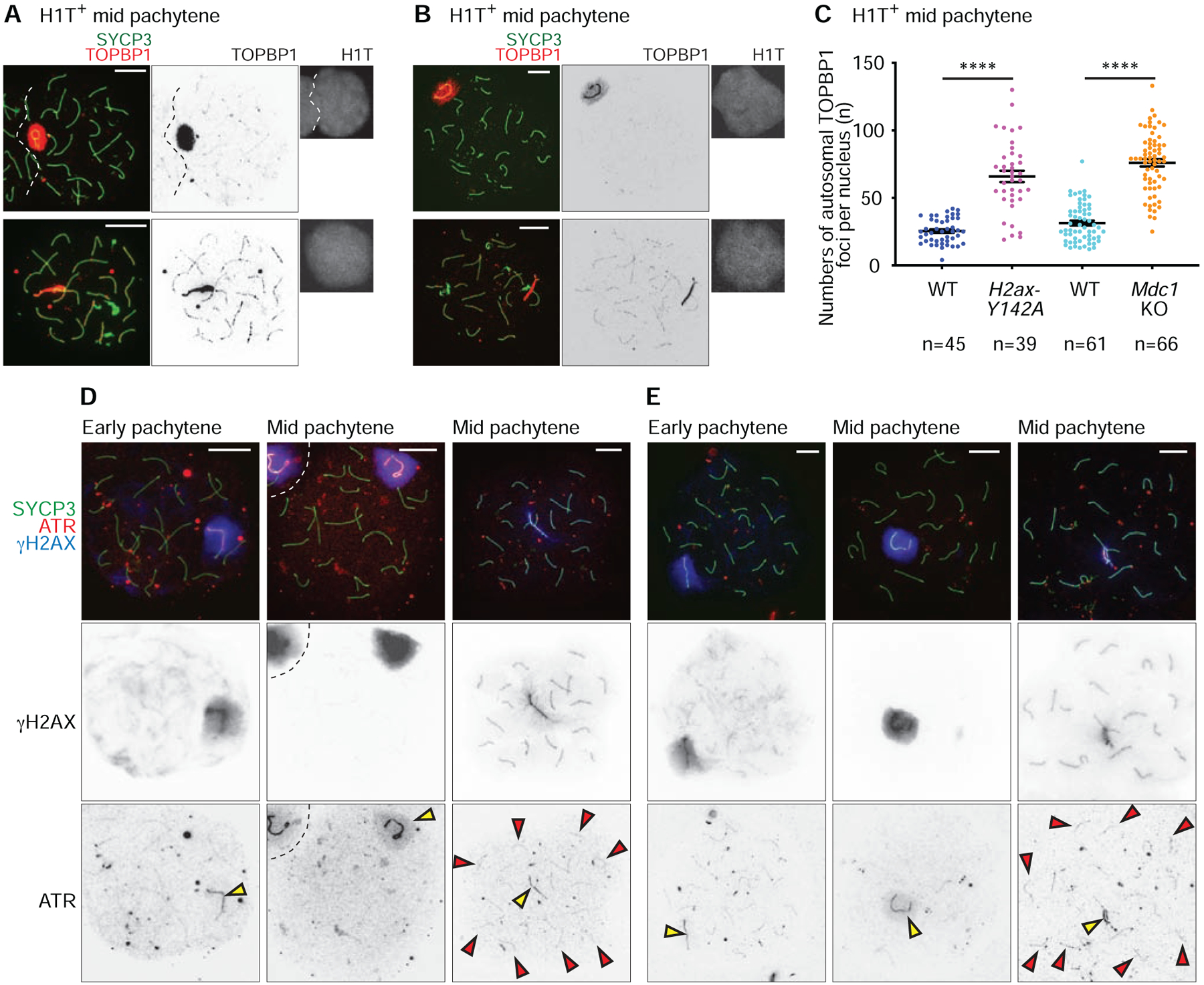
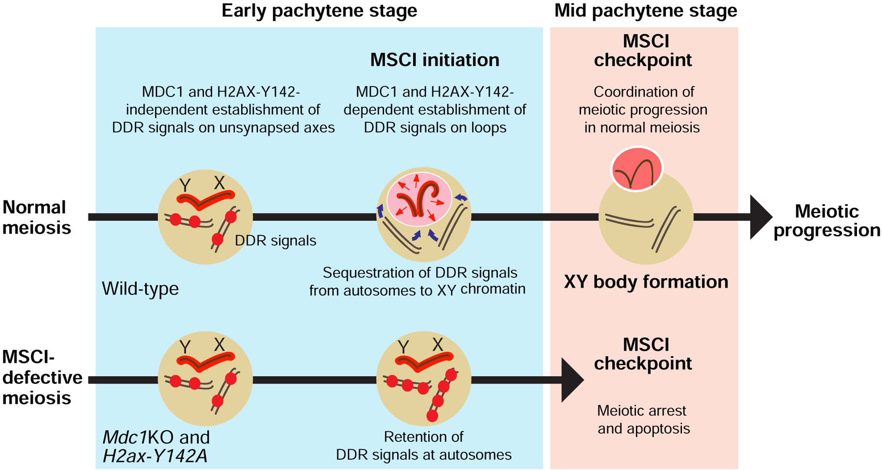
Meiotic sex chromosome inactivation (MSCI) is an essential event in the mammalian male germline. MSCI is directed by a DNA damage response (DDR) pathway centered on the phosphorylation of histone variant H2AX at serine 139 (termed γH2AX). The failure to initiate MSCI is linked to complete meiotic arrest and elimination of germ cells; however, the mechanisms underlying this arrest and elimination remain unknown. To address this question, we established a new separation-of-function mouse model for H2ax that shows specific and complete defects in MSCI. The genetic change is a point mutation in which another H2AX amino acid residue important in the DDR, tyrosine 142 (Y142), is converted to alanine (H2ax-Y142A). In H2ax-Y142A meiosis, the establishment of DDR signals on the chromosome-wide domain of the sex chromosomes is impaired. The initiation of MSCI is required for stage progression, which enables crossover formation, suggesting that the establishment of MSCI permits the timely progression of male meiosis. Our results suggest that normal meiotic progression requires the removal of ATR-mediated DDR signaling from autosomes. We propose a novel biological function for MSCI: the initiation of MSCI sequesters DDR factors from autosomes to the sex chromosomes at the onset of the pachytene stage, and the subsequent formation of an isolated XY nuclear compartment-the XY body-sequesters DDR factors to permit meiotic progression from the mid-pachytene stage onward. VIDEO ABSTRACT.

Keywords: DNA damage signaling; checkpoint; germline; meiosis; spermatogenesis.


Conflict of interest statement

Declaration of Interests The authors declare no competing interests.

Figures

Figure 1.
Figure 1.. H2AX-Y142 is an essential amino residue for completing spermatogenesis.
(A) Schematic: Induction of a point mutation and resulting sequence alteration. The introduction of an AflII target site was used to screen mutant mice. (B) Wild-type (WT) and H2ax-Y142A mice at 36 days post-partum (dpp). Body weights are shown as mean ± s.e.m. from 4 independent pairs of WT and H2ax-Y142A mice. ** p < 0.01, unpaired t test. (C) Testes from WT and H2ax-Y142A mice at 87 dpp. Scale bar: 1 cm. Ratios of testis weight (mg) to body weight (g) shown as mean ± s.e.m. for 4 independent pairs of WT and H2ax-H142A mice. **** p < 0.0001, unpaired t test. (D) Western blot of WT and H2ax-Y142A lysates from whole mouse testes with anti-H2AX-pY142 antibody. 20 μg protein samples were loaded in each lane. Two independent samples for WT and H2ax-Y142A are shown. Loading control: Lamin B1. (E) Chromosome spreads of WT and H2ax-Y142A pachytene spermatocytes immunostained with antibodies raised against SYCP3 and H2AX-pY142. Dashed circles indicate the sex chromosomes. Scale bars: 10 μm. (F) Testis sections from WT and H2ax-Y142A mice immunostained with antibodies raised against H1T and γH2AX. Dashed squares are magnified in panels to the right. White arrowheads indicate γH2AX signals on sex chromosomes in mid pachytene (H1T-positive) spermatocytes. Scale bars in larger panels: 100 μm; scale bars in smaller panels: 10 μm. See also Figure S1.
Figure 2.
Figure 2.. H2AX-Y142 is required for the initiation of MSCI
(A, C–H) Chromosome spreads of wild-type (WT) and H2ax-Y142A pachytene spermatocytes immunostained with antibodies raised against the following proteins: SYCP3 (A, C–H), γH2AX (A), SYCP1 (C), ATR (D), TOPBP1 (E), BRCA1 (F), HORMAD2 (G), and MDC1 (H). Dashed squares are magnified in the panels to the right. (B) Model of the initiation of MSCI. ATR and its activator, TOPBP1, are recruited to unsynapsed axes in an MDC1- and Y142-H2AX-independent manner, resulting in phosphorylation of H2AX to generate γH2AX on axes (left). γH2AX then recruits MDC1, which facilitates progressive recruitment of ATR and TOPBP1, resulting in γH2AX and MDC1 spreading throughout loops (right). (C) Percentages of mid pachytene chromosome synapsis for autosome and sex chromosomes, shown as mean ± s.e.m.. Total numbers of analyzed nuclei obtained from 3 independent littermate pairs are indicated in the panel. n.s., not significant; * p < 0.05, unpaired t test. Scale bars: 10 μm unless otherwise described in the panels. See also Figure S2.
Figure 3.
Figure 3.. Impaired XY body formation in H2ax-Y142A mouse.
(A) Wild-type (WT) littermate control and H2ax-Y142A pachytene spermatocytes on 3D slides (see STAR METHODS) immunostained with antibodies raised against RNAPII and BRCA1. Although not shown in the panel, the spermatocytes were also immunostained with an anti-H1T antibody to determine their stage; the spermatocytes shown are H1T-positive. Dashed circles indicate the sex chromosomes. (B) WT littermate control and H2ax-Y142A testis sections immunostained with antibodies raised against BRCA1 and H1T. Dashed squares are magnified in the panels to the right. White arrowheads indicate the axes of sex chromosomes in H1T-positive pachytene spermatocytes. Nuclei were counterstained with DAPI. Scale bars: 100 μm and, in the panels to the right, 10 μm. (C) WT and H2ax-Y142A pachytene spermatocytes on 3D slides immunostained with an antibody raised against BRCA1. Although not shown in the panel, the spermatocytes were also immunostained with an anti-H1T antibody to determine their stage; the spermatocytes shown are H1T-positive. The relative distances are shown in a box-and-whisker plot: The central line is the median, the bottom edge of the box is the first quartile, the top edge of the box is the third quartile, and the whiskers encompass, from top to bottom, the first to ninth decile. Total numbers of analyzed nuclei, obtained from 3 independent wild-type mice and 4 independent H2ax-Y142A mice, are indicated in the panel. **** p < 0.0001, Mann-Whitney U test. Nuclei were counterstained with DAPI. Scale bars: 10 μm.
Figure 4.
Figure 4.. Initial steps of DSB repair take place normally on autosomes of MSCI defective mutants.
(A, B, E, F) Chromosome spreads of wild-type (WT) littermate control and H2ax-Y142A (A, B) or Mdc1KO (E, F) mid pachytene spermatocytes immunostained with antibodies raised against SYCP3 (A, B, E, F), HIT (A, B, E, F), RAD51 (A, E), or MLH3 (B, F). Sex chromosomes in dashed squares are magnified in the panels to the right (A, E). Autosomes in dashed squares are magnified in the panels to the right, and the dashed circles indicate the sex chromosomes (B, F). Dot plots indicate the numbers of autosome RAD51 foci (A, E), sex chromosome RAD51 foci (A, E), or MLH3 foci (B, F) per mid pachytene (HIT-positive) spermatocyte, shown as mean ± s.e.m. for 3 independent H2ax-Y142A littermate pairs (A, B) and 3 independent Mdc1KO littermate pairs (E, F). Total numbers of analyzed nuclei are indicated in the panels. n.s.: not significant; **** p < 0.0001, unpaired t tests. XY chr.: XY chromosomes. Scale bars: 10 μm. (C) Model of the MSCI checkpoint and its relationship to meiotic recombination. (D) DAPI counterstaining of a 3D slide (see STAR METHODS). The dashed circle indicates the XY body. Scale bar: 10 μm. See also Figures S3 and S4.
Figure 5.
Figure 5.. DDR factors centered on ATR signaling are sequestered from autosomes to the sex chromosomes at the onset of MSCI.
(A, B, D, E) Chromosome spreads of wild-type (WT) littermate control and H2ax-Y142A (A, D) or Mdc1KO (B, E) mid pachytene spermatocytes immunostained with antibodies raised against SYCP3 (A, B, D, E), TOPBP1 (A, B), H1T (A, B), γH2AX (D, E), and ATR (D, E). Yellow arrowheads indicate the sex chromosomes, and red arrowheads indicate ATR foci that persist on H2ax-Y142A autosomes (D, E). Scale bars: 10 μm. (C) Numbers of TOPBP1 foci on autosomes in mid pachytene (H1T-positive) spermatocytes, shown as mean ± s.e.m. for 3 independent H2ax-Y142A littermate pairs (left) and 3 independent Mdc1KO littermate pairs (right). Total numbers of analyzed nuclei are indicated in the panels. **** p < 0.0001, Mann-Whitney U test. See also Figure S5.
Figure 6.
Figure 6.. A model of the MSCI checkpoint: The physical sequestration of DDR factors from autosomes to the XY body is a critical checkpoint in meiosis progression and gamete development.
At the onset of MSCI, DDR factors (shown as red spheres) are sequestered from autosomes to the sex chromosomes. The physical sequestration of DDR factors on/at a sex chromosome-specific chromo-nuclear compartment, the XY body, is a critical step in the MSCI checkpoint in the mid pachytene stage of meiotic prophase I. While the MSCI checkpoint ensures meiotic stage progression in normal meiosis, the abolishment of MSCI enables the ectopic retention of DDR signals on/at autosomes; these, in turn, trigger complete meiotic arrest and cell death in response to the checkpoint. See also Figure S6.

Comment in

References

    1. Subramanian VV, and Hochwagen A (2014). The meiotic checkpoint network: step-by-step through meiotic prophase. Cold Spring Harb Perspect Biol 6, a016675. - PMC - PubMed
    1. Handel MA, and Schimenti JC (2010). Genetics of mammalian meiosis: regulation, dynamics and impact on fertility. Nat Rev Genet 11, 124–136. - PubMed
    1. Ichijima Y, Sin HS, and Namekawa SH (2012). Sex chromosome inactivation in germ cells: emerging roles of DNA damage response pathways. Cell Mol Life Sci 69, 2559–2572. - PMC - PubMed
    1. Turner JM (2015). Meiotic Silencing in Mammals. Annu Rev Genet 49, 395–412. - PubMed
    1. Fernandez-Capetillo O, Mahadevaiah SK, Celeste A, Romanienko PJ, Camerini-Otero RD, Bonner WM, Manova K, Burgoyne P, and Nussenzweig A (2003). H2AX is required for chromatin remodeling and inactivation of sex chromosomes in male mouse meiosis. Dev Cell 4, 497–508. - PubMed

Publication types