MRI depiction of fetal brain abnormalities
- PMID: 31903224
- PMCID: PMC6927204
- DOI: 10.1177/2058460119894987
MRI depiction of fetal brain abnormalities
Abstract
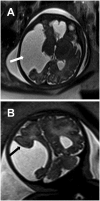
Intracranial abnormalities are commonly suspected findings on antenatal ultrasound that require evaluation by magnetic resonance imaging. This review depicts multiple abnormalities imaged as a means to guide clinicians in proper diagnosis.
Keywords: Congenital brain malformations; fetal magnetic resonance imaging; midline abnormalities.
© The Foundation Acta Radiologica 2019.
Figures



Similar articles
-
Usefulness of additional fetal magnetic resonance imaging in the prenatal diagnosis of congenital abnormalities.Arch Gynecol Obstet. 2012 Dec;286(6):1443-52. doi: 10.1007/s00404-012-2474-4. Epub 2012 Aug 9. Arch Gynecol Obstet. 2012. PMID: 22875047
-
Magnetic resonance imaging of the fetal central nervous system in Singapore.Ann Acad Med Singap. 2009 Sep;38(9):774-81. Ann Acad Med Singap. 2009. PMID: 19816636
-
Fetal Neurosonogaphy: Ultrasound and Magnetic Resonance Imaging in Competition.Ultraschall Med. 2016 Dec;37(6):555-557. doi: 10.1055/s-0042-117142. Epub 2016 Dec 15. Ultraschall Med. 2016. PMID: 27978593 English.
-
[Prenatal imaging of the fetal brain--indications and developmental implications of fetal MRI].Harefuah. 2008 Jan;147(1):65-70, 93. Harefuah. 2008. PMID: 18300627 Review. Hebrew.
-
Fetal magnetic resonance imaging: a review.Curr Opin Obstet Gynecol. 2007 Apr;19(2):151-6. doi: 10.1097/GCO.0b013e32809bd978. Curr Opin Obstet Gynecol. 2007. PMID: 17353684 Review.
Cited by
-
Clinical Applications of Fetal MRI in the Brain.Diagnostics (Basel). 2022 Mar 21;12(3):764. doi: 10.3390/diagnostics12030764. Diagnostics (Basel). 2022. PMID: 35328317 Free PMC article. Review.
References
-
- Victoria T, Jaramillo D, Roberts TPL, et al. Fetal magnetic resonance imaging: jumping from 1.5 to 3 tesla (preliminary experience). Pediatr Radiol 2013; 44:376–386. - PubMed
-
- Sans Cortes M, Bargallo N, Arranz A, et al. Feasibility and success rate of a fetal MRI and MR spectroscopy research protocol performed at term using a 3.0-tesla scanner. Fetal Diagn Ther 2017; 41:127–135. - PubMed
-
- Santo S, D’Antonio F, Homfray T, et al. Counseling in fetal medicine: agenesis of the corpus callosum. Ultrasound Obstet Gynecol 2012; 40:513–521. - PubMed
-
- Paul LK, Brown WS, Adolphs R, et al. Agenesis of the corpus callosum: genetic, developmental and functional aspects of connectivity. Nat Rev Neurosci 2007; 8:287–299. - PubMed
LinkOut - more resources
Full Text Sources

