

A new tool for diagnosing parathyroid lesions: angio plus ultrasound imaging
- PMID: 31903273
- PMCID: PMC6940251
- DOI: 10.21037/jtd.2019.11.29
A new tool for diagnosing parathyroid lesions: angio plus ultrasound imaging
Abstract
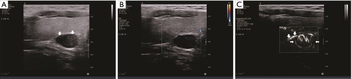
Background: Our aim was to examine the potential value of angio plus ultrasound imaging in diagnosing parathyroid lesions. Angio plus ultrasound imaging uses a new paradigm in Doppler performance, which allows for a better detection of flow in small vessels while maintaining workflow of conventional color flow imaging characteristics.
Methods: Thirty parathyroid lesions, composed of 26 histopathologically diagnosed adenoma and 4 hyperplasia (two hyperplasia in the same patient), from 29 consecutive patients (6 men and 23 women; median age: 53 years, range: 25-78 years) were evaluated using both color Doppler and angio plus ultrasound imaging. The polar vessel (visible or invisible), number and distribution (peripheral, central, and both) of blood flow signals were compared between the two techniques.
Results: On color Doppler, the polar vessel was visible in 6 (20%) lesions. The median number of blood flow signals was 6 [3-23]. The distribution of peripheral, central and both was shown in 4 (13.3%), 9 (30%), and 15 (50%) lesions respectively, and no blood signals was shown in 2 (6.7%) lesions. On angio plus ultrasound imaging, the polar vessel was visible in 16 (53.3%) lesions. The median number of blood flow signals was 3 [0-18]. The distribution of peripheral, central and both was shown in 1 (3.3%), 4 (13.3%), and 25 (83.3%) lesions respectively. There was significant difference between color Doppler and angio plus ultrasound imaging in the detection of the polar vessel, number and distribution of blood flow signals.
Conclusions: The typical polar vessel and increased vascularity commonly associated with parathyroid lesions may be obtained with angio plus ultrasound imaging. Angio plus ultrasound imaging can improve the detection rate and distribution characteristics of blood flow signals of parathyroid lesions. Further research is needed to clarify the clinical meaning of increased blood flow information, such as the differentiation of parathyroid lesions from other lesions.
Keywords: Hyperparathyroidism; angio plus; color Doppler; ultrasound.
2019 Journal of Thoracic Disease. All rights reserved.
Conflict of interest statement
Conflicts of Interest: The authors have no conflicts of interest to declare.
Figures
Similar articles
-
Ultrasound-Based Noncontrast Microvascular Imaging for Evaluation of Breast Lesions: Imaging Techniques and Review of Diagnostic Criteria.Indian J Radiol Imaging. 2024 Mar 17;34(4):702-713. doi: 10.1055/s-0044-1782162. eCollection 2024 Oct. Indian J Radiol Imaging. 2024. PMID: 39318571 Free PMC article. Review.
-
[Possibilities and limits of a new color technique: ultrasound angiography--results of the "Heidelberg Round Table Discussion"].Bildgebung. 1995 Mar;62(1):53-63. Bildgebung. 1995. PMID: 7538838 German.
-
Contrast-Enhanced Ultrasound Qualitative and Quantitative Characteristics of Parathyroid Gland Lesions.Medicina (Kaunas). 2021 Dec 21;58(1):2. doi: 10.3390/medicina58010002. Medicina (Kaunas). 2021. PMID: 35056309 Free PMC article.
-
Using ultrafast angio planewave ultrasensitive and conventional doppler imaging techniques to assess intramuscular blood perfusion in older adults.BMC Med Imaging. 2024 Nov 29;24(1):324. doi: 10.1186/s12880-024-01495-y. BMC Med Imaging. 2024. PMID: 39614195 Free PMC article.
-
The role of color Doppler in assisted reproduction: A narrative review.Int J Reprod Biomed. 2019 Dec 26;17(11):779-788. doi: 10.18502/ijrm.v17i10.5484. eCollection 2019 Dec. Int J Reprod Biomed. 2019. PMID: 31911960 Free PMC article. Review.
Cited by
-
Testicular ultrasonic microvascular density in assessing spermatogenesis and predicting successful sperm retrieval.Quant Imaging Med Surg. 2024 Jul 1;14(7):4903-4912. doi: 10.21037/qims-24-26. Epub 2024 Jun 21. Quant Imaging Med Surg. 2024. PMID: 39022271 Free PMC article.
-
Current status and advances in ultrasound evaluation of neovascularization within carotid artery plaques: a systematic review.Cardiovasc Ultrasound. 2025 Sep 1;23(1):19. doi: 10.1186/s12947-025-00356-0. Cardiovasc Ultrasound. 2025. PMID: 40887612 Free PMC article. Review.
-
Ultrasound diagnosis of parathyroid adenomas mimicking normal lymph nodes: microvascular clues.Quant Imaging Med Surg. 2025 Mar 3;15(3):2222-2231. doi: 10.21037/qims-24-2057. Epub 2025 Feb 12. Quant Imaging Med Surg. 2025. PMID: 40160669 Free PMC article.
-
The Monocyte-to-Lymphocyte Ratio Was Associated With Intraplaque Neovascularization of the Carotid Artery on AngioPLUS.Brain Behav. 2024 Oct;14(10):e70058. doi: 10.1002/brb3.70058. Brain Behav. 2024. PMID: 39344357 Free PMC article.
-
Association between fibrinogen-to-albumin ratio and carotid intraplaque neovascularization on AngioPLUS in patients with asymptomatic carotid stenosis.Clinics (Sao Paulo). 2025 Jul 9;80:100710. doi: 10.1016/j.clinsp.2025.100710. Online ahead of print. Clinics (Sao Paulo). 2025. PMID: 40639300 Free PMC article.
References
LinkOut - more resources
Full Text Sources