Dual Plating of the Distal Femur: Indications and Surgical Techniques
- PMID: 31903313
- PMCID: PMC6935741
- DOI: 10.7759/cureus.6483
Dual Plating of the Distal Femur: Indications and Surgical Techniques
Abstract
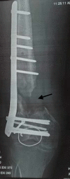
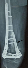
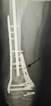
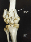
Dual-plating of the distal femur is required in some cases to achieve stable fixation. The indications of a medial plate in addition to the lateral plate are medial supracondylar bone loss, low trans-condylar bicondylar fractures, medial Hoffa fracture, peri-prosthetic distal femur fractures, non-union after failed fixation with single lateral plate, poor bone quality and comminuted distal femur fractures (AO type C3). We recommend orthogonal plate configuration with locked plates by a single incision or dual incision approach as per surgeon choice.
Keywords: distal femur; dual-plating; medial plate fixation.
Copyright © 2019, Sain et al.
Conflict of interest statement
The authors have declared that no competing interests exist.
Figures



References
-
- Double plating of intra-articular multifragmentary C3-type distal femoral fractures through the anterior approach. Imam MA, Torieh A, Matthana A. Eur J Orthop Surg Traumatol. 2018;28:121–130. - PubMed
-
- A double-plating approach to distal femur fracture: a clinical study. Steinberg E, Elis J, Steinberg Y, Salai M, Ben-Tov T. Injury. 2017;48:2260–2265. - PubMed
-
- Modern retrograde intramedullary nails versus periarticular locked plates for supracondylar femur fractures after total knee arthroplasty. Meneghini RM, Keyes BJ, Reddy KK, Maar DC. J Arthroplasty. 2014;29:1478–1481. - PubMed
-
- Double plating of distal femur fractures: indications and technique. Chapman J, Henley M. Tech Orthop. 1994;9:210–216.
Publication types
LinkOut - more resources
Full Text Sources
Miscellaneous
