

World Health Organization Grade III Supratentorial Extraventricular Ependymomas in Adults: Case Series and Review of Treatment Modalities
- PMID: 31903356
- PMCID: PMC6896608
- DOI: 10.4103/ajns.AJNS_239_18
World Health Organization Grade III Supratentorial Extraventricular Ependymomas in Adults: Case Series and Review of Treatment Modalities
Abstract
Context: Supratentorial ependymomas and their anaplastic variants are relatively uncommon central nervous system neoplasms that afflict both adults and children.
Aims: Discuss the clinical and pathological features of patients with anaplastic ependymomas involving an extraventricular supratentorial location and review modalities and options of treatment for those rare tumors.
Settings and design: Whereas the treatment algorithm in the pediatric population is well established, however, treatment in the adult population is less defined. Treatment options are exposed through the author's cases and review of the literature.
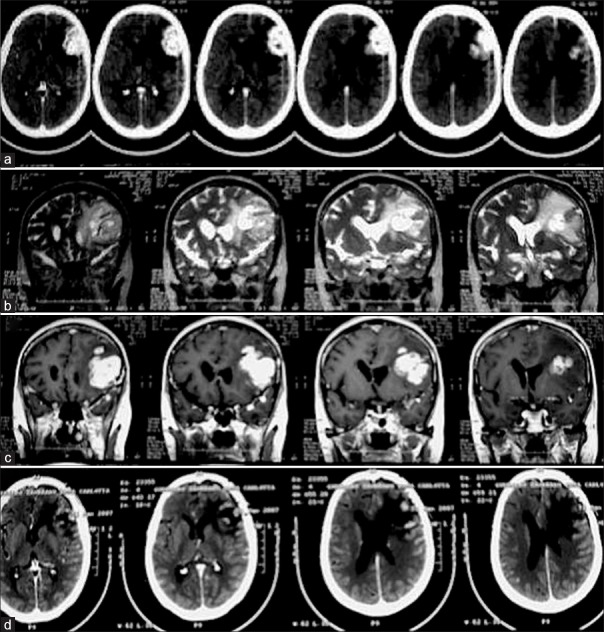
Subjects and methods: In our case series of two adult patients with supratentorial ependymomas World Health Organization (WHO) Grade III (anaplastic variant), patients presented in both cases in the emergency room after having a generalized tonic-clonic seizure at home the first case, and mild hemiparesis the second case.
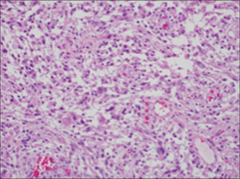
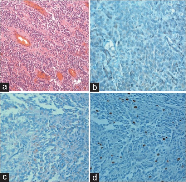
Results: Patients underwent surgical treatment, and a gross total resection was achieved in both cases. The histopathological examination revealed a diagnosis of anaplastic ependymoma (WHO Grade III). Both patients had additional radiotherapy, and in the first case, adjuvant platinum-based chemotherapy was administered due to leptomeningeal gliomatosis.
Conclusion: In our experience, gross total resection was achieved in all patients with supratentorial extraventricular ependymomas WHO Grade III with additional radiotherapy and platinum-based chemotherapy. Patients require initial close serial imaging follow-up. The role of chemotherapy is still uncertain but may be necessary in younger patients and in tumors that behave more like the pediatric ependymomas.
Keywords: Anaplastic ependymomas; extraventricular; supratentorial; surgery; treatment.
Copyright: © 2019 Asian Journal of Neurosurgery.
Conflict of interest statement
There are no conflicts of interest.
Figures

References
-
- Metellus P, Barrie M, Figarella-Branger D, Chinot O, Giorgi R, Gouvernet J, et al. Multicentric French study on adult intracranial ependymomas: Prognostic factors analysis and therapeutic considerations from a cohort of 152 patients. Brain. 2007;130:1338–49. - PubMed
-
- Barone BM, Elvidge AR. Ependymomas. A clinical survey. J Neurosurg. 1970;33:428–38. - PubMed
-
- Coulon RA, Till K. Intracranial ependymomas in children: A review of 43 cases. Childs Brain. 1977;3:154–68. - PubMed
-
- Mork SJ, Loken AC. Ependymoma: A follow-up study of 101 cases. Cancer. 1977;40:907–15. - PubMed