. 2019 Dec 20;5(12):eaay2118.
doi: 10.1126/sciadv.aay2118. eCollection 2019 Dec.

Fidelity of translation initiation is required for coordinated respiratory complex assembly

Affiliations

Fidelity of translation initiation is required for coordinated respiratory complex assembly

Danielle L Rudler et al. Sci Adv. .

Abstract

Mammalian mitochondrial ribosomes are unique molecular machines that translate 11 leaderless mRNAs; however, it is not clear how mitoribosomes initiate translation, since mitochondrial mRNAs lack untranslated regions. Mitochondrial translation initiation shares similarities with prokaryotes, such as the formation of a ternary complex of fMet-tRNAMet, mRNA and the 28S subunit, but differs in the requirements for initiation factors. Mitochondria have two initiation factors: MTIF2, which closes the decoding center and stabilizes the binding of the fMet-tRNAMet to the leaderless mRNAs, and MTIF3, whose role is not clear. We show that MTIF3 is essential for survival and that heart- and skeletal muscle-specific loss of MTIF3 causes cardiomyopathy. We identify increased but uncoordinated mitochondrial protein synthesis in mice lacking MTIF3, resulting in loss of specific respiratory complexes. Ribosome profiling shows that MTIF3 is required for recognition and regulation of translation initiation of mitochondrial mRNAs and for coordinated assembly of OXPHOS complexes in vivo.


Figures

Fig. 1
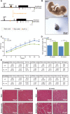
Fig. 1. Heart and skeletal muscle conditional knockout of mouse Mtif3 causes cardiomyopathy.
(A) Schematic showing the homologous recombination at the Mtif3 locus to generate conditional knockout mice. LoxP sites were introduced to allow the deletion of exon 3 by Cre recombinase. (B) Development of embryos of Mtif3+/+ and constitutive Mtif3−/− mice at embryonic day 8.5 (E8.5). (C) Weight differences between control (L/L) and knockout (L/L, cre) mice from 3 to 25 weeks of age. (D) Heart weight–to–tibia length ratio in control (L/L) and knockout (L/L, cre) mice at 10 and 25 weeks. (E) Echocardiographic parameters for control (L/L), and knockout (L/L, cre), 10- and 25-week-old mice. LVIDd, left ventricular end diastolic diameter; LVIDs, left ventricular end systolic diameter; FS, fractional shortening; LVPWd, left ventricular posterior wall in diastole; LVPWs, left ventricular posterior wall in systole; IVSd, intraventricular septum in diastole; IVSs, intraventricular septum in systole; HR, heart rate. (F) Heart and skeletal muscle sections cut to 5-μm thickness from 10-week-old and (G) 25-week-old L/L and L/L, cre mice were stained with H&E; yellow arrows show centralized nuclei in the skeletal muscle. Scale bars, 100 μm. All values are means ± SEM of n = 5. *P < 0.05, **P < 0.01, ***P < 0.001, Student’s t test.
Fig. 2
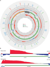
Fig. 2. Transcriptome analysis of mitochondrial transcripts by RNA-Seq.
(A) Complete map of changes in mitochondrial transcript abundance determined by RNA-Seq coverage from control (L/L) and knockout (L/L, cre) mice on heavy (outer track) and light (inner track) strands. Increases are shown in red, and decreases are shown in blue (log2[RPMKO/RPMWT]; scale, −1.0 to 2.0). The mitochondrial genome is displayed in the central track, with the nucleotide position in base pairs displayed across the exterior; rRNAs are displayed in orange, mRNAs are in green, tRNAs are in blue, and the noncoding region (NCR) is in gray. (B) Genome browser view of the mean RNA-Seq coverage (log2[RPMKO/RPMWT] of Co1 and Atp8/6 mRNAs (scale, −0.6 to 2.1).
Fig. 3
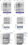
Fig. 3. Loss of MTIF3 results in uncoordinated mitochondrial protein synthesis.
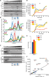
(A) Levels of de novo protein synthesis were measured in heart mitochondria from control (L/L) and knockout (L/L, cre) 10-week-old mice by pulse and chase incorporation of 35S-labeled cysteine and methionine. Mitochondrial protein was separated by SDS–polyacrylamide gel electrophoresis (SDS-PAGE), stained with Coomassie, and visualized by autoradiography. Representative gels of five independent biological experiments are shown. (B) Levels of de novo protein synthesis in heart mitochondria from 25-week-old control (L/L) and knockout (L/L, cre) mice, determined as in (A). Mitochondrial proteins from isolated heart mitochondria of control (L/L) and knockout (L/L, cre) 10-week-old (C) and 25-week-old (D) mice were resolved on 4 to 20% SDS-PAGE gels and immunoblotted using antibodies to investigate the steady-state levels of OXPHOS proteins. SDHA was used as a loading control. Levels of mitoribosomal proteins, proteases, and MTIF2 proteins from isolated heart mitochondria of control (L/L) and knockout (L/L, cre) 10-week-old (E) and 25-week-old (F) mice. Porin was used as a loading control. All values are means ± SEM. *P < 0.05, **P < 0.01, ***P < 0.001, Student’s t test.
Fig. 4
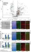
Fig. 4. Loss of MTIF3 affects the stability and assembly of the mitochondrial OXPHOS complexes.
(A) Quantitative proteomic analysis of mitochondrial proteins from control (L/L) and knockout (L/L, cre) 25-week-old mice. (B) Isolated heart mitochondria from 25-week-old mice were treated with 1% n-dodecyl-β-d-maltoside, resolved on 4 to 16% native bis-tris gels, and immunoblotted with the blue native OXPHOS cocktail antibody. (C) Isolated heart mitochondria from 25-week-old mice were treated with 1% digitonin, resolved on 4 to 16% native bis-tris gels, and immunoblotted to show complexes I and III (upper panels), complexes I and IV (middle panels), and complexes I and V (lower panels). (D) Oxygen consumption through the N-pathway and S-pathway using either pyruvate, glutamate, malate, or succinate as substrates in the absence or presence of mitochondrial inhibitors, and 2 μM carbonyl cyanide p-trifluoromethoxyphenylhydrazone (FCCP) was measured for leak (L), OXPHOS capacity (P), and ET capacity (ET) states in heart mitochondria from control (L/L) and knockout (L/L, cre) 25-week-old mice using an Oroboros oxygen electrode. All values are means ± SEM. *P < 0.05, **P < 0.01, Student’s t test.
Fig. 5
Fig. 5. MTIF3 does not affect mitochondrial ribosome assembly but alters the interactions between mitoribosomes and mt-mRNAs.
(A) Mitochondrial ribosome subunits and translating ribosomes were resolved on 10 to 30% sucrose gradients in control (L/L) and knockout (L/L, cre) 25-week-old mice. MTIF2 and the mitochondrial ribosomal protein markers of the small (MRPS34 and MRPS16) and large (MRPL44 and MRPL37) ribosomal subunits were detected by immunoblotting. The data are representative of results from four independent biological experiments. (B) The distributions of the 12S and 16S rRNAs and mRNAs in sucrose gradients were analyzed by qRT-PCR. The data are expressed as a percentage total of RNA abundance and show results from three independent biological replicates. (C) Mitochondrial ribosome subunits and translating ribosomes were resolved on 10 to 30% sucrose gradients in control (L/L) and knockout (L/L, cre) 25-week-old mice. MTIF3, MTIF2, TACO1, and LRPPRC were detected by immunoblotting. The data are representative of results from four independent biological experiments. (D) The normalized read length profile (reads per million) of the mitoribosome and its subunits on mRNAs is altered in the absence of MTIF3. (E) The association of tRNA-Met with the mitoribosome and its subunits is increased in the absence of MTIF3. (F) Enriched peaks at the 5′ end of mt-Atp8/6, mt-Nd4l/4, and mt-Nd5 mRNAs associating with the translating ribosome indicate ribosomal stalling. Peaks were considered 5′ or 3′ if they partially or fully overlapped a 100-nt window centered on the transcript 5′ or 3′ ends.
Fig. 6
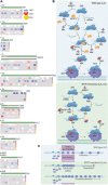
Fig. 6. The mechanism of MTIF3 and its role in mRNA recognition and translation initiation.
(A) Mitochondrial ribosome profiling shows the distribution of footprints on mt-mRNAs in the SSU (first two tracks), large subunit (middle two tracks), and mitoribosome (bottom two tracks) that are enriched (red) or depleted (blue) in the knockout compared to control mice. Double tracks per ribosomal fraction were used to distinguish partially overlapping footprints on the mRNAs. (B) Schematic showing the proposed roles of MTIF3 in mitochondrial translation (wild type, L/L). MTIF3 prevents the translation initiation complex formation if it is bound by a tRNA in the absence of mRNA. Only small ribosomal subunits that have bound mRNA before the recruitment of tRNA and MTIF2 are able to proceed from translation initiation to elongation. In the absence of MTIF3 (MTIF3 knockout, L/L,cre) preinitiation complexes cannot remove tRNAMet and positioning of the mRNA and its start codon cannot be monitored, so these complexes are able to participate in mature initiation complex formation. GDP, guanosine diphosphate; GTP, guanosine triphosphate. (C) Without the molecular proofreading steps performed by MTIF3, translation initiation proceeds at an accelerated rate but at the expense of fidelity. When fidelity of initiation is compromised, initiation complexes can stall at the 5′ ends of mRNAs, leaving the remainder of the mRNA prone to degradation by 3′-5′ exoribonucleases.

References

    1. Rackham O., Mercer T. R., Filipovska A., The human mitochondrial transcriptome and the RNA-binding proteins that regulate its expression. WIREs RNA 3, 675–695 (2012). - PubMed
    1. Greber B. J., Bieri P., Leibundgut M., Leitner A., Aebersold R., Boehringer D., Ban N., The complete structure of the 55S mammalian mitochondrial ribosome. Science 348, 303–308 (2015). - PubMed
    1. Amunts A., Brown A., Toots J., Scheres S. H. W., Ramakrishnan V., The structure of the human mitochondrial ribosome. Science 348, 95–98 (2015). - PMC - PubMed
    1. Suzuki T., Proteomic analysis of the mammalian mitochondrial ribosome. Identification of protein components in the 28 S small subunit. J. Biol. Chem. 276, 33181–33195 (2001). - PubMed
    1. Lee R. G., Rudler D. L., Rackham O., Filipovska A., Is mitochondrial gene expression coordinated or stochastic? Biochem. Soc. Trans. 46, 1239–1246 (2018). - PubMed

Publication types

MeSH terms