Review
. 2019 Feb;1(2):189-200.
doi: 10.1038/s42255-018-0021-8. Epub 2019 Jan 21.

Metabolic adaptation and maladaptation in adipose tissue

Affiliations
Review

Metabolic adaptation and maladaptation in adipose tissue

Edward T Chouchani et al. Nat Metab. 2019 Feb.

Abstract

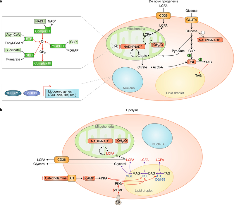
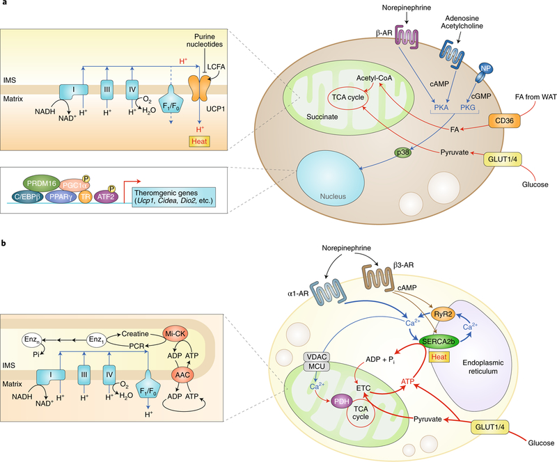
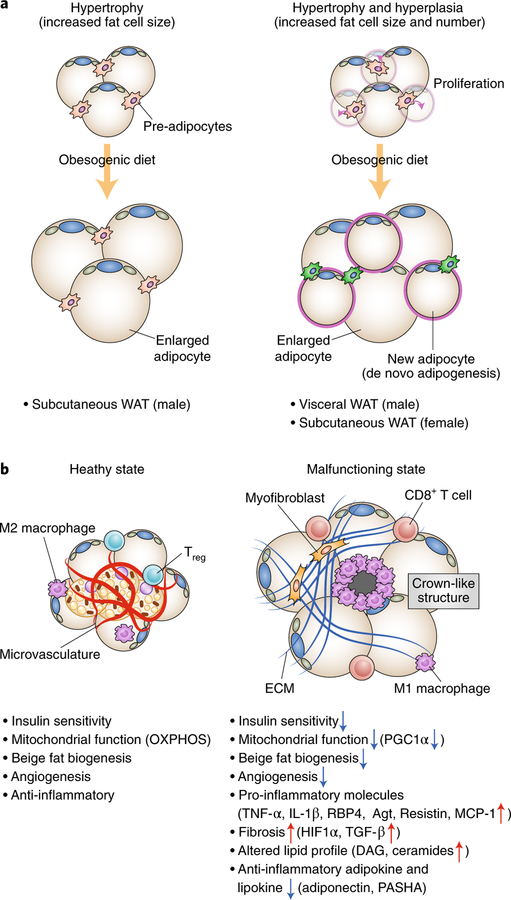
Adipose tissue possesses the remarkable capacity to control its size and function in response to a variety of internal and external cues, such as nutritional status and temperature. The regulatory circuits of fuel storage and oxidation in white adipocytes and thermogenic adipocytes (brown and beige adipocytes) play a central role in systemic energy homeostasis, whereas dysregulation of the pathways is closely associated with metabolic disorders and adipose tissue malfunction, including obesity, insulin resistance, chronic inflammation, mitochondrial dysfunction, and fibrosis. Recent studies have uncovered new regulatory elements that control the above parameters and provide new mechanistic opportunities to reprogram fat cell fate and function. In this Review, we provide an overview of the current understanding of adipocyte metabolism in physiology and disease and also discuss possible strategies to alter fuel utilization in fat cells to improve metabolic health.


Conflict of interest statement

Competing interests The authors declare no competing interests.

Figures

Fig. 1 |
Fig. 1 |. Control of fatty acid storage and oxidation.
a, Adipocytes access long-chain fatty acids (LCFAs) and glucose from the circulation. Fatty acids can be oxidized by mitochondrial beta-oxidation, which elevates the reduction state of the mitochondrial ubiquinone and NADH/NAD+ pool. Imported glucose drives maintenance of an elevated NADPH/NADP+ ratio (1), which is essential for supporting both TAG synthesis (2) and thiol redox state in the cell. Oxidized glucose also generates G3P, which is required to initiate DNL as the glycerol backbone (3). Citrate, generated by either glucose or fatty-acid-linked metabolism is exported by mitochondria to form cytosolic Acetyl-CoA, which is the building block of the elongated acyl chains of TAG upon DNL. Importantly, high mitochondrial Q and NADH/NAD+ reduction will feedback to inhibit mitochondrial citrate metabolism, and these may act as a metabolic signal to promote citrate export to the cytosol and DNL. Additionally, a high Q reduction state will inhibit oxidation of cytosolic G3P by the mitochondrial glycerophosphate dehydrogenase and similarly may promote DNL (2). An expanded schematic on the left (4) summarizes the major metabolic pathways that share electrons with the mitochondrial Q pool and that therefore are subject to feedback regulation on the basis of its reduction state (that is, the QH2/Q ratio). These include mitochondrial complex I-mediated oxidation of mitochondrial NADH; mitochondrial oxidation of fatty acids through the electron transferring flavoprotein (ETF); succinate oxidation by succinate dehydrogenase (SDH); and oxidation of cytosolic G3P by the mitochondrial glycerol-phosphate dehydrogenase (mGPDH). In the nucleus, the transcription factors ChREBP and SREBP1c control genes involved in DNL. b, Adipocytes are engaged to generate high levels of LCFAs locally via triglyceride lipolysis. This process is initiated by external stimulation, for example by catecholamines followed by elevation in cytosolic cAMP and PKA activation, or by NP followed by elevation in cGMP and PKG signalling. PKA or PKG signalling stimulates liberation of free fatty acids from TAG through endogenous lipases, such as ATGL, HSL, and MGL. Upon liberation, LCFAs are either oxidized locally or released into the circulation to supply fuel to other cells.
Fig. 2 |
Fig. 2 |. Cellular metabolism in thermogenic fat cells.
a, UCP1-dependent thermogenesis in brown and beige adipocytes involves active free fatty acid and glucose oxidation. In response to thermogenic stimuli, such as norepinephrine, adenosine, acetylcholine, or NP, the intracellular cAMP or cGMP level is elevated, which triggers PKA or PKG signalling, respectively. p38MAPK (p38) phosphorylates transcriptional regulators, including PGC1a and ATF2, and promote the expression of BAT-specific thermogenic genes (Ucp1, Cidea, and Dio2) and mitochondrial biogenesis. Free fatty acid and glucose are actively imported from the circulation through CD36 and glucose transporter 1 or 4 (Glut1/4), respectively, and are oxidized in the mitochondria for UCP1-dependent thermogenesis. UCP1 activity is inhibited by purine nucleotides, while long-chain free fatty acids bind to UCP1 and stimulate proton uncoupling. b, UCP1-independent thermogenesis in beige adipocytes involves Ca2+ cycling through the SERCA2b–RyR2 pathway in the sarco/endoplasmic reticulum and subsequent activation of PDH in the mitochondria. In the mitochondria, creatine substrate cycling also generates heat independently of UCP1. Both pathways require active ATP synthesis in the mitochondria (ATP-dependent thermogenesis) and glucose oxidation. FA, fatty acid; P, phosphorylation; Pi, inorganic phosphate.
Fig. 3 |
Fig. 3 |. Adaptation and maladaptation in adipose tissue.
a, Adipose tissue expansion occurs through hypertrophy and hyperplasia. Left, Adipose tissue hypertrophy involves increased cell size of existing adipocytes. Subcutaneous WAT in mice is increased in mass by hypertrophy. Right, Adipose tissue hyperplasia involves an increase in the number of adipocytes by adipocyte precursor proliferation and de novo adipogenesis. Visceral WAT of mice and subcutaneous WAT of female mice increase in mass through the combination of hypertrophy and hyperplasia. b, Hallmarks of healthy adipose tissue (left) and malfunctioning adipose tissue (right). Healthy adipose tissues are insulin sensitive with active mitochondrial biogenesis and OXPHOS. They also possess thermogenic beige adipocytes, increased angiogenesis, and anti-inflammatory M2 macrophages and regulatory T cells (Tregs). By contrast, malfunctioning ‘unhealthy’ adipose tissues are insulin resistant, and mitochondrial biogenesis and OXPHOS, beige adipocyte biogenesis, and angiogenesis are impaired. Adipose tissue expresses many pro-inflammatory cytokines (M1 macrophages and CD8+ T cells) and produces excess ECM from myofibroblasts, leading to the recruitment of pro-inflammatory immune cells and the formation of adipose tissue fibrosis and crown-like structures. Adipocytes also display altered lipid and adipokine and lipokine profiles, including increased DAG and ceramides as well as reduced secretion of adiponectin and palmitic acid hydroxystearic acids (PASHA).

Similar articles

Cited by

References

    1. Guarente L Mitochondria—a nexus for aging, calorie restriction, and sirtuins? Cell 132, 171–176 (2008). - PMC - PubMed
    1. Anderson RM & Weindruch R Metabolic reprogramming, caloric restriction and aging. Trends Endocrinol. Metab 21, 134–141 (2010). - PMC - PubMed
    1. Kajimura S, Spiegelman BM & Seale P Brown and beige fat: physiological roles beyond heat generation. Cell Metab 22, 546–559 (2015). - PMC - PubMed
    1. Sepa-Kishi DM, Sotoudeh-Nia Y, Iqbal A, Bikopoulos G & Ceddia RB Cold acclimation causes fiber type-specific responses in glucose and fat metabolism in rat skeletal muscles. Sci. Rep 7, 15430 (2017). - PMC - PubMed
    1. Jeffery E, Church CD, Holtrup B, Colman L & Rodeheffer MS Rapid depot-specific activation of adipocyte precursor cells at the onset of obesity. Nat. Cell Biol 17, 376–385 (2015). - PMC - PubMed

Publication types