. 2020 Feb;19(2):e13099.
doi: 10.1111/acel.13099. Epub 2020 Jan 5.

Intrinsically altered lung-resident γδT cells control lung melanoma by producing interleukin-17A in the elderly

Affiliations

Intrinsically altered lung-resident γδT cells control lung melanoma by producing interleukin-17A in the elderly

Min Cheng et al. Aging Cell. 2020 Feb.

Abstract

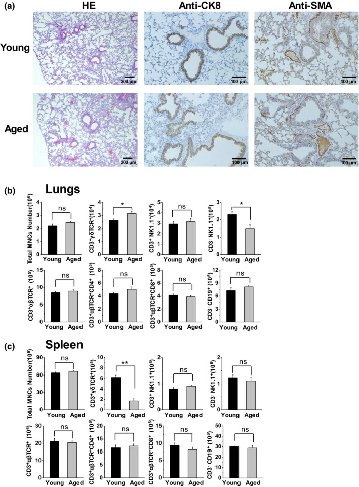
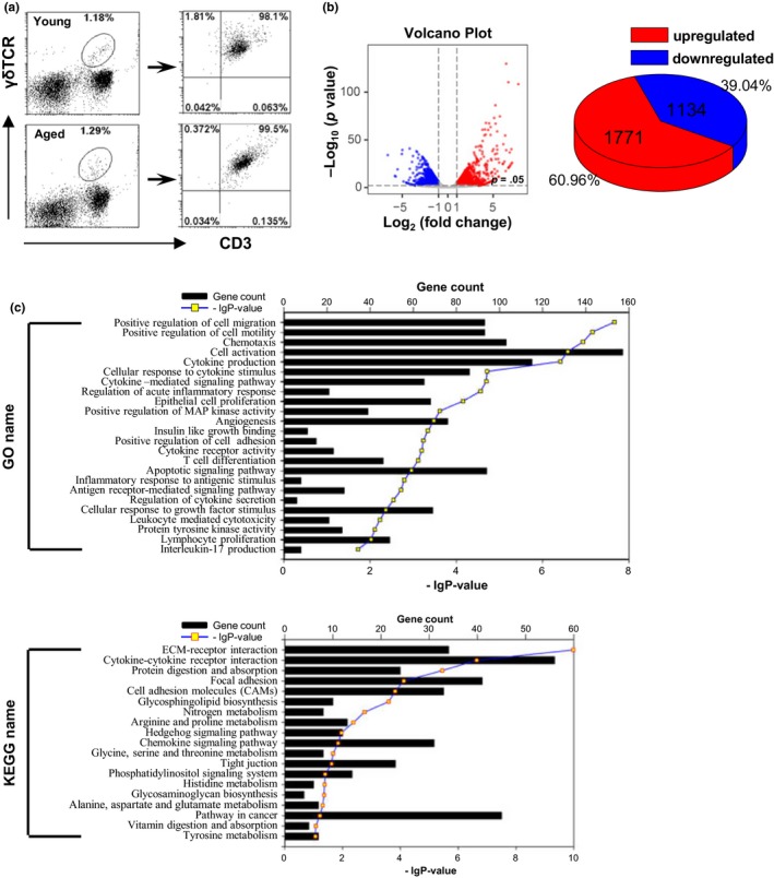
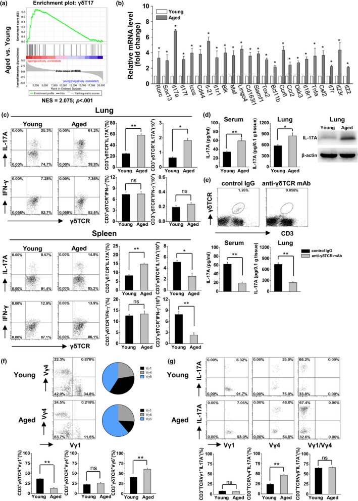
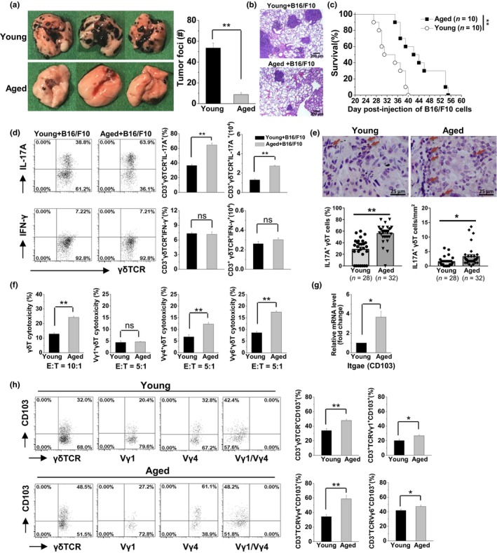
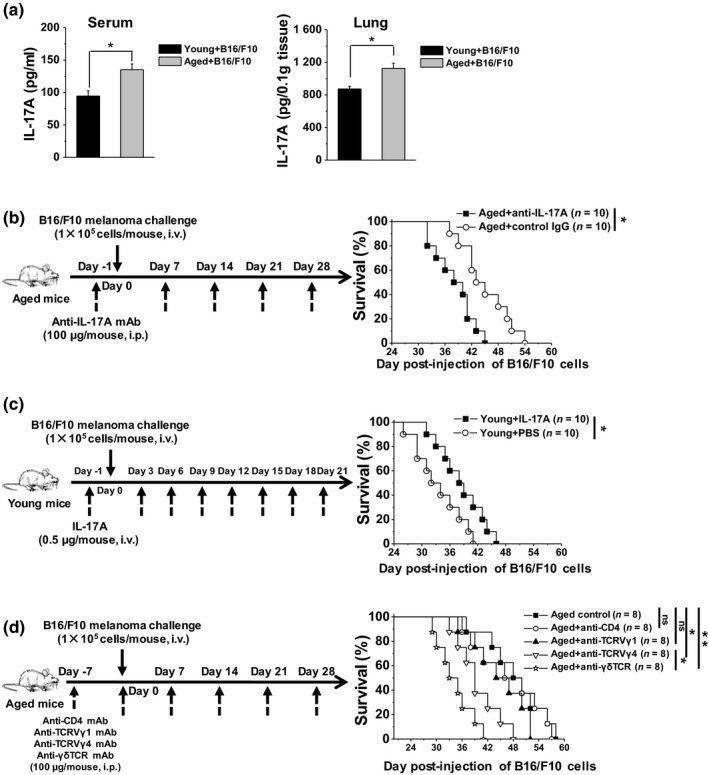
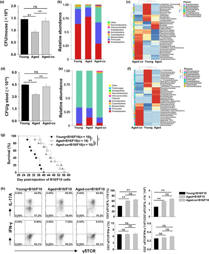
Cancer is an age-associated disease, potentially related to the altered immune system of elderly individuals. However, cancer has gradually decreased incidence in the eldest globally such as the most common lung cancer, the mechanisms of which remain to be elucidated. In this study, it was found that the number of lung-resident γδT cells was significantly increased with altered gene expression in aged mice (20-24 months) versus young mice (10-16 weeks). Aged lung Vγ4+ and Vγ6+ γδT cells predominantly produced interleukin-17A (IL-17A), resulting in increased levels in the serum and lungs. Moreover, the aged mice exhibited smaller tumors and reduced numbers of tumor foci in the lungs after challenge with intravenous injection of B16/F10 melanoma cells compared with the young mice. Aged lung Vγ4+ and Vγ6+ γδT cells were highly cytotoxic to B16/F10 melanoma cells with higher expression levels of CD103. The markedly longer survival of the challenged aged mice was dependent on γδT17 cells, since neutralization of IL-17A or depletion of indicated γδT cells significantly shortened the survival time. Consistently, supplementation of IL-17A significantly enhanced the survival time of young mice with lung melanoma. Furthermore, the anti-tumor activity of aged lung γδT17 cells was not affected by alterations in the load and composition of commensal microbiota, as demonstrated through co-housing of the aged and young mice. Intrinsically altered lung γδT17 cells underlying age-dependent changes control lung melanoma, which will help to better understand the lung cancer progression in the elderly and the potential use of γδT17 cells in anti-tumor immunotherapy.

Keywords: aging; commensal microbiota; interleukin-17A; lung cancer; lung-resident γδT cell.


Conflict of interest statement

The authors have declared no conflict of interests.

Figures

Figure 1
Figure 1
Increased number of γδT cells in the lungs of aged mice compared with young mice. (a) Lung samples were collected from young mice (10–16 weeks) and aged mice (20–24 months) for hematoxylin and eosin staining, and histochemical analysis using anti‐α‐SMA and anti‐CK8 antibodies. (b&c) The MNCs were isolated and analyzed using FACS. Lymphocytes were gated through FSC and SSC. The total number of MNCs and the absolute number of each lymphocyte subset in the lungs (b) and spleen (c) are shown (n = 6). The data are shown as the mean ± SEM. Student's t test was used. *p < .05, **p < .01. CK8, cytokeratin 8; FACS, fluorescence‐activated cell sorting; FSC, forward scatter; MNCs, mononuclear cells; SEM, standard error of the mean; SSC, side scatter; α‐SMA, alpha‐smooth muscle actin
Figure 2
Figure 2
The gene expression of lung γδT cells was distinguished between aged and young mice. (a) γδT cells (CD3+ γδTCR+) were purified from lung MNCs (20 mice/sample) and detected using FACS. Purified γδT cells were analyzed through mRNA sequencing. (b) The Volcano plots based on the fold change and p value showed the differential expression of the indicated genes. The two vertical lines correspond to a twofold change in expression. The horizontal line indicates p = .05. Red plots represent the upregulated genes. Blue plots represent the downregulated genes. The pie chart shows the distribution of DEGs in the aged group compared with young group. (c) The list of DEGs was converted into Entrez‐IDs for GO and KEGG analyses with r 3.2.3 using the library gostats 2.34.0 and the R bioconductor genomewide mouse annotations from the package org.Mm.eg.db (version 3.3.0). DEGs, differentially expressed genes; FACS, fluorescence‐activated cell sorting; GO, Gene Ontology; KEGG, Kyoto Encyclopedia of Genes and Genomes; MNCs, mononuclear cells
Figure 3
Figure 3
γδT17 cells were predominant in the lungs with high levels of IL‐17A production in aged mice. (a) According to the DEGs in lung γδT cells, GSEA from the aged versus young mice with normalized enrichment scores for γδT17 cell gene signatures. NES and p value are shown. (b) The mRNA expression levels of selected DEGs in the purified γδT cells (CD3+ γδTCR+) were measured using real‐time PCR (n = 3). (c) The MNCs were isolated from lung and spleen and analyzed using FACS. The CD3+ γδTCR+ cells were gated to analyze the frequency and number of CD3+ γδTCR+ IL‐17A+ cells (γδT17) and CD3+ γδTCR+ IFN‐γ+ cells (γδT1). There were six mice in each group. (d) The expression levels of IL‐17A protein in the serum were detected using ELISA (n = 3). In lung tissue, these levels were detected through ELISA and Western blotting. The results are representative of three independent experiments. (e) Inhibition of γδT cells was performed through injection of anti‐γδTCR mAb (UC7‐13D5, 200 μg/mouse, i.p.) twice every 3 days. Three days after the treatment, CD3+ γδTCR+ cells stained positively with a different monoclonal antibody against γδTCR (GL3) were analyzed using FACS. The expression levels of IL‐17A protein in the serum and lung tissue were detected using ELISA (n = 3). (f) Usage of the Vγ chain (Vγ1, Vγ4, and Vγ6) was analyzed for the lung γδT cells (CD3+ γδTCR+). Vγ6+ γδT cells were considered as Vγ1Vγ4 in the lungs. (g) Each γδT‐cell subset (CD3+ γδTCR+ Vγ1+, CD3+ γδTCR+ Vγ4+, and CD3+ γδTCR+ Vγ1Vγ4) was gated to analyze the production of IL‐17A. There were six mice in each group. The data are shown as the mean ± SEM. Student's t test was used. *p < .05, **p < .01. DEGs, differentially expressed genes; ELISA, enzyme‐linked immunosorbent assay; FACS, fluorescence‐activated cell sorting; GSEA, gene set enrichment analysis; mAb, monoclonal antibody; NES, normalized enrichment scores
Figure 4
Figure 4
Higher anti‐tumor activity of γδT17 cells was associated with resistance to B16 melanoma metastasis in the lungs of aged mice. Young and aged mice were challenged with B16/F10 cells (1 × 105 cells/mouse, i.v.). On day 21 after the B16/F10 challenge, the lungs were analyzed. (a) The graphs showed the tumor nodules in the lungs and the total number of tumor foci was calculated. Data are shown as mean ± SEM. Student's t test was used. **p < .01. (b) The lung samples were collected for hematoxylin and eosin staining. (c) The survival rate of B16/F10 challenged mice was calculated and analyzed using the Kaplan–Meier method. (d) On day 21 after the B16/F10 challenge, the CD3+ γδTCR+ cells were gated to analyze the frequency and number of CD3+ γδTCR+ IL‐17A+ cells (γδT17) and CD3+ γδTCR+ IFN‐γ+ cells (γδT1) in the lungs. There were six mice in each group. (e) Infiltrated γδT 17 cells in the lung cancer tissues in the aged patients compared with younger patients through histochemical analysis. γδTCR+ cells were shown as brown, IL‐17A+ cells were shown as red, γδTCR+ IL‐17A+ double‐positive cells were shown as brown‐red. The double‐positive cells of γδTCR+IL‐17A+ were indicated by the arrows. The percentages and numbers of γδTCR+IL‐17A+cells were calculated in each sample. (f) The cytotoxicity of purified lung γδT cells (CD3+ γδTCR+) against B16/F10 cells (E:T = 10:1), purified lung Vγ1+γδT cells (CD3+ γδTCR+ Vγ1+), Vγ4+γδT cells (CD3+ γδTCR+ Vγ4+), and Vγ6+γδT cells (CD3+ γδTCR+ Vγ1 Vγ4) against B16/F10 cells (E:T = 5:1). Data are shown as the mean ± SEM from triplicates of one of the three independent experiments. (g) The mRNA expression levels of CD103 in the purified γδT cells (CD3+ γδTCR+) were measured using real‐time PCR. (h) The expression levels of CD103 on each lung γδT‐cell subset were detected through flow cytometry analysis. The data are shown as the mean ± SEM. Student's t test was used. **p < .01. *p < .05. PCR, polymerase chain reaction; TCR, T‐cell antigen receptor
Figure 5
Figure 5
Aged mice exhibited higher anti‐tumor activity dependent on IL‐17A in the lungs. (a) On day 21 after the B16/F10 challenge (1 × 105 cells/mouse, i.v.), the expression levels of IL‐17A protein in the serum and lung tissue were detected using ELISA (n = 3). The data are shown as the mean ± SEM. Student's t test was used. *p < .05. (b) Anti‐IL‐17A mAb was injected into the aged mice 1 day prior to injection of B16/F10 cells (1 × 105 cells/mouse, i.v.). Additional injections were performed every 7 days. (c) IL‐17A was injected into young mice 1 day prior to injection of B16/F10 (1 × 105 cells/mouse, i.v.). Additional injections were performed every 3 days. (d) Anti‐CD4 mAb, anti‐TCRVγ1, anti‐TCRVγ4, and anti‐γδTCR antibodies were injected i.p. into the aged mice 7 days prior to injection of B16/F10 cells (1 × 105 cells/mouse, i.v.). Additional injections were performed every 7 days. The survival rate of B16/F10‐challenged mice was calculated and analyzed using the Kaplan–Meier method. *p < .05, **p < .01. ELISA, enzyme‐linked immunosorbent assay; mAb, monoclonal antibody: TCR, T‐cell antigen receptor
Figure 6
Figure 6
γδT cells showed intrinsic anti‐tumor activity with higher levels of IL‐17 production in the co‐housed aged mice independent of the alterations in the load and composition of commensal microbiota. In the co‐culture group, the aged mice were co‐housed with the young mice for 4 weeks. Bacterial loads were measured using BAP culture in the upper respiratory tract (a) and stool (d) from the co‐cultured aged mice compared with the control mice (n = 3/group). The data are shown as the mean ± SEM. Analysis of variance (one‐way ANOVA) was used. **p < .01. Relative abundance (b) and a clustering map (c) for the bacteria in the upper respiratory tract were determined through 16S rRNA analysis (3 samples/group, 10 mice/sample). Relative abundance (e) and a clustering map (f) for the bacteria in the stool were determined through 16S rRNA analysis (3 mice/group). (g) The co‐housed mice were challenged with B16/F10 cells (1 × 105 cells/mouse, i.v.). The survival rates were calculated and analyzed using the Kaplan–Meier method. **p < .01. (h) On day 21 after the B16/F10 challenge, the MNCs isolated from the lungs were analyzed using FACS. The CD3+ γδTCR+ cells were gated to analyze the frequency and number of IL‐17A+ CD3+ γδTCR+ cells (γδT17) and IFN‐γ+ CD3+ γδTCR+ cells (γδT1). The data are shown as the mean ± SEM (n = 6). Analysis of variance (one‐way ANOVA) was performed. **p < .01. BAP, blood agar plate; FACS, fluorescence‐activated cell sorting; MNCs, mononuclear cells; TCR, T‐cell antigen receptor

Similar articles

Cited by

References

    1. Bai, H. , Gao, X. , Zhao, L. , Peng, Y. , Yang, J. , Qiao, S. , … Yang, X. (2017). Respective IL‐17A production by gammadelta T and Th17 cells and its implication in host defense against chlamydial lung infection. Cellular & Molecular Immunology, 14, 850–861. - PMC - PubMed
    1. Boe, D. M. , Boule, L. A. , & Kovacs, E. J. (2017). Innate immune responses in the ageing lung. Clinical and Experimental Immunology, 187, 16–25. - PMC - PubMed
    1. Bourke, W. , Milstein, D. , Giura, R. , Donghi, M. , Luisetti, M. , Rubin, A. H. , & Smith, L. J. (1992). Lung cancer in young adults. Chest, 102, 1723–1729. - PubMed
    1. Bray, F. , Ferlay, J. , Soerjomataram, I. , Siegel, R. L. , Torre, L. A. , & Jemal, A. (2018). Global cancer statistics 2018: GLOBOCAN estimates of incidence and mortality worldwide for 36 cancers in 185 countries. CA: A Cancer Journal for Clinicians, 68(6), 394–424. - PubMed
    1. Chen, W. , Zheng, R. , Baade, P. D. , Zhang, S. , Zeng, H. , Bray, F. , … He, J. (2016a). Cancer statistics in China, 2015. CA: A Cancer Journal for Clinicians, 66(2), 115–132. - PubMed

Publication types

MeSH terms