. 2020 Feb;28(2):303-314.
doi: 10.1002/oby.22683. Epub 2020 Jan 5.

Transendothelial Insulin Transport is Impaired in Skeletal Muscle Capillaries of Obese Male Mice

Affiliations

Transendothelial Insulin Transport is Impaired in Skeletal Muscle Capillaries of Obese Male Mice

Ian M Williams et al. Obesity (Silver Spring). 2020 Feb.

Abstract

Objective: The continuous endothelium of skeletal muscle (SkM) capillaries regulates insulin's access to skeletal myocytes. Whether impaired transendothelial insulin transport (EIT) contributes to SkM insulin resistance (IR), however, is unknown.

Methods: Male and female C57/Bl6 mice were fed either chow or a high-fat diet for 16 weeks. Intravital microscopy was used to measure EIT in SkM capillaries, electron microscopy to assess endothelial ultrastructure, and glucose tracers to measure indices of glucose metabolism.

Results: Diet-induced obesity (DIO) male mice were found to have a ~15% reduction in EIT compared with lean mice. Impaired EIT was associated with a 45% reduction in endothelial vesicles. Despite impaired EIT, hyperinsulinemia sustained delivery of insulin to the interstitial space in DIO male mice. Even with sustained interstitial insulin delivery, DIO male mice still showed SkM IR indicating severe myocellular IR in this model. Interestingly, there was no difference in EIT, endothelial ultrastructure, or SkM insulin sensitivity between lean female mice and female mice fed a high-fat diet.

Conclusions: These results suggest that, in male mice, obesity results in ultrastructural alterations to the capillary endothelium that delay EIT. Nonetheless, the myocyte appears to exceed the endothelium as a contributor to SkM IR in DIO male mice.


Conflict of interest statement

Disclosure: While this study was being conducted, F.A.V. and J.S.B were employees of Eli Lilly and Company, a pharmaceutical company. Eli Lilly generously provided the fluorescent insulin probe (INS-647) used in the study. The authors declare no other competing financial interests.

Figures

Figure 1:
Figure 1:. Skeletal muscle capillaries of DIO male mice contain fewer endothelial vesicles than lean mice.
A) Representative electron micrographs of the capillary endothelium in the gastrocnemius of lean and DIO male mice. B) Volume of vesicles relative to total endothelial volume in lean (n=5) and DIO (n=4) male mice. C) Frequency distribution of endothelial vesicle densities in all capillaries grouped from lean (n=64) and DIO male mice (n=66). D) The average diameter of all endothelial vesicles in lean (n=227) and DIO (n=116) male mice. E) The circularity of vesicles in lean (n=146) and DIO (n=88) male mice. Circularity values of 1 and 0 indicate perfect circles and very elongated shapes, respectively. F) Frequency distribution of the localization of vesicles in the capillary endothelium. G) Average basement membrane thickness in capillaries from lean (n=31) and DIO male mice (n=31). In the box and whisker blots, the box extends from the 25th to the 75th percentiles and the whiskers indicate the range. H) The number of test grid points counted within each capillary segment in lean (n=184) and DIO (n=181) male mice, an index of endothelial volume. Groups were compared using Student’s t-test. C – capillary lumen, EC – endothelial cell, BM – basement membrane, DIO – diet-induced obese.
Figure 2:
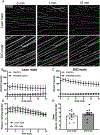
Figure 2:. Obese male mice have impaired trans-endothelial insulin transport in skeletal muscle capillaries.
A) Representative INS-647 images (maximum intensity projections) in lean (n=12) and DIO (n=7) male mice. B-C) Capillary, interstitial, and total extravascular INS-647 in the field of view as a function of time following the beginning of imaging in B) lean and C) DIO male mice. T = 0 min indicates the beginning of imaging which occurs ~15 seconds after injection of INS-647. The interstitial space is defined as the region emanating 1–3μm from the capillary wall. D) The ratio of plasma to interstitial INS-647 as a function of time following INS-647 injection, normalized to the ratio at t = 0 min. E) Decay constant of the plasma / interstitial INS-647 ratio, a measure of trans-endothelial insulin transport kinetics. Groups were compared using Student’s t-test. INS-647 – insulin-647.
Figure 3:
Figure 3:. DIO male mice display skeletal muscle insulin resistance.
A) Glucose excursions in anesthetized lean (n=5) and DIO (n=7) male mice following a 4U/kg intravenous insulin bolus. B) Area under the glucose excursion curves in A. C) Plasma insulin excursions in lean (n=4) and DIO (n=7) male mice following a 4U/kg intravenous insulin bolus. D) Area under the insulin excursion curves in C. E) Clearance of 2[14C]deoxyglucose during the insulin tolerance tests by the soleus, gastrocnemius, and vastus muscles. Groups were compared using Student’s t-test.
Figure 4:
Figure 4:. Capillary trans-endothelial insulin transport in skeletal muscle does not involve the insulin receptor or endothelial insulin accumulation.
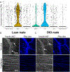
A) Violin plots of insulin receptor mRNA expression in tissue-specific endothelial cells as determined by single-cell RNA sequencing. B,C) Intravital microscopy images of INS-647 and rhodamine-labeled 2MDa dextran (Rho-dex) in venules (top panels) and capillaries (bottom panels) from B) lean and C) DIO male mice. Both INS-647 and rho-dex can be seen accumulating in the endothelium of venules but not capillaries. Arrows indicate regions of insulin accumulation in the venular endothelium. Rho-dex – 2MDa tetramethylrhodamine-dextran, CPM – counts per million.
Figure 5:
Figure 5:. No difference in albumin equilibration between lean and DIO male mice.
A) Representative Alb-647 images (maximum intensity projections) in lean (n=11) and DIO (n=13) male mice. B-C) Capillary and interstitial Alb-647 intensity as a function of time following injection in B) lean and C) DIO male mice. The interstitial space is defined as the region emanating 1–3μm from the capillary wall. D) The ratio of plasma to interstitial Alb-647 as a function of time following Alb-647 injection. E) Mean plasma to interstitial Alb-647 ratio over the course of the experiment. Groups were compared using Student’s t-test. Alb-647 – albumin-647.
Figure 6:
Figure 6:. No effect of HFD on endothelial vesicles in female mice.
A) Representative electron micrographs of the capillary endothelium in the gastrocnemius of chow and HFD-fed female mice. B) Volume of vesicles relative to total endothelial volume in chow (n=5) and HFD-fed (n=4) female mice. C) Frequency distribution of endothelial vesicular densities in all capillaries pooled from chow (n=49) and HFD-fed female mice (n=40). Groups were compared by Student’s t-test.
Figure 7:
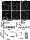
Figure 7:. HFD does not alter trans-endothelial insulin transport in females.
A) Representative INS-647 images (maximum intensity projections) in chow (n=6) and HFD-fed (n=8) female mice. B-C) Capillary, interstitial, and total extravascular INS-647 in the field of view as a function of time following injection in B) chow and C) HFD-fed female mice. The interstitial space is defined as the region emanating 1–3μm from the capillary wall. D) The ratio of plasma to interstitial INS-647 as a function of time following INS-647 injection, normalized to the ratio at t = 0 min. E) The gradient decay constant of the plasma to interstitial INS-647 ratio as a function of time. Groups were compared using Student’s t-test.

Similar articles

Cited by

References

    1. Rabinowitz D, and Zierler KL (1962) Forearm metabolism in obesity and its response to intra-arterial insulin. Characterization of insulin resistance and evidence for adaptive hyperinsulinism. J. Clin. Invest 41, 2173–81 - PMC - PubMed
    1. Laakso M, Edelman SV, Brechtel G, and Baron AD (1990) Decreased effect of insulin to stimulate skeletal muscle blood flow in obese man. A novel mechanism for insulin resistance. J. Clin. Invest 85, 1844–52 - PMC - PubMed
    1. DeFronzo RA, and Tripathy D (2009) Skeletal muscle insulin resistance is the primary defect in type 2 diabetes. Diabetes Care. 32 Suppl 2, S157–63 - PMC - PubMed
    1. Chen YL, and Messina EJ (1996) Dilation of isolated skeletal muscle arterioles by insulin is endothelium dependent and nitric oxide mediated. Am. J. Physiol 270, H2120–4 - PubMed
    1. Barrett EJ, Wang H, Upchurch CT, and Liu Z (2011) Insulin regulates its own delivery to skeletal muscle by feed-forward actions on the vasculature. AJP Endocrinol. Metab 301, E252–E263 - PMC - PubMed

Publication types