. 2020 Jan 31;40(1):BSR20192666.
doi: 10.1042/BSR20192666.

The novel testicular enrichment protein Cfap58 is required for Notch-associated ciliogenesis

Affiliations

The novel testicular enrichment protein Cfap58 is required for Notch-associated ciliogenesis

Zheng-Zheng Li et al. Biosci Rep. .

Abstract

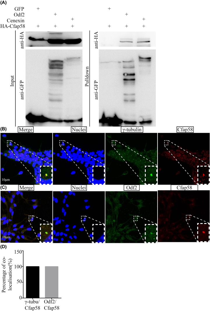
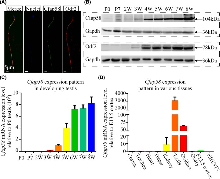
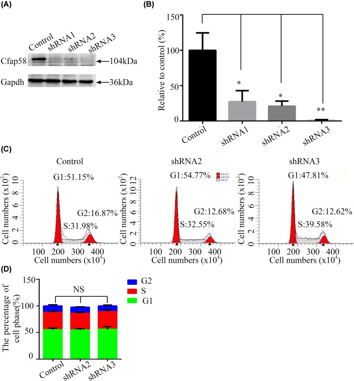
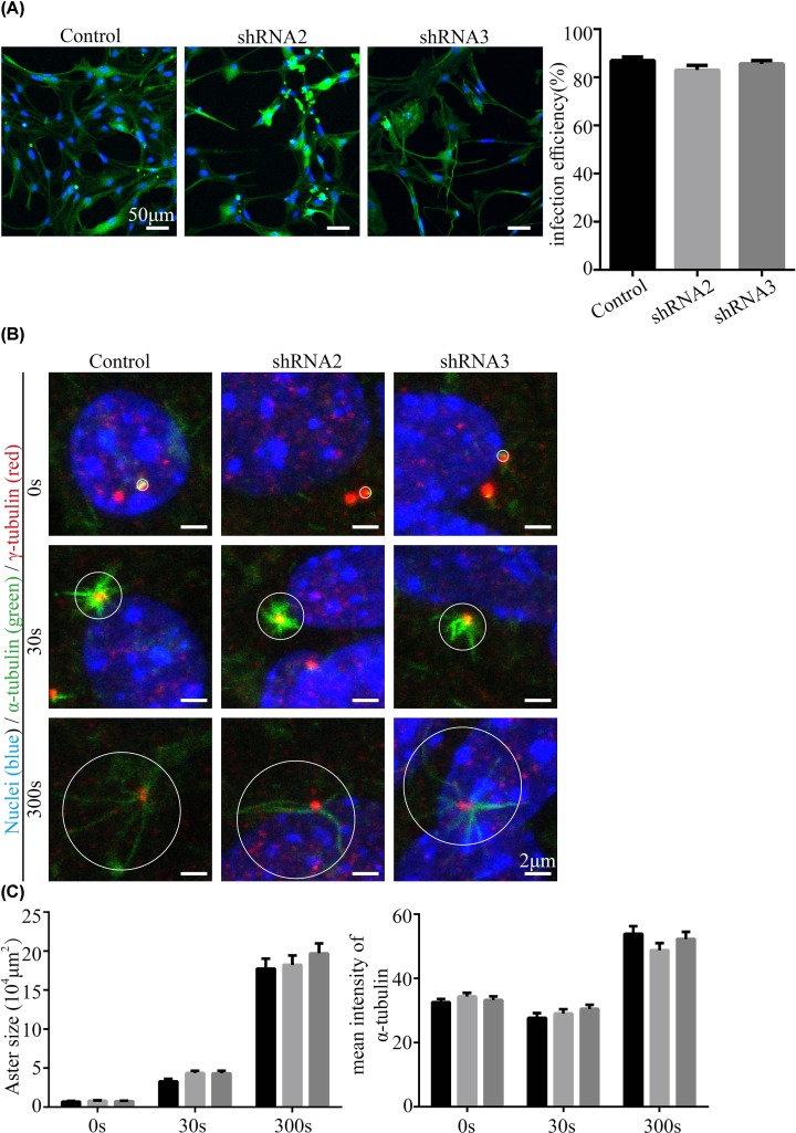
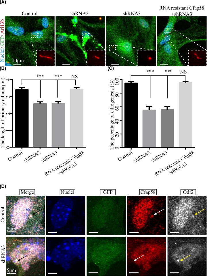
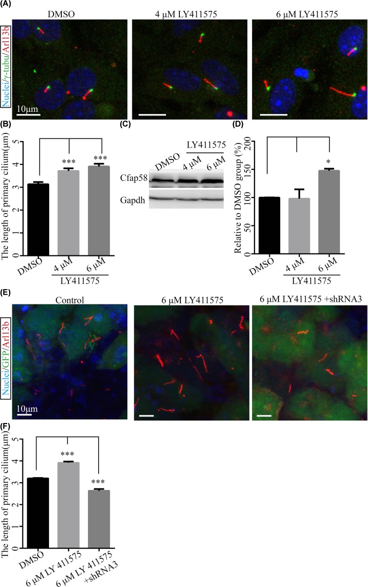
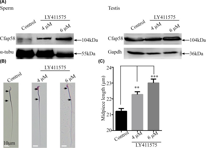
Cilia and flagella are critical organelles with conserved internal structures and diverse developmental and physiological processes according to cell type. Although the core components of structures are shared with thousands of associated proteins involved in cilia or flagella formation, we hypothesized that some unknown proteins, such as outer dense fiber 2 (Odf2/Cenexin) perform distinct functions in these organelles. In the present study, we identified several uncharacterized proteins through mass spectrometry interactome analysis of Odf2/Cenexin proteins. We further examined the expression patterns and functions of a protein named cilia and flagella associated protein 58 (Cfap58) in cultured astrocytes and sperm flagella. The results of a combination of biochemical analyses and drug administration studies reveal that Cfap58 is a testis-enrichment protein that exhibits similar localization to Odf2/Cenexin proteins and is required for the elongation of the primary cilium and sperm midpiece via modulation of the Notch signaling pathway. However, the cell cycle-related functions and localization of Odf2/Cenexin in the mother centriole were not altered in Cfap58 knockdown cells. These findings indicate that Cfap58 may be partially recruited by Odf2/Cenexin proteins and is indispensable for the cilia and flagellar assembly. These data provide us with a better understanding of ciliogenesis and flagellar elongation and may aid in identifying new targets for diseases caused by Notch-mediated ciliopathies and flagellar abnormalities.

Keywords: centrosomes; cilia; notch signalling pathway.


Conflict of interest statement

The authors declare that there are no competing interests associated with the manuscript.

Figures

Figure 1
Figure 1. Cfap58 interacts with Odf2/Cenexin and localizes in centrosome/basal body and sperm flagellum abundantly
(A) Western blot analysis shows the interaction between Cfap58 and Odf2/Cenexin in vitro. (B) Localization of γ-tubulin (green) and Cfap58 (red) in the centrosome in mouse astrocytes. Nuclei (blue) are stained with Hoechst34580. Scale bar = 10 μm. (C) Co-localization of Odf2/Cenexin (green) and Cfap58 (red) in mouse astrocytes. Nuclei (blue) are stained with Hoechst34580. Scale bar = 10 μm. (D) Statistical results the percentage of localization of γ-tubulin/Cfap58 and Odf2/Cfap58 for (B,C) (n=50 dots/group). Data are shown as the means.
Figure 2
Figure 2. Cfap58 is a testis-enriched protein
(A) Localization of Cfap58 (green) and Odf2 (red) in mouse sperm flagellum. Nuclei (blue) are stained with Hoechst34580. Scale bar = 5 μm. (B) Western blot analysis were performed that the expression patterns of Cfap58 and Odf2 in mouse developing testes. (C,D) qRT-PCR analysis of Cfap58 mRNA in and developing testes (C) and mouse adult tissues (D). Mouse Gapdh mRNA level was an internal control. Data are shown as the means ± SEM.
Figure 3
Figure 3. No significant alteration in cell growth and cell cycle was observed after down-regulation of Cfap58 in astrocytes
(A) Western blot analysis exhibits validation of shRNA vectors targeted to mouse HA-Cfap58 in HEK293T cells. (B) Statistical result shows the silence efficiency of Cfap58 shRNA vectors. (C,D) Representative FACS results (C) and statistical results (D) show no significant difference in the proportion of G0/G1, G2/M and S phase between negative control and Cfap58 knockdown groups. Data are shown as the mean ± SEM. NS, P>0.05; *, P≤0.05; **, P≤0.01.
Figure 4
Figure 4. MTOC activity of the centriole does not change after the down-regulation of Cfap58 expression in astrocytes
(A) Immunostaining data (left panel) and statistical results (right panel) show the infection efficiency of control and Cfap58 shRNAs virus. (B) Centrioles stained by γ-tubulin antibody (red) show regrowth of microtubules stained by α-tubulin antibody (green) in time course. Nuclei (blue) are stained with Hoechst34580. Scale bar = 2 μm. (C) Statistical results show the areas and mean α-tubulin intensities of asters in (B) after nocodazole treatment in the scramble, Cfap58 shRNA2 and shRNA3 groups. (n≥50 asters, the white cycles indicated asters and measurements). Data are shown as the means ± SEM. NS, P>0.05
Figure 5
Figure 5. Silencing Cfap58 in astrocytes causes ciliogenesis deficiency without the mislocalization of Odf2/Cenexin
(A) Immunostaining analysis shows that the number and length of cilia are reduced in Cfap58 KD cells, and the RNAi-resistant Cfap58 + shRNA3 group compared with the control group. Cells were stained with Arl13b antibody (red), GFP antibody (green) or Hoechst34580 indicating the primary cilia marker, infected cells marker and nuclei, respectively. Scale bar = 5 μm. (B) Statistical results show the percentages of ciliated cells in control, Cfap58 shRNA2, shRNA3 and rescue groups. (C) Statistical results show the length of cilia in the scramble, Cfap58 KD and rescue groups. (D) Triple immunostaining with Odf2/Cenexin (white) antibody and Cfap58 (red) antibody in control cells and Cfap58 KD cells shows that the localization of Odf2/Cenexin is not altered in astrocytes. GFP (green) indicates infected cells. n=25 and 30 cells for control and Cfap58 KD cells. Scale bar = 5 μm. Data are shown as the means ± SEM. NS, P>0.05; ***, P≤0.001.
Figure 6
Figure 6. Inhibition of Notch signaling activity facilitates ciliogenesis by increasing Cfap58 protein expression in astrocytes
(A) Representative immunofluorescent images of mouse astrocytes cultured for 3 days with 0, 4 or 6 μM LY411575 stained with Hocesht34580 (blue), γ-tubulin (green) and Arl13b antibody (red) as nuclei, basal body and primary cilia, respectively. Scale bar = 10 μm. (B) Statistical results show a significant increase in the length of cilia after inactivation of Notch signaling in astrocytes. (C) Western blot analysis shows up-regulation of Cfap58 in astrocytes treated with LY411575 in a dose-dependent manner for 3 days. The Gapdh protein level was as a loading control. (D) Histogram indicates the statistical results of Cfap58 expression level (three times). (E) Representative immunofluorescent images of mouse astrocytes treated with LY411575 for a day post-infection 3 days with the control and Cfap58 shRNA3 lentivirus. The nuclei stained with Hoechst34580 (blue), GFP (infected cells, green) and Arl13b (primary cilia). (F) Statistical results exhibit the length of cilia for (E). Data are shown as the means ± SEM. *, P≤0.05, ***, P≤0.001.
Figure 7
Figure 7. LY411575 enhances Cfap58 expression and the length of the midpiece in mouse sperm in vivo
(A) Cfap58 expression is increased in mouse sperms but not adult testis. (B) Representative images of sperms stained by Giemsa dye to highlight the length of sperm flagellum. Black arrows indicate the region of the midpiece. (C) The histogram shows that the length of the midpiece is increased after LY411575 treatment for 35 days. Data are shown as the means ± SEM. **, P≤0.01; ***, P≤0.001

Similar articles

Cited by

References

    1. Gerdes J.M., Davis E.E. and Katsanis N. (2009) The vertebrate primary cilium in development, homeostasis, and disease. Cell 137, 32–45 10.1016/j.cell.2009.03.023 - DOI - PMC - PubMed
    1. Lindemann C.B. and Lesich K.A. (2016) Functional anatomy of the mammalian sperm flagellum. Cytoskeleton (Hoboken) 73, 652–669 10.1002/cm.21338 - DOI - PubMed
    1. Lee M.S., Hwang K.S., Oh H.W. et al. . (2015) IFT46 plays an essential role in cilia development. Dev. Biol. 400, 248–257 10.1016/j.ydbio.2015.02.009 - DOI - PMC - PubMed
    1. Zhao H., Zhu L., Zhu Y. et al. . (2013) The Cep63 paralogue Deup1 enables massive de novo centriole biogenesis for vertebrate multiciliogenesis. Nat. Cell Biol. 15, 1434–1444 10.1038/ncb2880 - DOI - PubMed
    1. Wu J., Bao J., Kim M. et al. . (2014) Two miRNA clusters, miR-34b/c and miR-449, are essential for normal brain development, motile ciliogenesis, and spermatogenesis. Proc. Natl. Acad. Sci. U.S.A. 111, E2851–E2857 10.1073/pnas.1407777111 - DOI - PMC - PubMed

Publication types