. 2020 Apr:326:113168.
doi: 10.1016/j.expneurol.2019.113168. Epub 2020 Jan 2.

Cuprizone-induced demyelination under physiological and post-stroke condition leads to decreased neurogenesis response in adult mouse brain

Affiliations

Cuprizone-induced demyelination under physiological and post-stroke condition leads to decreased neurogenesis response in adult mouse brain

Fucheng Luo et al. Exp Neurol. 2020 Apr.

Abstract

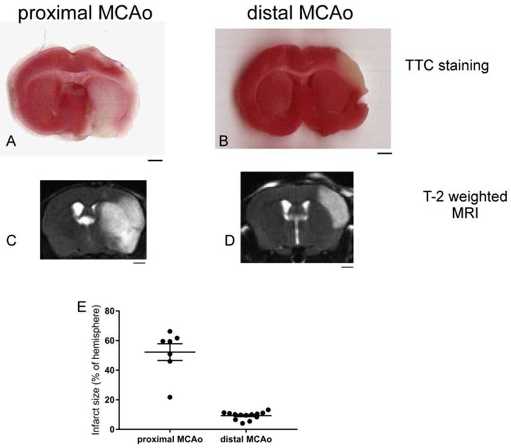
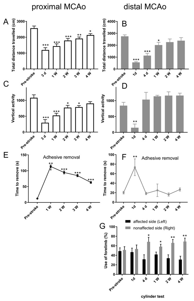
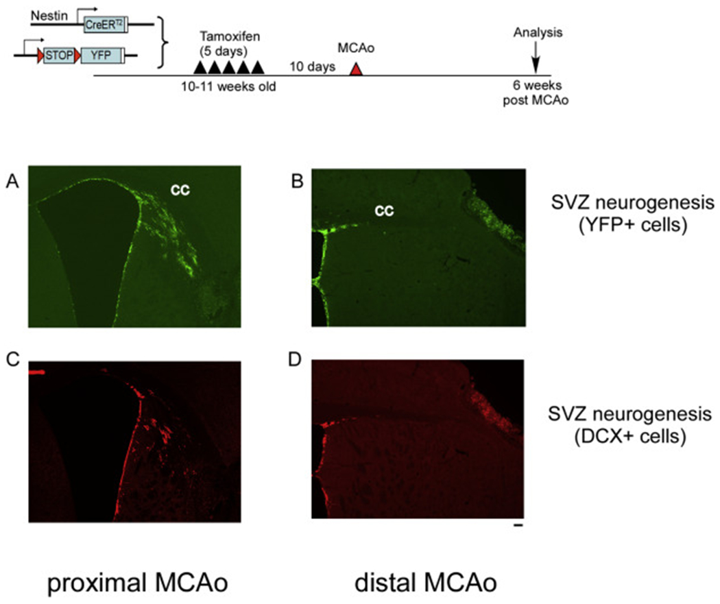
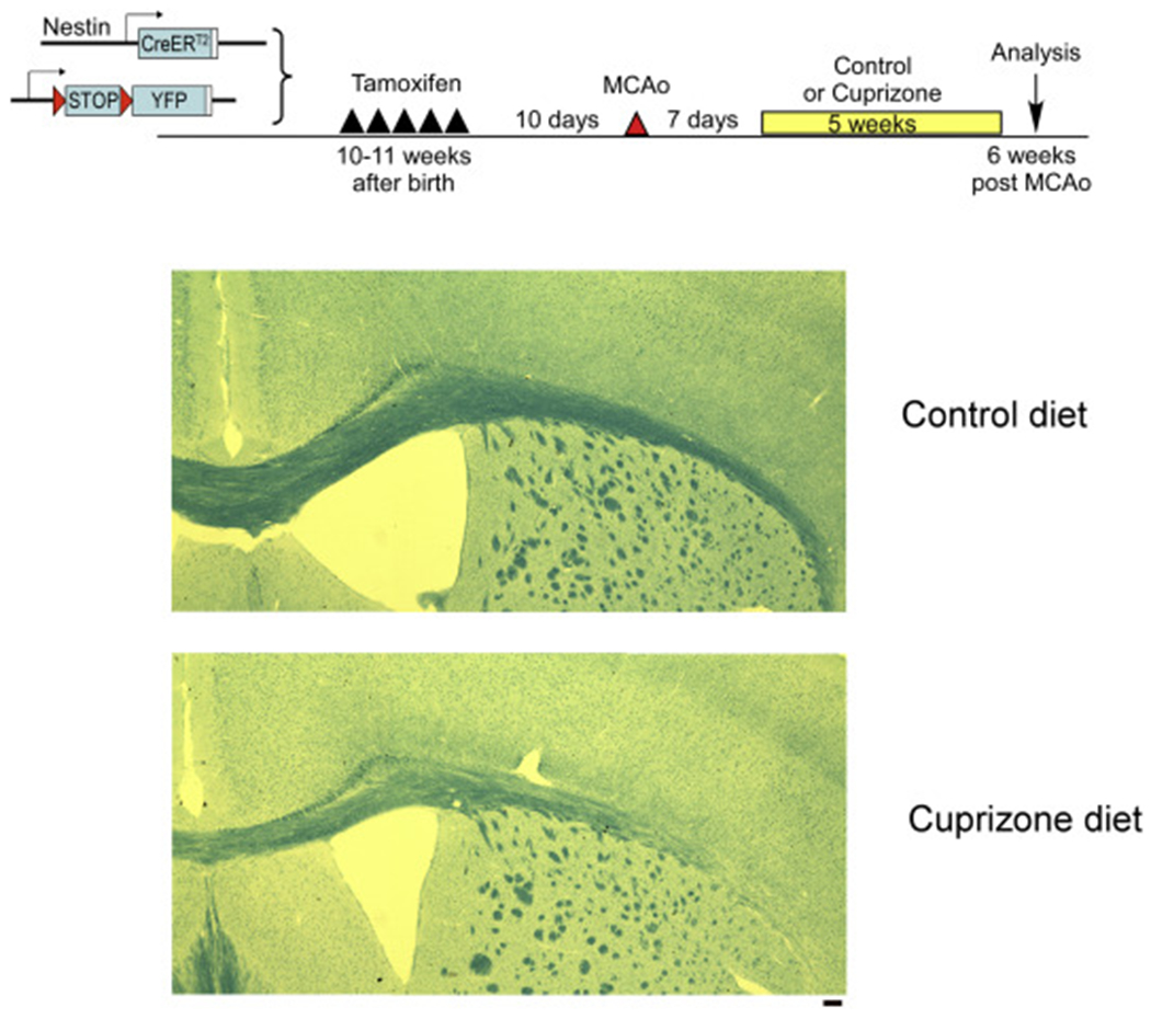
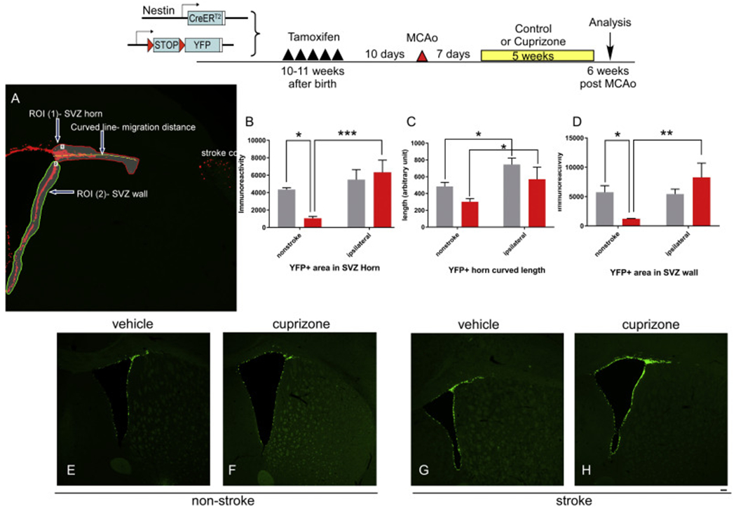
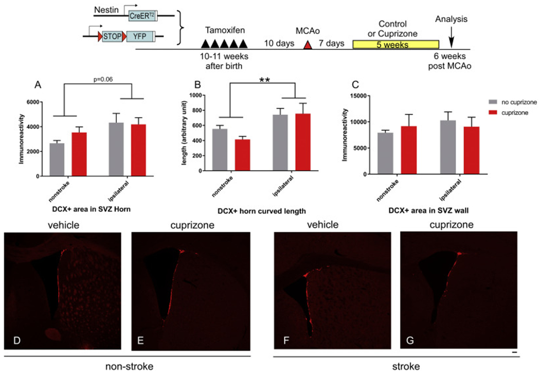
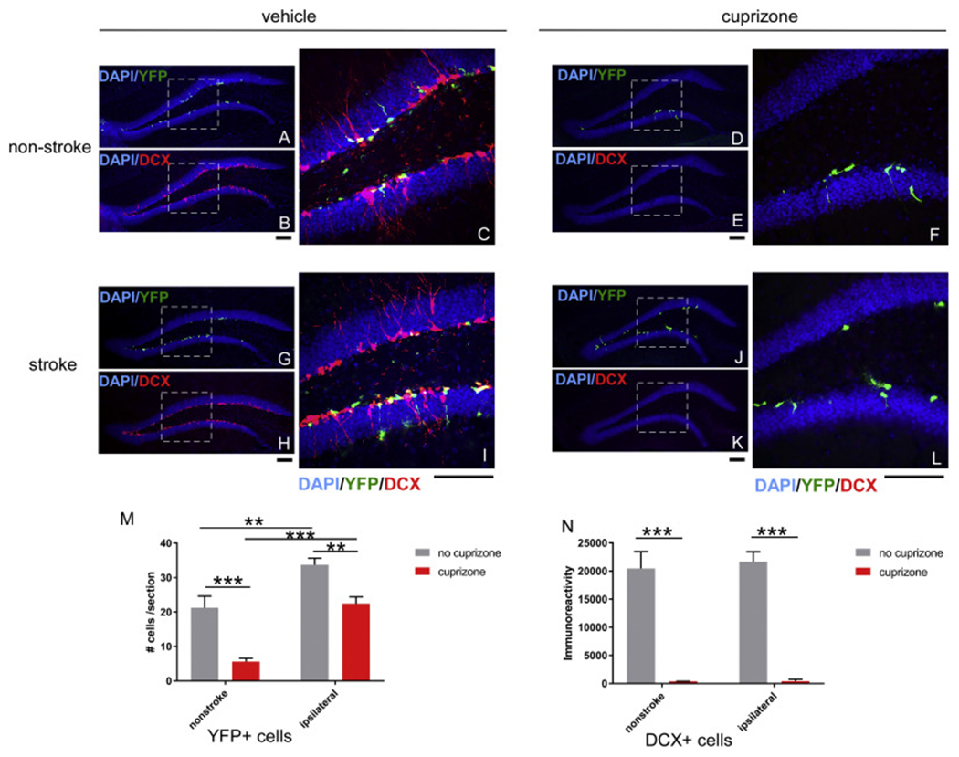
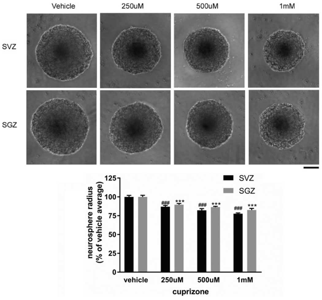
Due to the limitation in treatment window of the rtPA (recombinant tissue plasminogen activator), the development of delayed treatment for stroke is needed. We previously reported that there is a difference in neurogenesis and neuroblast migration patterns in different mouse stroke models (proximal and distal middle cerebral artery occlusion models, pMCAo or dMCAo). Specifically, compared to robust neurogenesis and substantial migration of newly born neuroblasts in pMCAo model, dMCAo only illicit limited neurogenesis and migration of neuroblasts towards ischemic area. One potential reason for this difference is the relative location of ischemic area to white matter and the neurogenic niche (subventricular zone, SVZ). Specifically, white matter could serve as a physical barrier or inhibitory factor to neurogenesis and migration in the dMCAo model. Given that a major difference in human and rodent brains is the content of white matter in the brain, in this study, we further characterize these two models and test the important hypothesis that white matter is an important contributing inhibitory factor for the limited neurogenesis in the dMCAo model. We utilized a genetically inducible NSC-specific reporter mouse line (nestin-CreERT2-R26R-YFP) to label and track NSC proliferation, survival and differentiation in ischemic brain. To test whether myelin is inhibitory to neurogenesis in dMCAo model, we demyelinated mouse brains using cuprizone treatment after stroke and examined whether there is enhanced neurogenesis or migration of neuroblasts cells in stroke mice treated with cuprizone. Our data suggests that demyelination of the brain does not result in enhanced neurogenesis or migration of neuroblasts, supporting that myelin is not a major inhibitory factor for stroke-induced neurogenesis. In addition, our results suggest that in non-stroke mice, demyelination causes decreased neurogenesis in adult brain, indicating a potential positive role of myelin in maintenance of adult neural stem cell niche.

Keywords: Distal MCAo; Neurogenesis; Proximal MCAo; Stroke; White matter.


Figures

Fig. 1.
Fig. 1.
Proximal MCAo (pMCAo) and distal MCAo (dMCAo) induces different infarct size and locations in mouse brain. (A-B) TTC staining, (C-D) T2-weighted MRI images of pMCAo and dMCAo after 24 h. (E) quantification of infarct size using T2-weighted MRI imaging. The data are presented as MEAN± SEM (n = 7 for pMCAo and n = 13 for dMACo). Scale bar = 1 mm.
Fig. 2.
Fig. 2.
Behavioral outcomes in pMCAo and dMCAo mice. (A-B) Total horizontal distance traveled, (C-D) Vertical activity, (E-F) Adhesive removal test and (G) Cylinder test results at pre- and post-stroke days. The data are presented as MEAN± SEM. * p < .05, ** p < .01, and ***p < .001, or. ANOVA. (n = 7 for pMCAo and n = 13 for dMCAo).
Fig. 3.
Fig. 3.
pMCAo induces more substantial neurogenesis and migration of double cortin (DCX) positive neuroblasts compared to dMCAo. Top panel, experimental timeline. (A-B) YFP+ cells at 6 weeks after pMCAo or dMCAo. (C-D) DCX+ neuroblasts at 6 weeks after pMCAo or dMCAo. Scale bar =100um
Fig. 4.
Fig. 4.
Demylination of corpus collosum following 5-week cuprizone treatment. Luxol fast blue staining in vehicle or cuprizone treated brains. Scale bar =100um. (For interpretation of the references to colour in this figure legend, the reader is referred to the web version of this article.)
Fig. 5.
Fig. 5.
Effects of Cuprizone treatment on YFP+ cells expansion and migration in non-stroke and stroke mice. Top panel, experimental timeline. A. Illustration of quantification parameters. (B) YFP+ immunoreactive positive cells in the SVZ horn area. (C) migration length of YFP+ cells in SVZ horn area and (D) YFP+ immunoreactive positive cells in the SVZ wall area in different group of mice. (E-H) representative images from each group quantified in B-D. The data are presented as MEAN± SEM. * p < .05, ** p < .01, and ***p < .001. ANOVA (n = 4–5 for each group). Scale bar =100um.
Fig. 6.
Fig. 6.
Effects of Cuprizone treatment on double cortin (DCX) + neuroblast cells expansion and migration in non-stroke and stroke mice. Top panel, experimental timeline. (A) DCX+ immunoreactive positive cells in the SVZ horn area. (B) migration length of DCX+ cells in SVZ horn area and (C) DCX+ immunoreactive positive cells in the SVZ wall area in different group of mice. (D-G) representative images from each group quantified in B-D. The data are presented as MEAN± SEM. * p < .05, ** p < .01, and ***p < .001. ANOVA (n = 4–5 for each group). Scale bar =100um.
Fig. 7.
Fig. 7.
Effects of Cuprizone treatment on subgranular zone (SGZ) YFP+ and double cortin (DCX) + neuroblast cells expansion and migration in non-stroke and stroke mice. Representative images of YFP+ or DCX+ cells in SGZ from each group (A-C, non-stroke+vehicle; D-F, non-stroke+cuprizone; G-I, stroke+vehicle and J-L, stroke+cuprizone. Dashed square indicate the area that is enlarged in the right panel with higher magnification. All groups are quantified in panel M (YFP+ cells) and panel N (DCX+ cells). The data are presented as MEAN+ SEM. * p < .05, ** p < .01, and ***p < .001. ANOVA (n = 4–5 for each group). Scale bar =100um.
Fig. 8.
Fig. 8.
Direct effects of Cuprizone treatment on SVZ and SGZ neurosphere growth. Representative neurosphere images from different Cuprizone concentration or vehicle treatment initiated at 48-h after cell plating and harvested at 7 days after plating. Quantification of neurosphere radius shown in lower panel. The data are presented as MEAN± SEM. ***p < .001. ANOVA (n = 6 for each group). Scale bar =100um.

References

    1. Abe H, Tanaka T, Kimura M, Mizukami S, Saito F, Imatanaka N, Akahori Y, Yoshida T, Shibutani M, 2015. Cuprizone decreases intermediate and late-stage progenitor cells in hippocampal neurogenesis of rats in a framework of 28-day oral dose toxicity study. Toxicol. Appl. Pharmacol 287 (3), 210–221. 10.1016/j.taap.2015.06.005. - DOI - PubMed
    1. An J, Yin JJ, He Y, Sui RX, Miao Q, Wang Q, Yu JZ, Yu JW, Shi FD, Ma CG, Xiao BG, 2019. Temporal and spatial dynamics of astroglial reaction and immune response in cuprizone-induced demyelination. Neurotox. Res 10.1007/s12640-019-00129-4. - DOI - PubMed
    1. Arvidsson A, Collin T, Kirik D, Kokaia Z, Lindvall O, 2002. Neuronal replacement from endogenous precursors in the adult brain after stroke. Nat. Med 8 (9), 963–970. 10.1038/nm747. - DOI - PubMed
    1. Benardais K, Kotsiari A, Skuljec J, Koutsoudaki PN, Gudi V, Singh V, Vulinovic F, Skripuletz T, Stangel M, 2013. Cuprizone [bis(cyclohexylidenehydrazide)] is selectively toxic for mature oligodendrocytes. Neurotox. Res 24 (2), 244–250. 10.1007/s12640-013-9380-9. - DOI - PubMed
    1. Bond AM, Ming GL, Song H, 2015. Adult mammalian neural stem cells and neuro-genesis: five decades later. Cell Stem Cell 17 (4), 385–395. 10.1016/j.stem.2015.09.003. - DOI - PMC - PubMed

Publication types

MeSH terms