. 2020 Mar;28(3):324-333.
doi: 10.1016/j.joca.2019.12.005. Epub 2020 Jan 2.

The molecular profile of synovial fluid changes upon joint distraction and is associated with clinical response in knee osteoarthritis

Affiliations

The molecular profile of synovial fluid changes upon joint distraction and is associated with clinical response in knee osteoarthritis

F E Watt et al. Osteoarthritis Cartilage. 2020 Mar.

Abstract

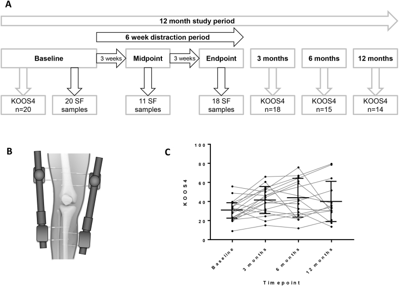
Objective: Surgical knee joint distraction (KJD) leads to clinical improvement in knee osteoarthritis (OA) and also apparent cartilage regeneration by magnetic resonance imaging. We investigated if alteration of the joint's mechanical environment during the 6 week period of KJD was associated with a molecular response in synovial fluid, and if any change was associated with clinical response.

Method: 20 individuals undergoing KJD for symptomatic radiographic knee OA had SF sampled at baseline, midpoint and endpoint of distraction (6 weeks). SF supernatants were measured by immunoassay for 10 predefined mechanosensitive molecules identified in our previous pre-clinical studies. The composite Knee injury and OA Outcome Score-4 (KOOS4) was collected at baseline, 3, 6 and 12 months.

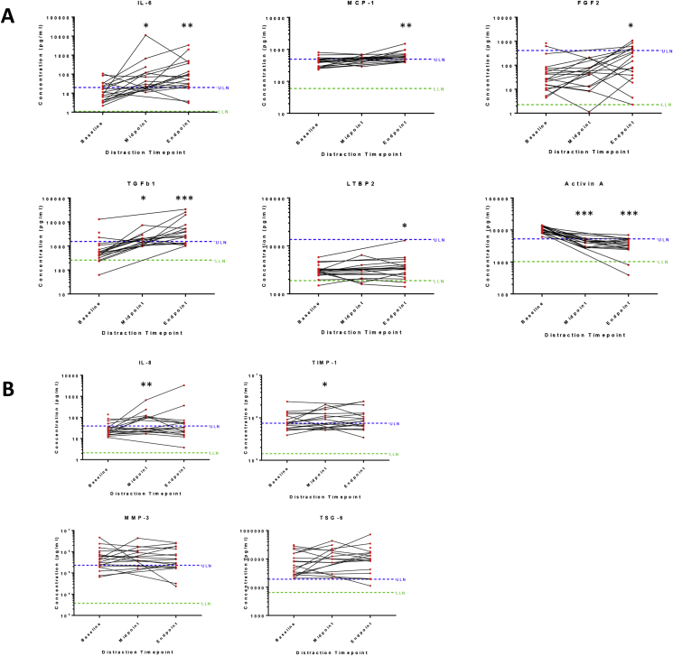
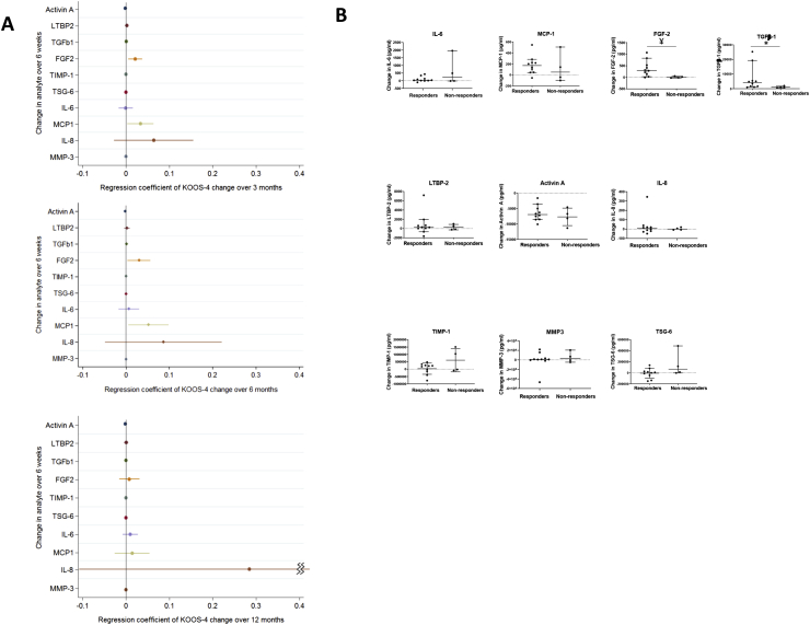
Results: 13/20 (65%) were male with mean age 54°±°5yrs. All had Kellgren-Lawrence grade ≥2 knee OA. 6/10 analytes showed statistically significant change in SF over the 6 weeks distraction (activin A; TGFβ-1; MCP-1; IL-6; FGF-2; LTBP2), P < 0.05. Of these, all but activin A increased. Those achieving the minimum clinically important difference of 10 points for KOOS4 over 6 months showed greater increases in FGF-2 and TGFβ-1 than non-responders. An increase in IL-8 during the 6 weeks of KJD was associated with significantly greater improvement in KOOS4 over 12 months.

Conclusion: Detectable, significant molecular changes are observed in SF following KJD, that are remarkably consistent between individuals. Preliminary findings appear to suggest that increases in some molecules are associated with clinically meaningful responses. Joint distraction may provide a potential opportunity in the future to define regenerative biomarker(s) and identify pathways that drive intrinsic cartilage repair.

Keywords: Biomarker; Cytokines; Distraction; Orthopaedic; Osteoarthritis; Synovial fluid.


Figures

Fig. 1
Fig. 1
Design and outcome measures of a proof-of-concept study to investigate synovial fluid analytes at time of knee joint distraction. A, Flow chart indicating timings of study visits, collection of synovial fluid samples and collection of KOOS from 20 participants, including completeness of sampling/data over the 12 month study period. A further 2 participants gave consent but no baseline SF could be aspirated so they were excluded from further analysis as per protocol. B, Illustration of distraction frame which is surgically placed on the knee joint for a 6 week period. C, KOOS4 measurements in participants at baseline (pre-distraction), 3 months, 6 months and 12 months after surgical knee joint distraction. Medians and inter-quartile ranges are shown (bar and line). Abbreviations: KOOS, Knee Injury and Osteoarthritis Outcome Score (KOOS4 is composite measure of 4 domains); SF, synovial fluid.
Fig. 2
Fig. 2
Measurement of synovial fluid analytes during knee joint distraction. Synovial fluid from study participants immediately prior to distraction (‘baseline’), after 3 weeks of knee joint distraction (‘midpoint’) and at 6 weeks after knee joint distraction (‘endpoint’) were assayed for pre-defined markers of interest by electrochemiluminescence or ELISA (see Table I). Measurements for each of 10 analytes are shown, with mean concentrations for each analyte plotted on a log 10 y axis. *P < 0.05,**P < 0.01,***P < 0.001 by Wilcoxon signed rank test, comparing paired levels at end point or midpoint vs baseline (individual P values are given in Supplementary Table 1). A, shows 6 analytes with change at endpoint vs baseline. B, shows 4 analytes without change at endpoint (although upper 2 showed change at midpoint). ULN and LLN of normal ranges were calculated for each analyte as described in methods and Table I. Abbreviations: LLOQ, lower limit of quantification; ULN, upper limit of normal; LLN, lower limit of normal; LTBP2, latent-transforming growth factor beta-binding protein 2; TGFβ-1, transforming growth factor beta 1; FGF-2, basic fibroblast growth factor; TIMP-1, tissue inhibitor of metalloproteinases 1; TSG-6, tumour necrosis factor-inducible gene 6 protein; IL-6, interleukin 6; MCP-1, monocyte chemoattractant protein 1; IL-8, interleukin 8; MMP3, matrix metalloproteinase-3.
Fig. 3
Fig. 3
Correlation between change of analytes in the synovial fluid of participants over period of knee joint distraction. Spearman rank tests were performed to determine correlations between the change in levels of synovial fluid analytes over the 6 week distraction period (concentration at 6 weeks-baseline concentrations). Correlation coefficients were calculated using all available participant data and the mean of 2 repeated (duplicate) measures for each synovial fluid sample. Strength of correlation by Spearman R coefficient is shown: * (Mid grey shading): Low positive (negative) correlation, 0.30 to 0.49 (−0.30 to −0.49). ** (Dark grey shading): Moderate positive (negative) correlation, 0.50 to 0.69 (−0.50 to −0.69). Abbreviations: LTBP2, latent-transforming growth factor beta-binding protein 2; TGFβ-1, transforming growth factor beta 1; FGF-2, basic fibroblast growth factor; TIMP-1, tissue inhibitor of metalloproteinases 1; TSG-6, tumour necrosis factor-inducible gene 6 protein; IL-6, interleukin 6; MCP-1, monocyte chemoattractant protein 1; IL-8, interleukin 8; MMP3, matrix metalloproteinase 3.
Fig. 4
Fig. 4
Association of change in synovial fluid analytes with the clinical outcome KOOS4.. A Linear regression models for the association of the change over the distraction period for each of 10 synovial fluid analytes (measured in pg/ml) with participants' change in KOOS4 over varying periods are shown: upper panel, change in KOOS4 over 3 months; middle panel, change in KOOS4 over 6 months and lower panel, change in KOOS4 over 12 months. Forest Plots of unadjusted (crude) results, including the regression coefficient for the effect on KOOS4 change over the specified period and 95% confidence intervals are shown (data also shown in Supplementary Table 2). B The change of concentration in each analyte over the 6 week distraction period is shown (6 week level – baseline level), for 2 subgroups: Responders (those whose change in KOOS4 over 6 months was ≥10 points, i.e., those achieving the MICD for KOOS4); and Non-Responders (those whose change in KOOS4 over 6 months was <10 points, i.e., those not achieving the MCID for KOOS4). The bars represent the median and 95% Confidence Intervals for each group. Between group comparisons were by Mann-Whitney U test, *P = 0.04; ¥P = 0.01. Abbreviations: MCID, minimal clinically important difference; LTBP2, latent-transforming growth factor beta-binding protein 2; TGFβ-1, transforming growth factor beta 1; FGF-2, basic fibroblast growth factor; TIMP-1, tissue inhibitor of metalloproteinases 1; TSG-6, tumour necrosis factor-inducible gene 6 protein; IL-6, interleukin 6; MCP-1, monocyte chemoattractant protein 1; IL-8, interleukin 8; MMP3, matrix metalloproteinase-3; KOOS, Knee Injury and Osteoarthritis Outcome Score (KOOS4 is composite measure of 4 domains).

Similar articles

Cited by

References

    1. Bijlsma J.W., Berenbaum F., Lafeber F.P. Osteoarthritis: an update with relevance for clinical practice. Lancet. 2011;377(9783):2115–2126. - PubMed
    1. Brandt K.D., Dieppe P., Radin E.L. Commentary: is it useful to subset "primary" osteoarthritis? A critique based on evidence regarding the etiopathogenesis of osteoarthritis. Semin Arthritis Rheum. 2009;39(2):81–95. - PubMed
    1. Burleigh A., Chanalaris A., Gardiner M.D., Driscoll C., Boruc O., Saklatvala J. Joint immobilization prevents murine osteoarthritis and reveals the highly mechanosensitive nature of protease expression in vivo. Arthritis Rheum. 2012;64(7):2278–2288. - PubMed
    1. Vincent T.L., Wann A.K.T. Mechanoadaptation: articular cartilage through thick and thin. J Physiol. 2019;597(5):1271–1281. - PMC - PubMed
    1. Benichou C., Wirotius J.M. Articular cartilage atrophy in lower limb amputees. Arthritis Rheum. 1982;25(1):80–82. - PubMed

Publication types

MeSH terms