. 2020 Mar:132:115220.
doi: 10.1016/j.bone.2019.115220. Epub 2020 Jan 2.

LPS-induced premature osteocyte senescence: Implications in inflammatory alveolar bone loss and periodontal disease pathogenesis

Affiliations

LPS-induced premature osteocyte senescence: Implications in inflammatory alveolar bone loss and periodontal disease pathogenesis

Ruben Aquino-Martinez et al. Bone. 2020 Mar.

Abstract

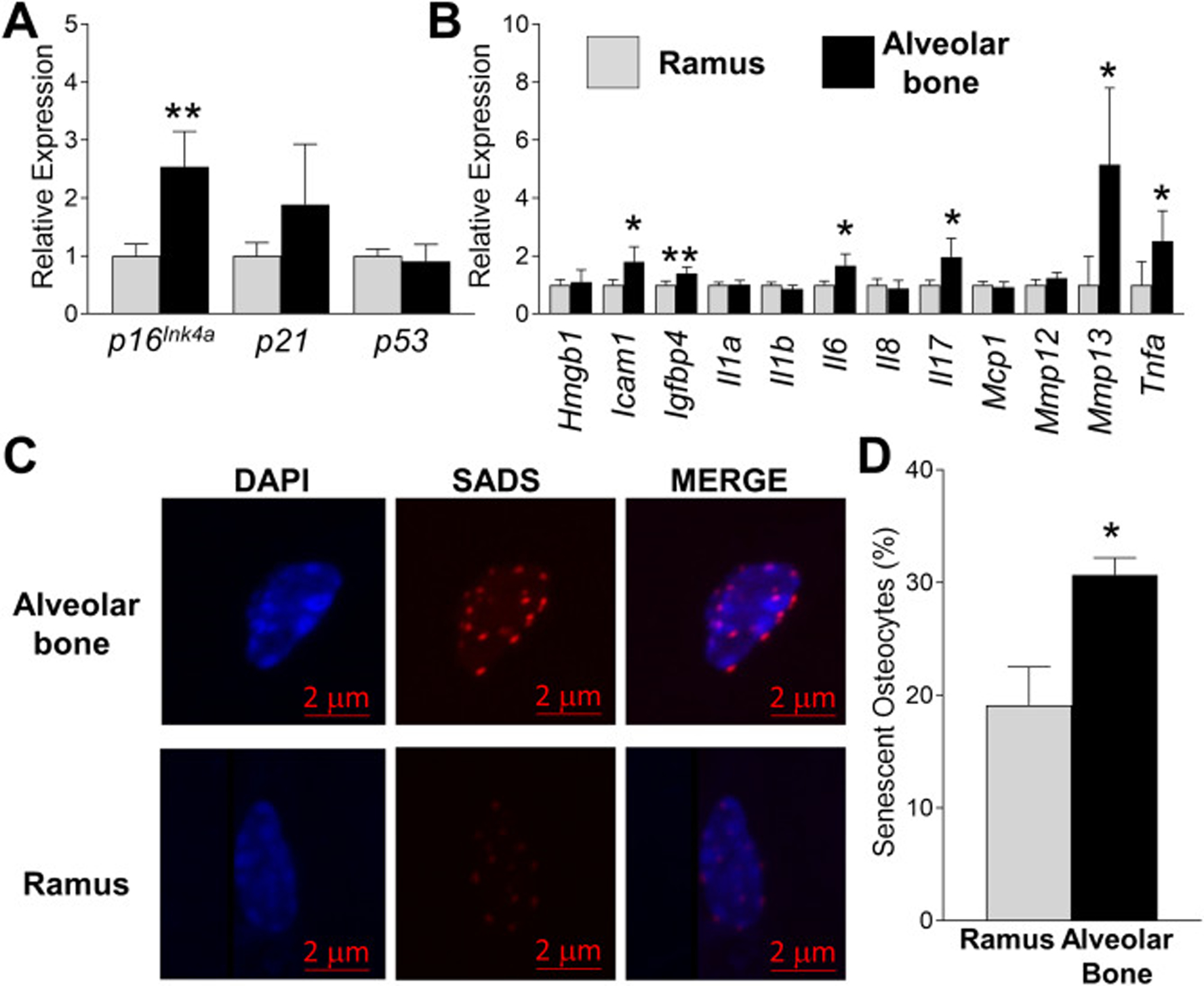
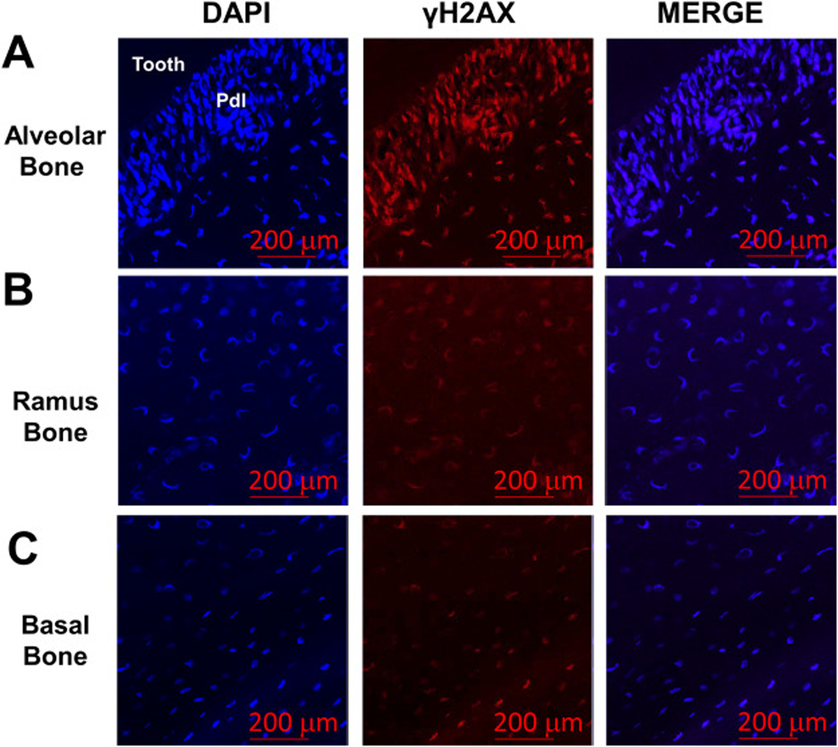
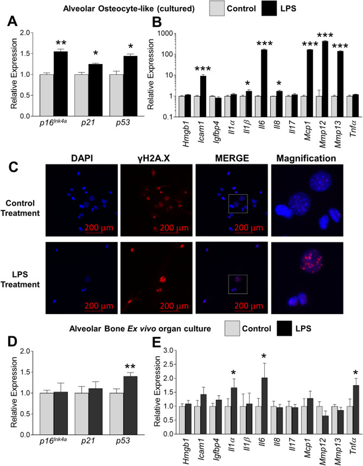
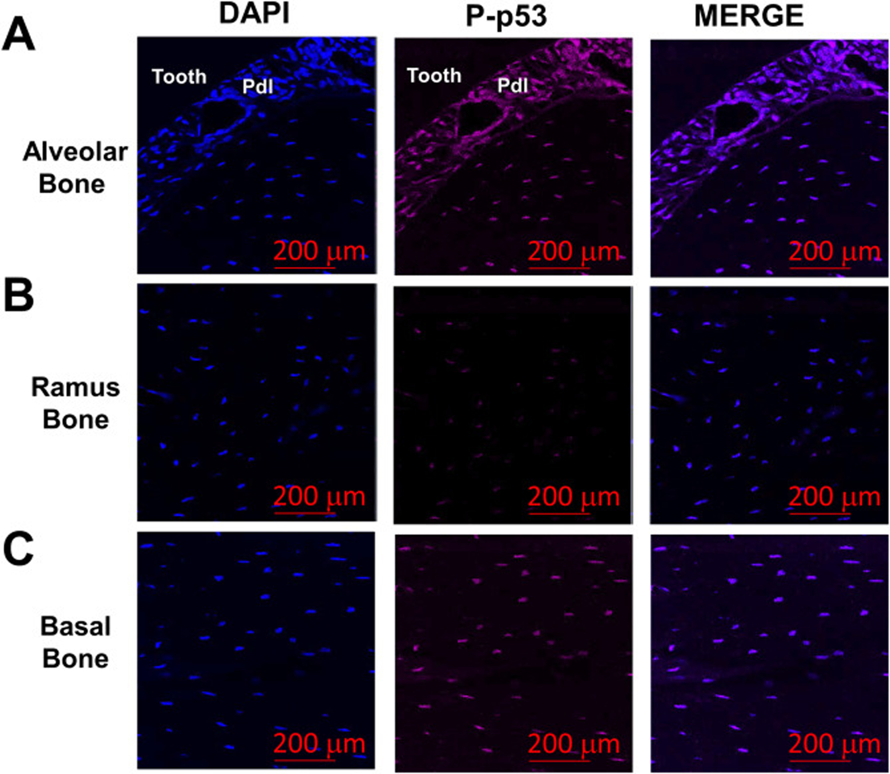
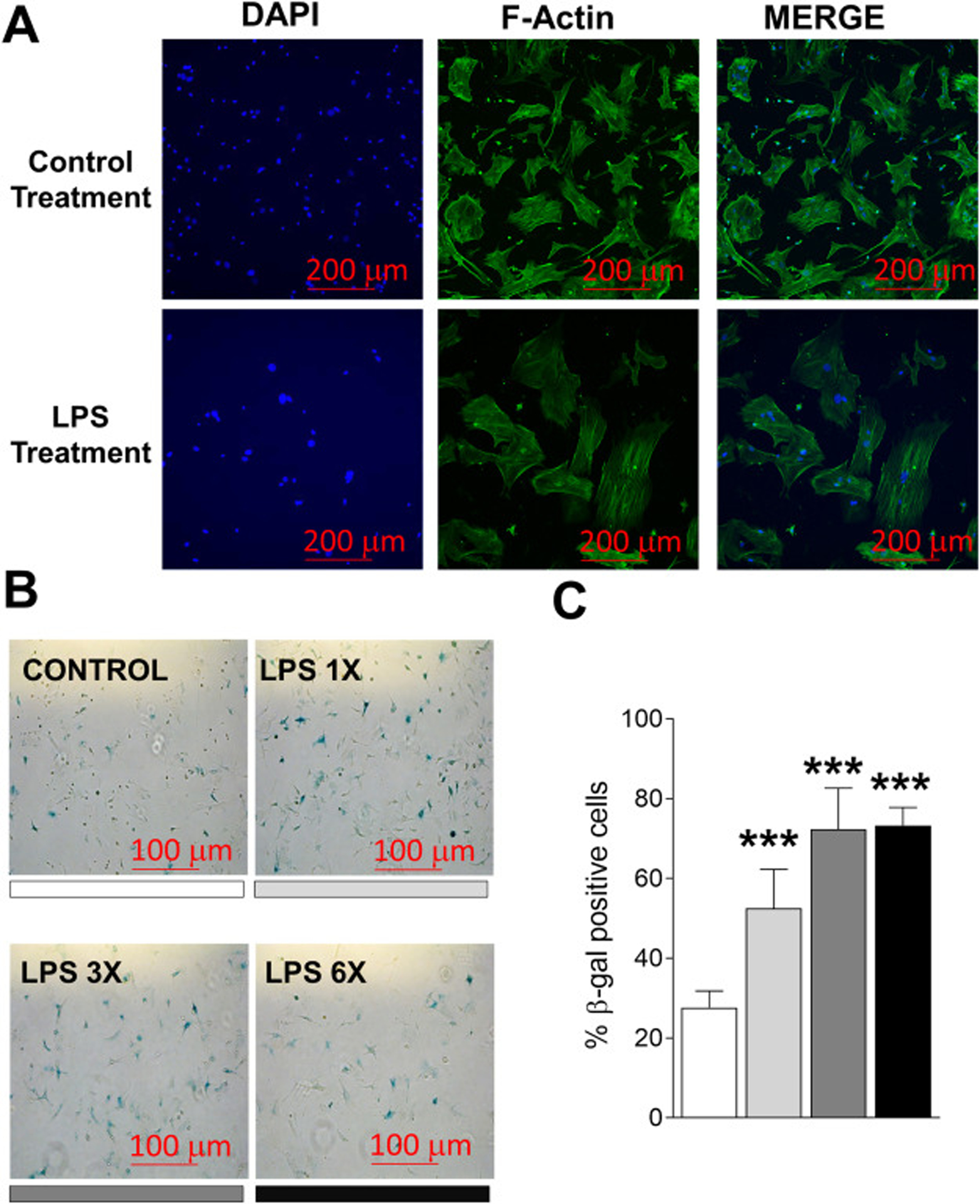
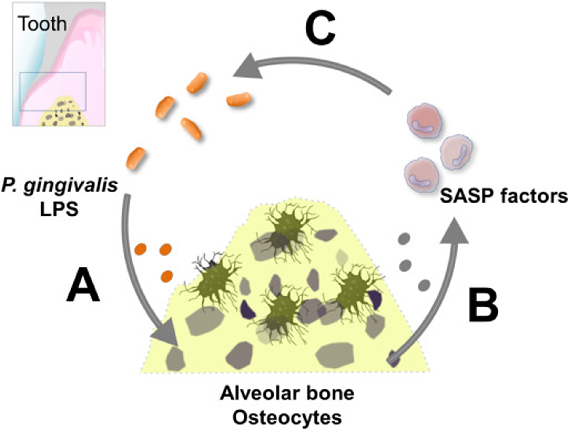
Cellular senescence is associated with inflammation and extracellular matrix tissue remodeling through the secretion of proteins termed the senescence-associated secretory phenotype (SASP). Although osteocyte senescence in older individuals in the skeleton is well recognized, whether young alveolar osteocytes can also become senescent is unknown. This is potentially important in the context of periodontal disease, which is an inflammatory condition caused by a gradual change from symbiotic to pathogenic oral microflora that can lead to tooth loss. Our aim was to identify whether senescent osteocytes accumulate in young alveolar bone and whether bacterial-derived lipopolysaccharide (LPS) can influence cellular senescence in alveolar bone. An osteocyte-enriched cell population isolated from alveolar bone expressed increased levels of the known senescence marker p16Ink4a, as well as select SASP markers known to be implicated alveolar bone resorption (Icam1, Il6, Il17, Mmp13 and Tnfα), compared to ramus control cells. Increased senescence of alveolar bone osteocytes was also observed in vivo using the senescence-associated distension of satellites (SADS) assay and increased γH2AX, a marker of DNA damage associated with senescent cells. To approximate a bacterial infection in vitro, alveolar osteocytes were treated with LPS. We found increased expression of various senescence and SASP markers, increased γH2AX staining, increased SA-β-Gal activity and the redistribution of F-actin leading to a larger and flattened cell morphology, all hallmarks of cellular senescence. In conclusion, our data suggests a model whereby bacterial-derived LPS stimulates premature alveolar osteocyte senescence, which in combination with the resultant SASP, could potentially contribute to the onset of alveolar bone loss.

Keywords: Alveolar bone; Bacteria; Inflammation; Osteocyte; Periodontal disease; SASP; Senescence.


Figures

Fig. 1.
Fig. 1.
Identification of senescent cells in young alveolar bone. (a-b) Alveolar bone and ramus osteocyte-enriched cell populations were prepared from 6 month-old WT female mice and immediately assayed (no culture) for expression of selected senescent marker and SASP genes using QPCR. Data represent Mean ± SEM (n=10). *P ≤ 0.05 and **P≤ 0.01 relative to ramus control. (c) Fluorescent in situ hybridization (FISH) was used to identify SADS-positive senescent cells in ramus and alveolar bone from 6 month-old WT female mice in vivo (a representative cell is shown). (d) Quantitation of SADS-positive cells in ramus and alveolar osteocyte-enriched cell populations (50 cells counted per sample). The red bar denotes the magnification scale (2 μm). Data represent Mean ± SEM (n=10). *P ≤ 0.05 relative to ramus control.
Fig. 2.
Fig. 2.
Levels of DNA damage are increased in alveolar bone osteocytes. (a) Presence of γH2AX, a marker of DNA damage, was evaluated by immunofluorescence in sections from alveolar bone from 6 month-old WT female mice in vivo. Stronger γH2AX (red signal) was observed both within the periodontal ligament (Pdl) and in neighboring alveolar osteocytes. Ramus (b) and underlying basal bone (c) displayed lower amounts of γH2AX signal. Cell nuclei were labeled with DAPI (blue signal). The red bars denote the magnification scale (200 μm).
Fig. 3.
Fig. 3.
Chronic LPS exposure increases several indices of cellular senescence in alveolar bone osteocytes. (a-b) Alveolar bone osteocyte-like cells were prepared from 6 month-old WT female mice, treated with either vehicle control or P. gingivalis LPS (10 ng/ml) for six days and assayed for expression of selected senescent marker and SASP genes using QPCR. Data represent Mean ± SEM (n=10). *P ≤ 0.05, **P ≤ 0.01 and ***P ≤ 0.001 relative to vehicle control. (c) Alveolar osteocyte-like cells were treated with control or LPS as in panels a-b and stained with an antibody against γH2AX (red signal). The boxed inset in the Merge panel is shown enlarged in the Magnification panel to observe single γH2AX-stained foci. Cell nuclei were labeled with DAPI (blue signal). The red bars denote the magnification scale (200 μm). (d-e) Intact (non-digested) left and right alveolar bone blocks were treated with vehicle control or LPS, respectively, for 6 days as assayed for expression of selected senescent marker and SASP genes using QPCR. Data represent Mean ± SEM (n=6). *P ≤ 0.05, and **P ≤ 0.01 relative to vehicle control.
Fig. 4.
Fig. 4.
P-p53 protein levels are increased in alveolar bone osteocytes in vivo. (a-c) Bone sections from alveolar, ramus and underlying basal bone from 6 month-old WT female mice were stained for Phospho(P)-p53 levels (magenta signal). Cell nuclei were labeled with DAPI (blue signal). Cell nuclei were labeled with DAPI (blue signal). The red bars denote the magnification scale (200 μm).
Fig. 5.
Fig. 5.
LPS treatment of alveolar bone osteocyte-like cells induces cytoskeletal alterations and the acquisition of senescence-associated SA-β-gal activity in vitro. (a) Alveolar bone osteocyte-enriched cells were prepared from 6 month-old WT female mice and treated with LPS and stained for F-Actin (green staining). (b-c) Alveolar bone osteocyte-enriched cells were cultured in supplemented α-MEM alone containing low serum (2% FBS) to reduce cell proliferation, or containing LPS (10 ng/ml). Cells were stimulated with LPS once, three times, and six times (for one day each treatment time; the shading under each image denotes the bar in panel c). The red bars denote the magnification scale (100 μm). Data represent Mean ± SEM (n=6). **P ≤ 0.001 relative to vehicle control.
Fig. 6.
Fig. 6.
Suggested model of the potential role of LPS induced osteocyte senescence in alveolar bone. (a) Anatomical proximity between periodontal bacterial infection and alveolar bone predisposes osteocytes to undergo LPS induced genotoxic stress, promoting premature senescence over time. (b) Senescent osteocytes release SASP factors to recruit host immune and inflammatory cells to promote tissue remodeling, including its own clearance. However, senescent osteocyte accumulation might exacerbate local inflammation and increase tissue breakdown products release, which fuels dysbiotic bacteria overgrowth. (c) A positive feedback loop between pathogenic bacteria and the senescent osteocytes might be established.

References

    1. Arigbede AO, Babatope BO, Bamidele MK, Periodontitis and systemic diseases: A literature review, J Indian Soc Periodontol 16 (2012) 487–491. - PMC - PubMed
    1. Loesche WJ, Grossman NS, Periodontal disease as a specific, albeit chronic, infection: diagnosis and treatment, Clin Microbiol Rev 14 (2001) 727–752, table of contents. - PMC - PubMed
    1. Berezow AB, Darveau RP, Microbial shift and periodontitis, Periodontol 2000 55 (2011) 36–47. - PMC - PubMed
    1. Hajishengallis G, Periodontitis: from microbial immune subversion to systemic inflammation, Nat Rev Immunol 15 (2015) 30–44. - PMC - PubMed
    1. Hajishengallis G, The inflammophilic character of the periodontitis-associated microbiota, Mol Oral Microbiol 29 (2014) 248–257. - PMC - PubMed

Publication types

Substances