Case Reports
. 2020 Apr 1;77(4):517-521.
doi: 10.1001/jamaneurol.2019.4509.

Tau Positron Emission Tomographic Findings in a Former US Football Player With Pathologically Confirmed Chronic Traumatic Encephalopathy

Affiliations
Case Reports

Tau Positron Emission Tomographic Findings in a Former US Football Player With Pathologically Confirmed Chronic Traumatic Encephalopathy

William G Mantyh et al. JAMA Neurol. .

Abstract

Importance: Biomarkers for chronic traumatic encephalopathy (CTE) are currently lacking. The radiotracer fluorine F 18-labeled (18F)-flortaucipir (FTP) detects tau pathology in Alzheimer disease, and positron emission tomography (PET) with FTP shows elevated binding in individuals at risk for CTE. No study, however, has assessed the correlation between in vivo FTP PET and postmortem tau in CTE.

Objective: To assess the regional association between in vivo FTP binding and postmortem tau pathology in a patient with pathologically confirmed CTE.

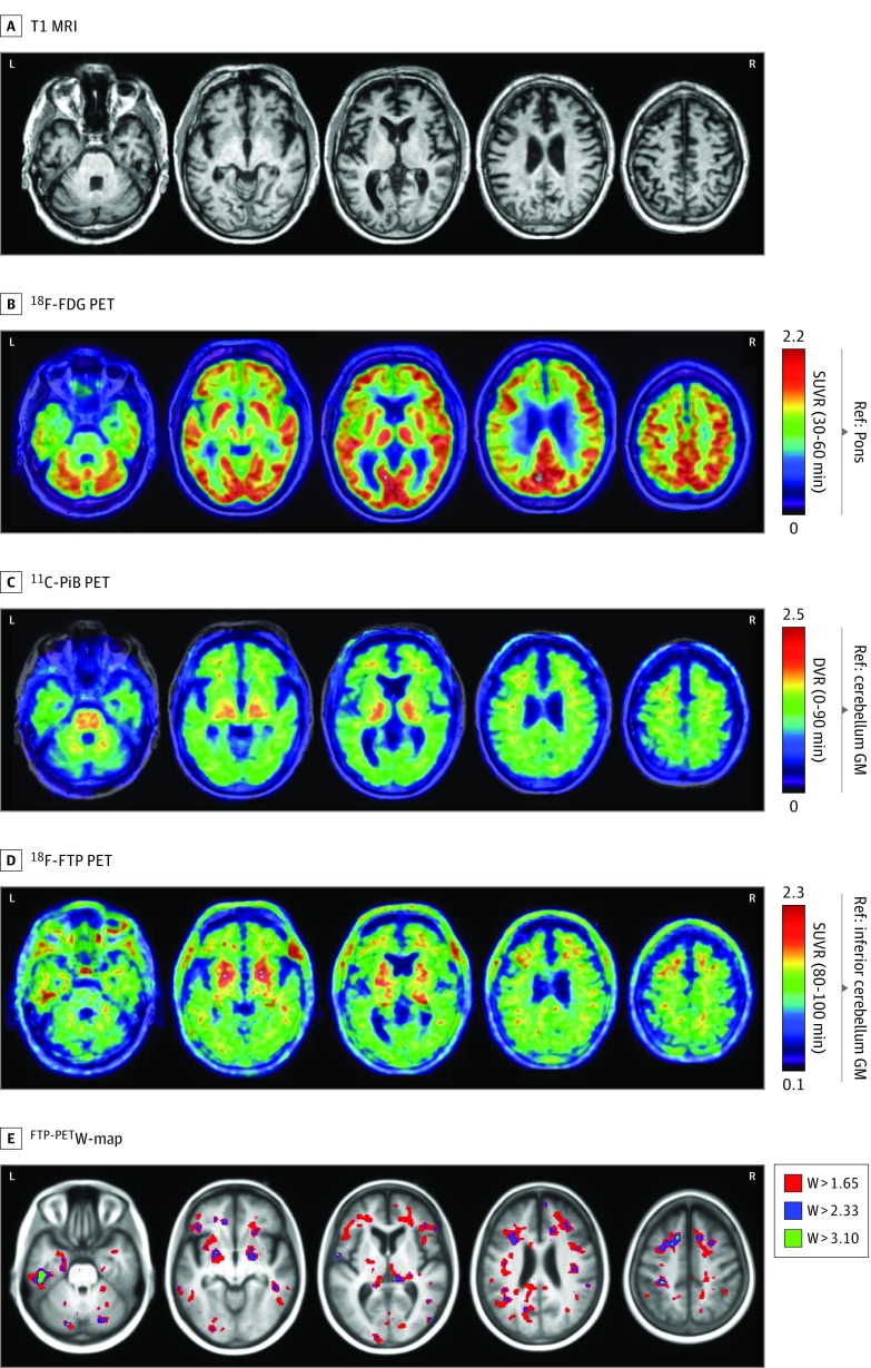
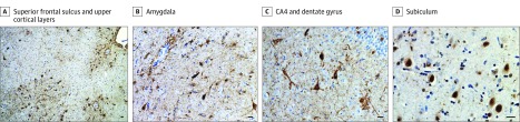
Design, setting, and participants: A white male former National Football League player with 17 years of US football exposure was clinically diagnosed with traumatic encephalopathy syndrome at a neurology tertiary referral center. 18F-Fludeoxyglucose, carbon 11-labeled Pittsburgh compound B, and FTP PET were performed 52 months prior to death, and magnetic resonance imaging, 50 months prior to death. Brain images were assessed qualitatively for abnormalities blinded to autopsy data. Autopsy was performed using a neurodegenerative research protocol. The FTP standardized uptake value ratios (inferior cerebellar gray reference region) and W-score (age-adjusted z-score) maps were compared with phosphorylated tau immunohistochemical analysis with monoclonal antibody CP13.

Main outcomes and measures: Qualitative and quantitative comparisons between antemortem FTP PET and tau pathology at autopsy.

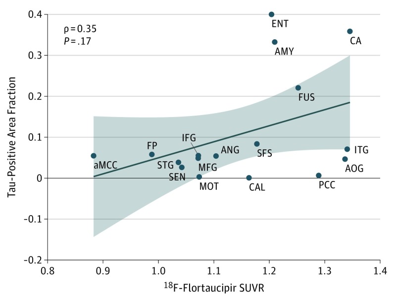
Results: Flortaucipir uptake was distributed in a patchy, frontotemporal-predominant pattern that overlapped with regions showing neurodegeneration on magnetic resonance imaging and hypometabolism on 18F-fludeoxyglucose PET. Pathological assessment revealed stage 4 CTE; limbic argyrophilic grain disease; stage 2 limbic-predominant, age-related transactive response DNA-binding protein 43 encephalopathy; and Braak neurofibrillary tangle stage 3. 18F-Flortaucipir W-maps matched areas of high postmortem tau burden in left fusiform and inferior temporal gyri and juxtacortical frontal white matter. High FTP W-scores with low tau burden were found in the basal ganglia, thalamus, motor cortex, and calcarine cortex. No regions with low FTP W-scores corresponded to areas with high pathological tau burden. A modest correlation, which did not reach statistical significance (ρ = 0.35, P = .17), was found between FTP standardized uptake value ratio and tau area fraction at the regional level.

Conclusions and relevance: In this patient, FTP PET findings during life showed a modest correspondence with postmortem pathology in CTE. These findings suggest that FTP may have limited utility as a tau biomarker in CTE.


Conflict of interest statement

Conflict of Interest Disclosures: Dr Soleimani-Meigooni reported receiving a grant from the Alzheimer’s Association during the conduct of the study. Dr Possin reported receiving grants from Global Brain Health Institute and the National Institute of Neurological Disorders and Stroke during the conduct of the study and grants from Quest Diagnostics, Merck Foundation, the National Institute on Aging, and the Centers for Medicare and Medicaid Innovation outside the submitted work. Dr Grinberg reported receiving grants from the National Institutes of Health (NIH), Avid Radiopharmaceuticals, and Eli Lilly and Company during the conduct of the study. Dr Seeley reported receiving grants from the NIH during the conduct of the study. Dr Rabinovici reported receiving grants from the NIH and from Tau Consortium during the conduct of the study; grants from Avid Radiopharmaceuticals, Eli Lilly and Company, GE Healthcare, and Life Molecular Imaging outside the submitted work; and personal fees from GE Healthcare, Axon Neurosciences, Merck, Eisai, Roche, and Genentech outside the submitted work. No other disclosures were reported.

Figures

Figure 1.
Figure 1.. Neuroimaging in Pathologically Proven Chronic Traumatic Encephalopathy
T1-weighted magnetic resonance (MRI) (top row) demonstrated mild medial temporal and frontal atrophy. Fluorine F 18–labeled (18F)–fludeoxyglucose (FDG) on positron emission tomography (PET) images (second row) demonstrated mild hypometabolism in the medial temporal, medial and dorsolateral frontal, and peri-insular regions, whereas carbon 11–labeled Pittsburgh compound B (PiB) PET scan (third row) was visually rated as negative for cortical retention. On 18F-flortaucipir PET imaging (fourth row), there was widespread patchy binding predominantly in the left greater than right frontotemporal areas. The 18F-flortaucipir PET W-map (bottom row) (thresholded at 3 different levels) showed that these focal increases were higher than what would be expected considering the patient’s age. DVR indicates distribution volume ratio; GM, gray matter; Ref, reference region; and SUVR, standardized uptake value ratio with acquisition windows in parentheses.
Figure 2.
Figure 2.. Neuropathologic Findings
Photomicrographs from immunohistochemical analyses with CP13 antibody for detection of phosphorylated tau (A-C) and TDP-43 antibody (D). Bars indicate 25 μm. A, Frequent perivascular and depth-of-sulci clusters of tau-immunoreactive astrocytes and neurons are seen in the superior frontal sulcus, together with frequent pretangles, fuzzy astrocytes, and dystrophic neurites in upper cortical layers. B, Numerous neurofibrillary tangles, dystrophic neurites, and perivascular astrocytic and neuronal clusters are present in the amygdala. C, Abundant perinuclear ringlike inclusions in the CA4 region of the hippocampus and frequent pretangles in the dentate gyrus are consistent with argyrophilic grain disease. D, Neuronal cytoplasmic inclusions of transactive response DNA-binding protein of 43 kDa (TDP-43) are seen in neurons lacking nuclear TDP-43 staining, and dystrophic neurites are seen in the subiculum.
Figure 3.
Figure 3.. Comparison Between Regional Flortaucipir Standardized Uptake Value Ratio (SUVR) and CP13-Labeled Tau Area Fraction
A modest correlation that did not reach statistical significance was found between regional fluorine F 18–labeled (18F)–flortaucipir SUVR and tau-positive area fraction at postmortem pathologic analysis. aMCC indicates anterior cingulate cortex; AMY, amygdala; ANG, angular gyrus; AOG, anterior orbital gyrus; CA, hippocampus; CAL, calcarine cortex; ENT, entorhinal cortex; FP, frontal pole; FUS, fusiform gyrus; IFG, inferior frontal gyrus; ITG, inferior temporal gyrus; MFG, middle frontal gyrus; MOT, primary motor cortex; PCC, posterior cingulate cortex; SEN, primary sensory cortex; SFS, superior frontal sulcus; and STG, superior/middle temporal gyrus.

Comment in

References

    1. McKee AC, Cairns NJ, Dickson DW, et al. ; TBI/CTE group . The first NINDS/NIBIB consensus meeting to define neuropathological criteria for the diagnosis of chronic traumatic encephalopathy. Acta Neuropathol. 2016;131(1):75-86. doi:10.1007/s00401-015-1515-z. doi:10.1007/s00401-015-1515-z - DOI - DOI - PMC - PubMed
    1. Stern RA, Adler CH, Chen K, et al. . Tau positron-emission tomography in former national football league players. N Engl J Med. 2019;380(18):1716-1725. doi:10.1056/NEJMoa1900757 - DOI - PMC - PubMed
    1. Lesman-Segev OH, La Joie R, Stephens ML, et al. . Tau PET and multimodal brain imaging in patients at risk for chronic traumatic encephalopathy. Neuroimage Clin. 2019;24:102025. doi:10.1016/j.nicl.2019.102025 - DOI - PMC - PubMed
    1. Kramer JH, Jurik J, Sha SJ, et al. . Distinctive neuropsychological patterns in frontotemporal dementia, semantic dementia, and Alzheimer disease. Cogn Behav Neurol. 2003;16(4):211-218. doi:10.1097/00146965-200312000-00002 - DOI - PubMed
    1. Spina S, Brown JA, Deng J, et al. . Neuropathological correlates of structural and functional imaging biomarkers in 4-repeat tauopathies. Brain. 2019;142(7):2068-2081. doi:10.1093/brain/awz122 - DOI - PMC - PubMed

Publication types