. 2020 Oct;16(10):1858-1870.
doi: 10.1080/15548627.2019.1709765. Epub 2020 Jan 8.

Crimean-Congo hemorrhagic fever virus replication imposes hyper-lipidation of MAP1LC3 in epithelial cells

Affiliations

Crimean-Congo hemorrhagic fever virus replication imposes hyper-lipidation of MAP1LC3 in epithelial cells

Marie Moroso et al. Autophagy. 2020 Oct.

Abstract

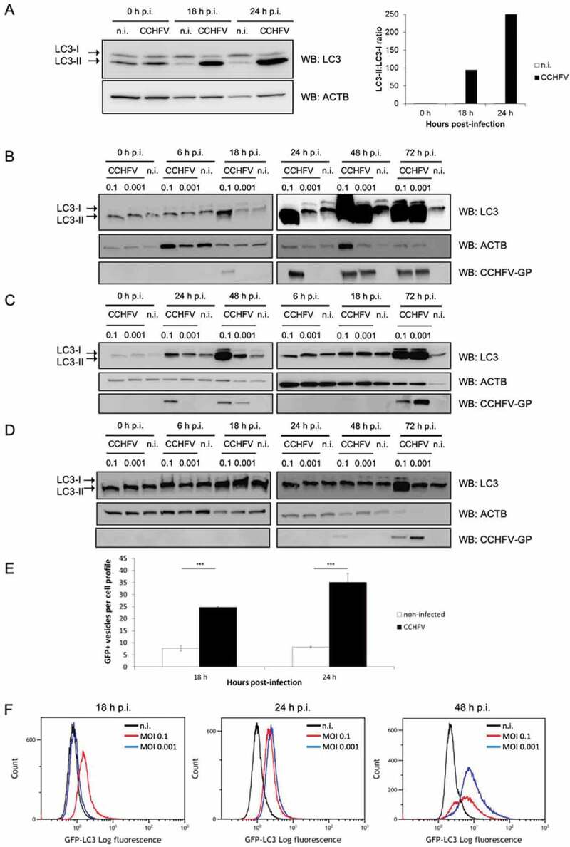
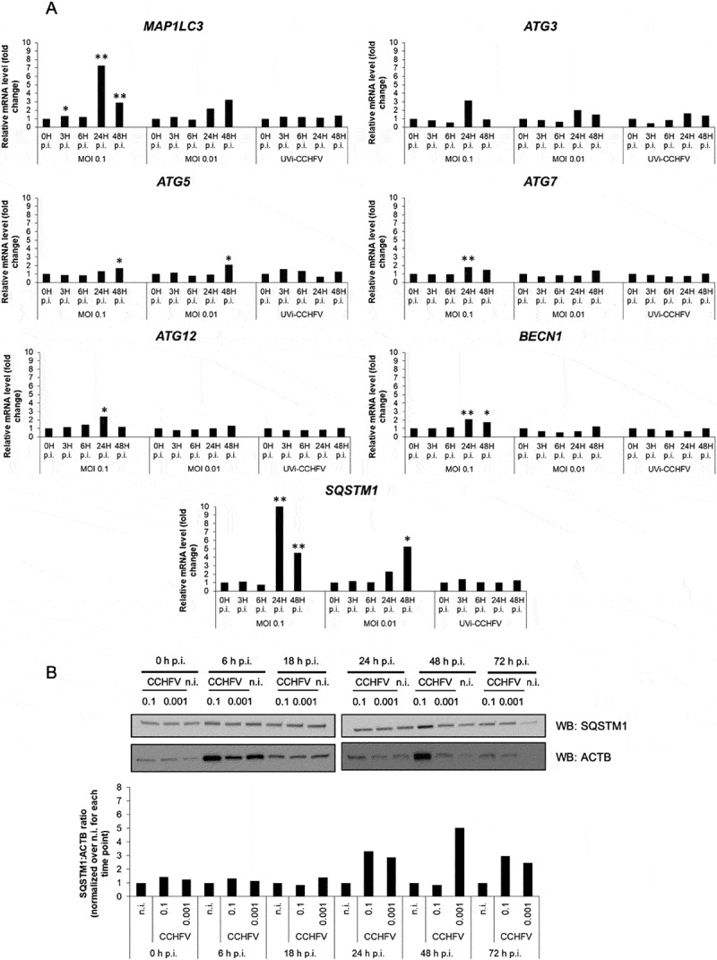
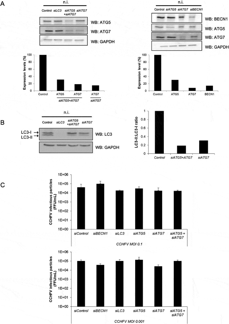
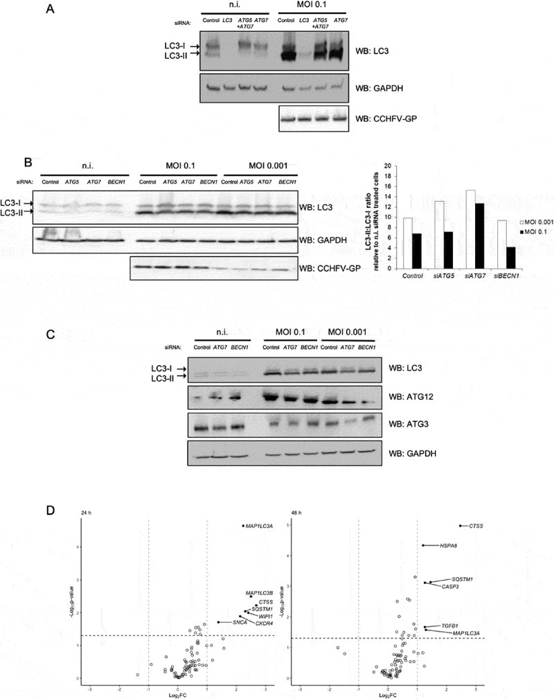
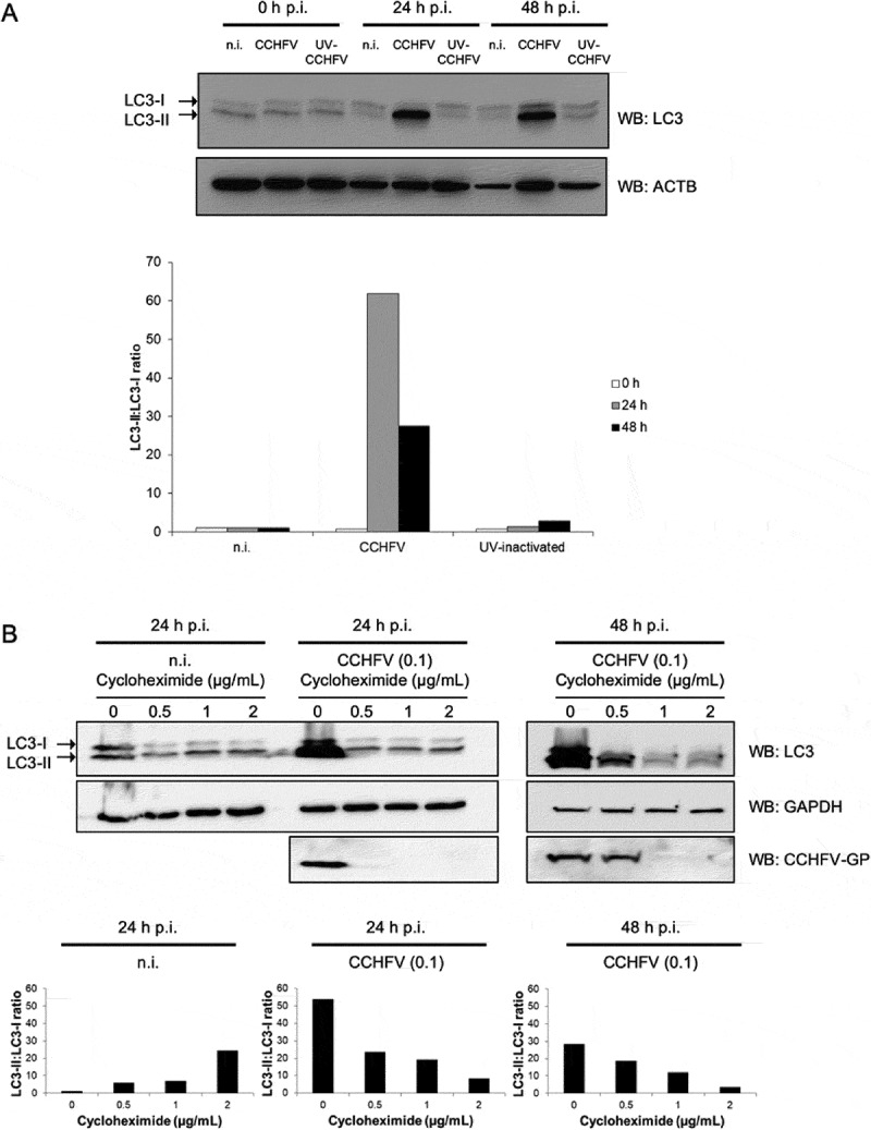
Crimean-Congo hemorrhagic fever virus (CCHFV) is a virus that causes severe liver dysfunctions and hemorrhagic fever, with high mortality rate. Here, we show that CCHFV infection caused a massive lipidation of LC3 in hepatocytes. This lipidation was not dependent on ATG5, ATG7 or BECN1, and no signs for recruitment of the alternative ATG12-ATG3 pathway for lipidation was found. Both virus replication and protein synthesis were required for the lipidation of LC3. Despite an augmented transcription of SQSTM1, the amount of proteins did not show a massive and sustained increase in infected cells, indicating that degradation of SQSTM1 by macroautophagy/autophagy was still occurring. The genetic alteration of autophagy did not influence the production of CCHFV particles demonstrating that autophagy was not required for CCHFV replication. Thus, the results indicate that CCHFV multiplication imposes an overtly elevated level of LC3 mobilization that involves a possibly novel type of non-canonical lipidation. Abbreviations: BECN1: Beclin 1; CCHF: Crimean-Congo hemorrhagic fever; CCHFV: Crimean-Congo hemorrhagic fever virus; CHX: cycloheximide; ER: endoplasmic reticulum; GFP: green fluorescent protein; GP: glycoproteins; MAP1LC3: microtubule associated protein 1 light chain 3; MOI: multiplicity of infection; n.i.: non-infected; NP: nucleoprotein; p.i.: post-infection; SQSTM1: sequestosome 1.

Keywords: Autophagy; CCHFV; LC3 lipidation; epithelial cells; viral infection.


Conflict of interest statement

No potential conflict of interest was reported by the authors.

Figures

Figure 1.
Figure 1.
Marked LC3 lipidation and induction of LC3-positive punctiform structures in epithelial cells infected with CCHFV. (A–D) Huh7 (A and B), HepG2 (C) and HeLa (D) cells were infected with CCHFV at MOI 0.1 (A) or MOI 0.1 and 0.001 (B), or left uninfected, and analyzed by western blot for LC3, ACTB/β-ACTIN and CCHFV-GP at 0, 18 and 24 h (A) or 0, 6, 18, 24, 48 and 72 h (B-D) after infection. Note that the order of sample loading might differ between cell lines. Quantification in A represents the LC3-II:LC3-I ratio. Both the massive LC3-II signal and the light LC3-I signal made difficult the calculation of an LC3-II:LC3-I ratio in panel B to D. (E) Fluorescence microscopy analysis of GFP-LC3-transfected Huh7 cells infected, or not, with CCHFV (MOI 0.1). GFP-LC3 puncta were quantified after 18 h and 24 h of infection. Results are expressed as the number of puncta per cell (mean ± SD). At least 150 cells were examined for each condition. Representative images are presented in Fig. S1C. (F) Non-infected or CCHFV-infected (MOI 0.1 and 0.001) GFP-LC3 HeLa cells were permeabilized by using saponin for evacuation of soluble forms of GFP-LC3 and analyzed by flow cytometry at 18, 24 and 48 h after infection. Histograms represent the log fluorescence intensity of non-soluble GFP-LC3 (x-axis) relative to cell number (y-axis). 50 000 events were collected per conditions. Experiments were carried out 3 times (A-D) or twice (F).
Figure 2.
Figure 2.
Status of autophagy-related gene expression and SQSTM1 in CCHFV-infected cells. (A) Transcripts of MAP1LC3A/B, ATG3, ATG5, ATG7, ATG12, BECN1 and SQSTM1 autophagy genes were quantified by RT-qPCR by using total RNA from Huh7 cells (histograms) infected with CCHFV (MOI 0.1 and 0.01) at 0, 3, 6, 24 and 48 h after infection. UV-inactivated CCHFV (MOI 0.1) was used in parallel as non-replicative control. GAPDH, HMBS and ACTB were used as house-keeping genes. Fold changes were calculated after deriving ΔΔCt. (B) Huh7 cells were infected with CCHFV at MOI 0.1 and 0.001 and analyzed by western blot for SQSTM1 and ACTB levels at 0, 6, 18, 24 and 72 h after infection. The companion histograms represent the SQSTM1:ACTB ratio normalized to non-infected cells (combining two independent experiments). The ACTB signals are the same as in Figure 1A due to simultaneous analysis of LC3 and SQSTM1. The quantification was not done for the 72 h condition due to faint ACTB signal. All experiments were carried out at least twice.
Figure 3.
Figure 3.
CCHFV particle production is independent of canonical autophagy. Huh7 cells were treated with siRNA targeting ATG5, ATG7, MAP1LC3A-C and BECN1 genes for 48 h. Cells were then infected (MOI 0.1 and 0.001), or not (n.i.), with CCHFV and analyzed for their capacity to release infectious viral particles after 48 h of infection. (A) Expression levels of ATG5, ATG7, BECN1 and GAPDH after 48 h of siRNA-mediated silencing (i.e. prior to infection with CCHFV). (B) Status of LC3 after 48 h of treatment with siRNA targeting MAP1LC3A-C, ATG7 and ATG5+ ATG7 in the absence of infection. Quantification represents the LC3-II:LC3-I ratio. (C) Measurement of the production of CCHFV viral particles by siRNA-treated Huh7 cells after 48 h of infection. The values represent the mean ± SD of 3 independent experiments. No differences reached statistical significance.
Figure 4.
Figure 4.
Neither ATG5, ATG7 and BECN1 core autophagy proteins, nor the ATG12–ATG3 complex are involved in CCHFV-induced LC3 lipidation. (A) Huh7 cells were treated with siRNA targeting MAP1LC3A-C, ATG5ATG7 or ATG7 genes for 48 h, infected or not with CCFV at MOI 0.1 and analyzed by western blot for LC3, CCHFV-GP and GAPDH levels 48 h after infection. (B) Huh7 cells were treated with siRNA targeting ATG5, ATG7 or BECN1 genes for 48 h, infected with CCHFV at MOI 0.1 vs 0.001, and analyzed by western blot for LC3, CCHFV-GP and GAPDH levels 48 h after infection. Quantification represents the LC3-II:LC3-I ratio normalized to that of the corresponding siRNA-treated, non-infected cells. (C) Huh7 cells were treated with siRNA targeting ATG7 or BECN1 genes for 48 h, infected with CCFV at MOI 0.1 vs 0.001, and analyzed by western blot for LC3 (17 and 15 kDa), ATG12 (55 kDa due to conjugation to ATG5), ATG3 (36 kDa) and GAPDH levels 48 h after infection. The presence of a 49 kDa complex could not be detected by either anti-ATG12 or anti-ATG3 antibodies. (D) Volcano plot representation of fold change of transcript level for 84 genes involved in autophagy execution or regulation (RT-Profiler PCR array) after infection of Huh7 cells with CCHFV (MOI 0.1). Changes are relative to non-infected control cells. Calculations and normalization were performed as described by SABiosciences using 5 house-keeping genes (ACTB, B2M [beta-2-microglobulin], GAPDH, RPLP0 (ribosomal protein lateral stalk subunit P0) and HPRT1 (hypoxanthine phosphoribosyltransferase 1)). Experiments were carried out twice (A-C) and 3 times (D).
Figure 5.
Figure 5.
Both CCHFV replicative capacity and protein synthesis are necessary for sustained LC3 lipidation in infected epithelial cells. (A) Huh7 cells were exposed to infectious CCHFV (CCHFV), UV-irradiated CCHFV (UV-CCHFV) (MOI 0.1) or left uninfected (n.i.) for 24 h and 48 h. Cell lysates were western blotted for detection of LC3 and ACTB. Quantification represents the LC3-II:LC3-I ratio. (B) Huh7 cells were treated with 0, 0.5, 1, or 2 μg/ml cycloheximide, infected or not with CCHFV (MOI 0.1), lysed at 24 h or 48 h after infection and analyzed by western blot for detection of LC3, CCHFV-GP and GAPDH. Quantification represents the LC3-II:LC3-I ratio. Experiments were carried out twice.

Similar articles

Cited by

References

    1. Ergonul O.Crimean-Congo haemorrhagic fever. Lancet Infect Dis. 2006;6(4):203–214. - PMC - PubMed
    1. Whitehouse CA.Crimean-Congo hemorrhagic fever. Antiviral Res. 2004;64(3):145–160. - PubMed
    1. Bente DA, Forrester NL, Watts DM, et al. Crimean-Congo hemorrhagic fever: history, epidemiology, pathogenesis, clinical syndrome and genetic diversity. Antiviral Res. 2013;100(1):159–189. - PubMed
    1. Vorou R, Pierroutsakos IN, Maltezou HC. Crimean-Congo hemorrhagic fever. Curr Opin Infect Dis. 2007;20(5):495–500. - PubMed
    1. Papa A, Mirazimi A, Köksal I, et al. Recent advances in research on Crimean-Congo hemorrhagic fever. J Clin Virol. 2015;64:137–143. - PMC - PubMed

Publication types

MeSH terms