

Molecular mechanism of an antagonistic antibody against glucose-dependent insulinotropic polypeptide receptor
- PMID: 31905038
- PMCID: PMC6973313
- DOI: 10.1080/19420862.2019.1710047
Molecular mechanism of an antagonistic antibody against glucose-dependent insulinotropic polypeptide receptor
Abstract
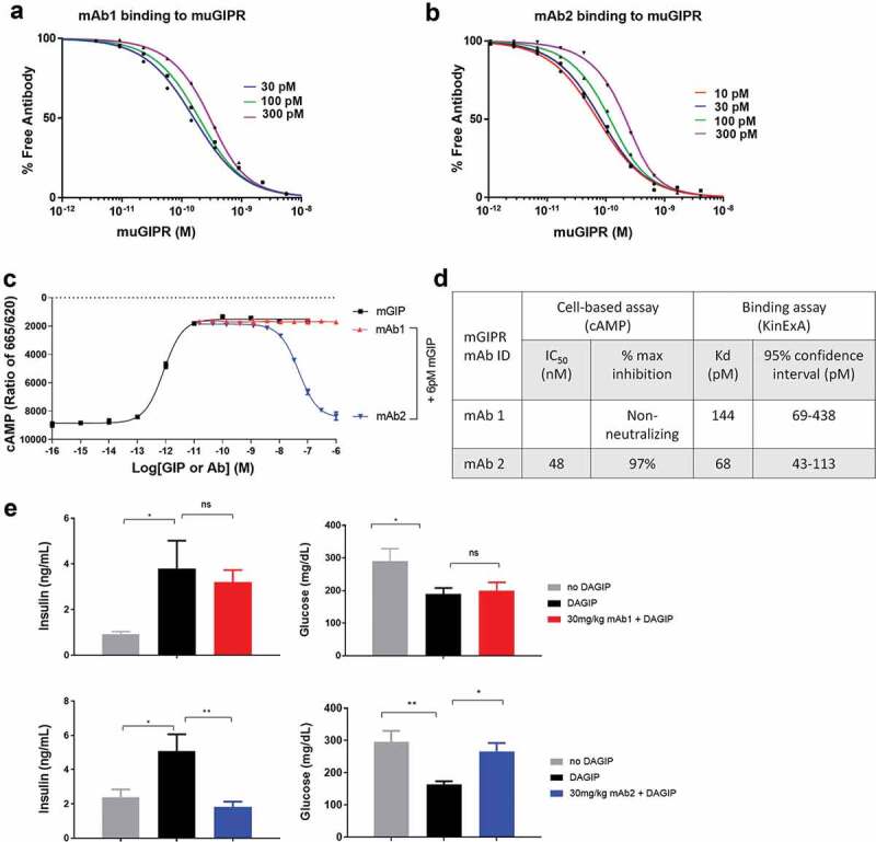
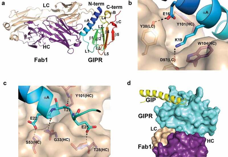
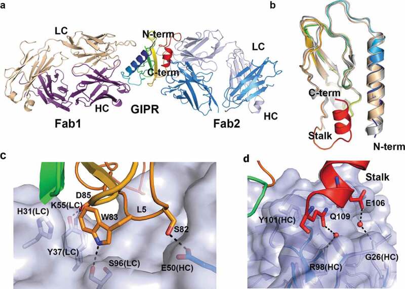
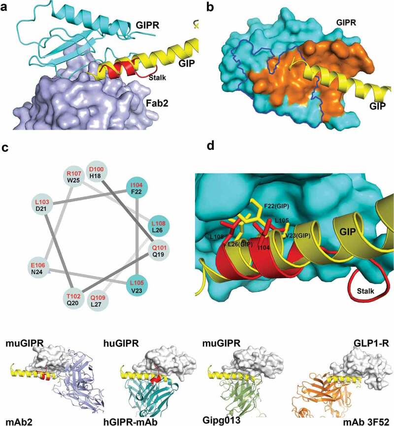
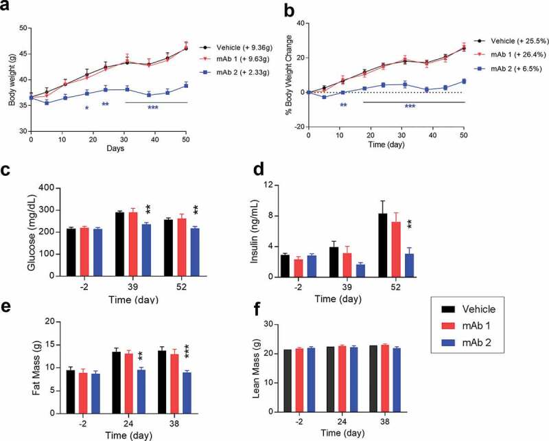
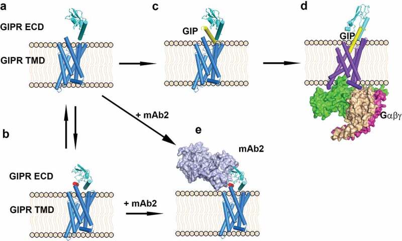
Glucose-dependent insulinotropic polypeptide (GIP) is an incretin hormone involved in regulating glucose and lipid metabolism. GIP receptor (GIPR) antagonism is believed to offer therapeutic potential for various metabolic diseases. Pharmacological intervention of GIPR, however, has limited success due to lack of effective antagonistic reagents. Previously we reported the discovery of two mouse anti-murine GIPR monoclonal antibodies (mAbs) with distinctive properties in rodent models. Here, we report the detailed structural and biochemical characterization of these two antibodies, mAb1 and mAb2. In vitro and in vivo characterizations demonstrated mAb2 is a full GIPR antagonistic antibody and mAb1 is a non-neutralizing GIPR binder. To understand the molecular basis of these two antibodies, we determined the co-crystal structures of GIPR extracellular domain in complex with mAb1 and with mAb2 at resolutions of 2.1 and 2.6 Å, respectively. While the non-neutralizing mAb1 binds to GIPR without competing with the ligand peptide, mAb2 not only partially occludes the ligand peptide binding, but also recognizes the GIPR C-terminal stalk region in a helical conformation that acts as a molecular mimic of the ligand peptide and locks GIPR in a novel auto-inhibited state. Furthermore, administration of mAb2 in diet-induced obesity mice for 7 weeks leads to both reduction in body weight gain and improvement of metabolic profiles. In contrast, mAb1 has no effect on body weight or other metabolic improvement. Together, our studies reveal the unique molecular mechanism of action underlying the superior antagonistic activity of mAb2 and signify the promising therapeutic potential of effective GIPR antagonism for the treatment of metabolic disorders.
Keywords: GIPR; antagonistic antibody; crystallography; structure.
Figures
Similar articles
-
Chronic treatment with anti-GIPR mAb alone and combined with DPP-4 inhibitor correct obesity, dyslipidemia and nephropathy in rodent animals.Life Sci. 2021 Mar 15;269:119038. doi: 10.1016/j.lfs.2021.119038. Epub 2021 Jan 13. Life Sci. 2021. PMID: 33453239
-
Chronic glucose-dependent insulinotropic polypeptide receptor (GIPR) agonism desensitizes adipocyte GIPR activity mimicking functional GIPR antagonism.Nat Commun. 2020 Oct 5;11(1):4981. doi: 10.1038/s41467-020-18751-8. Nat Commun. 2020. PMID: 33020469 Free PMC article.
-
Pharmacological antagonism of the incretin system protects against diet-induced obesity.Mol Metab. 2020 Feb;32:44-55. doi: 10.1016/j.molmet.2019.11.018. Epub 2019 Dec 3. Mol Metab. 2020. PMID: 32029229 Free PMC article.
-
Glucose-dependent insulinotropic polypeptide (GIP) receptor antagonists as anti-diabetic agents.Peptides. 2018 Feb;100:173-181. doi: 10.1016/j.peptides.2017.11.021. Peptides. 2018. PMID: 29412817 Review.
-
Blockade of gastric inhibitory polypeptide (GIP) action as a novel means of countering insulin resistance in the treatment of obesity-diabetes.Peptides. 2020 Mar;125:170203. doi: 10.1016/j.peptides.2019.170203. Epub 2019 Nov 13. Peptides. 2020. PMID: 31733230 Review.
Cited by
-
Loss of Function Glucose-Dependent Insulinotropic Polypeptide Receptor Variants Are Associated With Alterations in BMI, Bone Strength and Cardiovascular Outcomes.Front Cell Dev Biol. 2021 Oct 25;9:749607. doi: 10.3389/fcell.2021.749607. eCollection 2021. Front Cell Dev Biol. 2021. PMID: 34760890 Free PMC article.
-
360-Degree Perspectives on Obesity.Medicina (Kaunas). 2023 Jun 9;59(6):1119. doi: 10.3390/medicina59061119. Medicina (Kaunas). 2023. PMID: 37374323 Free PMC article. Review.
-
Targeting the GIPR for obesity: To agonize or antagonize? Potential mechanisms.Mol Metab. 2021 Apr;46:101139. doi: 10.1016/j.molmet.2020.101139. Epub 2020 Dec 5. Mol Metab. 2021. PMID: 33290902 Free PMC article. Review.
-
An integrated platform approach enables discovery of potent, selective and ligand-competitive cyclic peptides targeting the GIP receptor.Chem Sci. 2022 Feb 24;13(11):3256-3262. doi: 10.1039/d1sc06844j. eCollection 2022 Mar 16. Chem Sci. 2022. PMID: 35414877 Free PMC article.
-
The role of GIPR in food intake control.Front Endocrinol (Lausanne). 2025 Mar 17;16:1532076. doi: 10.3389/fendo.2025.1532076. eCollection 2025. Front Endocrinol (Lausanne). 2025. PMID: 40166681 Free PMC article. Review.
References
Publication types
MeSH terms
Substances
LinkOut - more resources
Full Text Sources
Other Literature Sources
Molecular Biology Databases