. 2020 Apr;236(4):630-659.
doi: 10.1111/joa.13134. Epub 2020 Jan 6.

Computed tomography analysis of the cranium of Champsosaurus lindoei and implications for the choristoderan neomorphic ossification

Affiliations

Computed tomography analysis of the cranium of Champsosaurus lindoei and implications for the choristoderan neomorphic ossification

Thomas W Dudgeon et al. J Anat. 2020 Apr.

Abstract

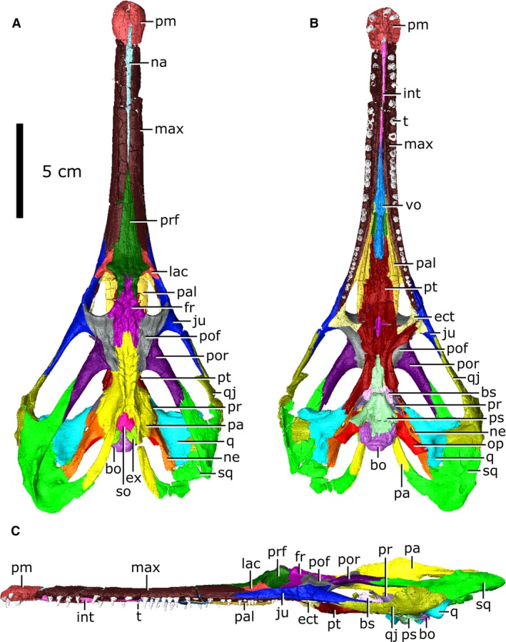
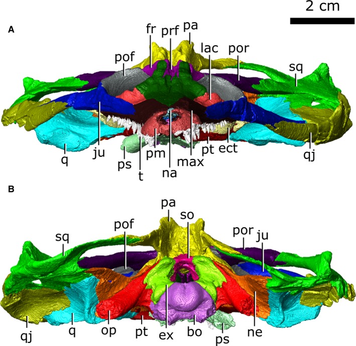
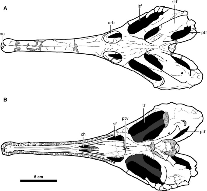
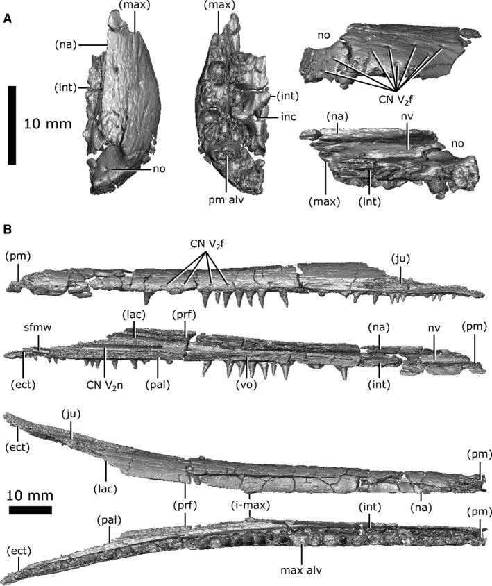
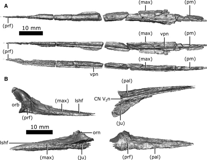
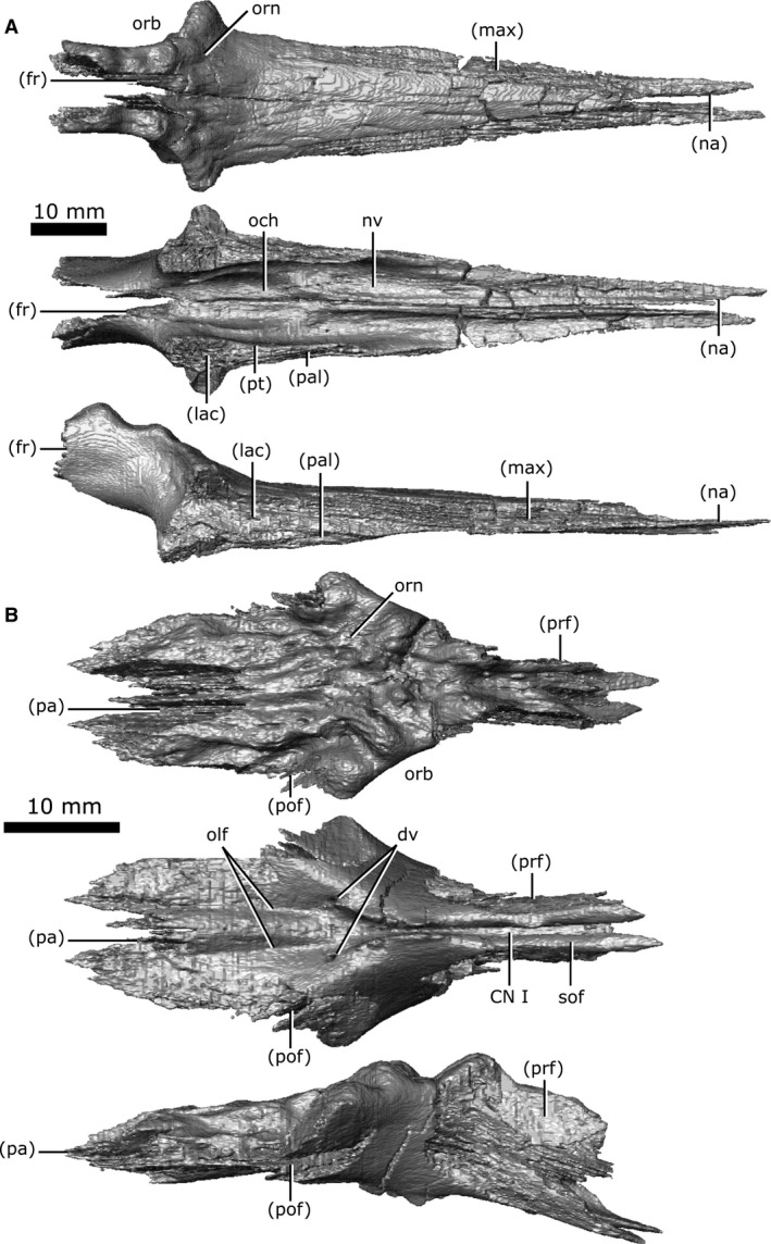
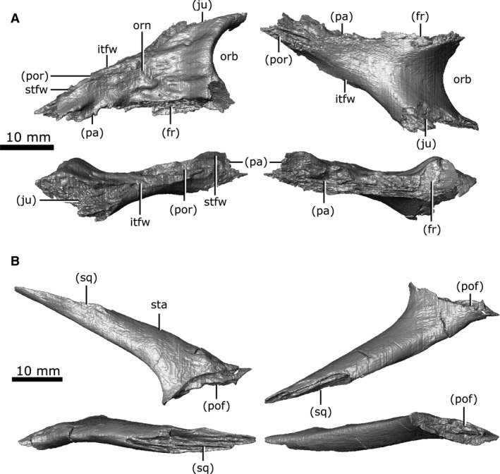
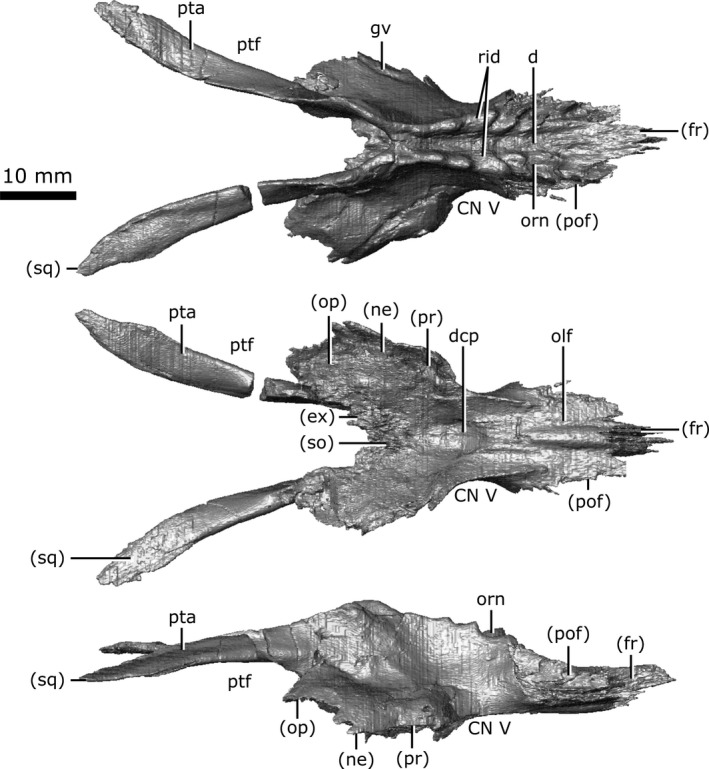
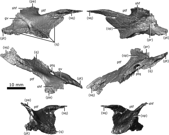
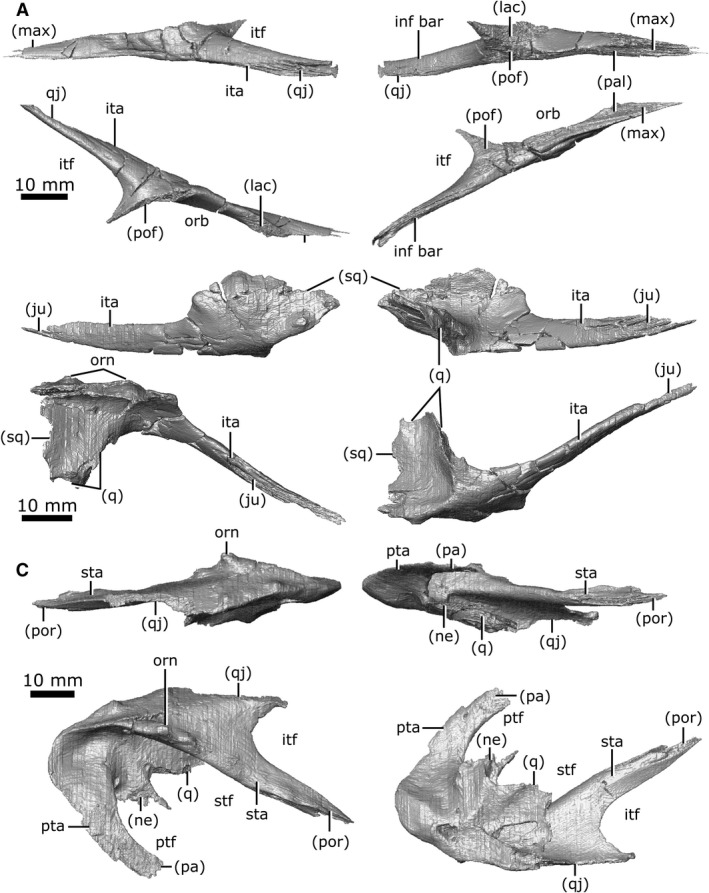
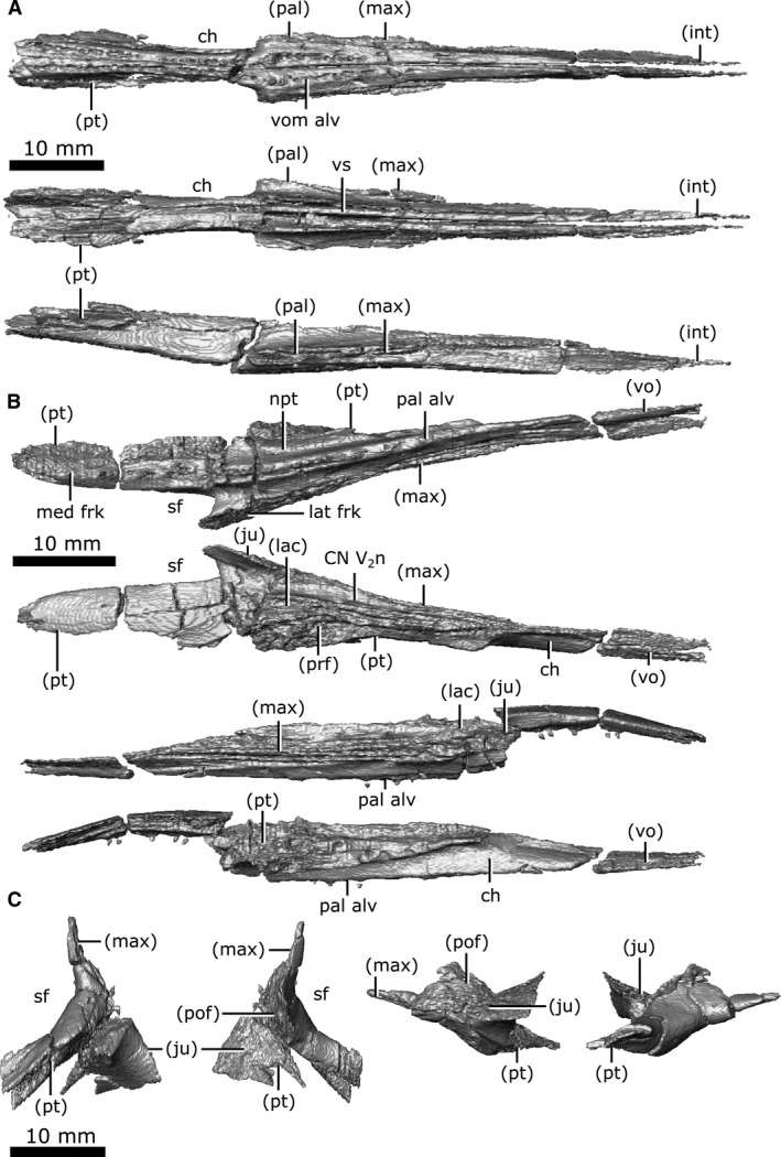
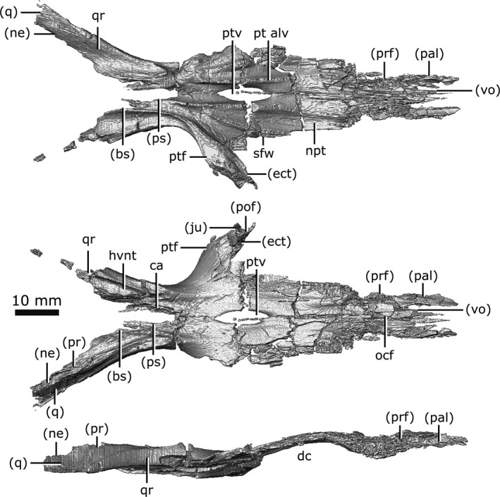
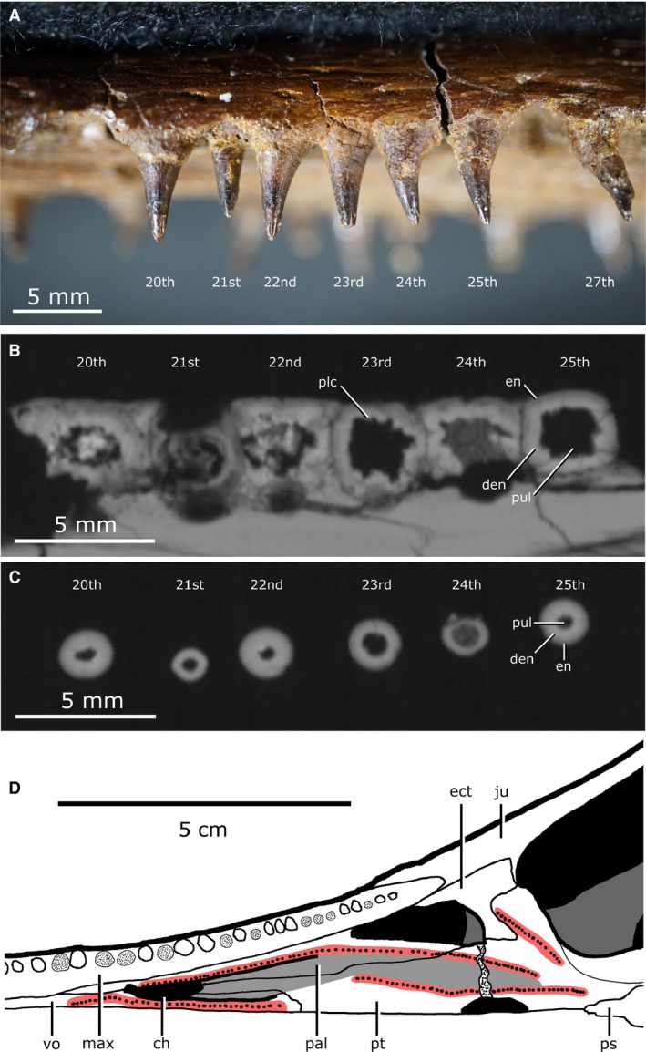
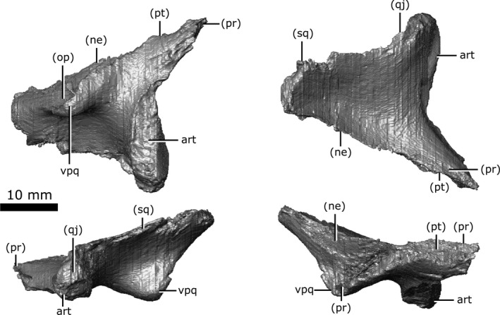
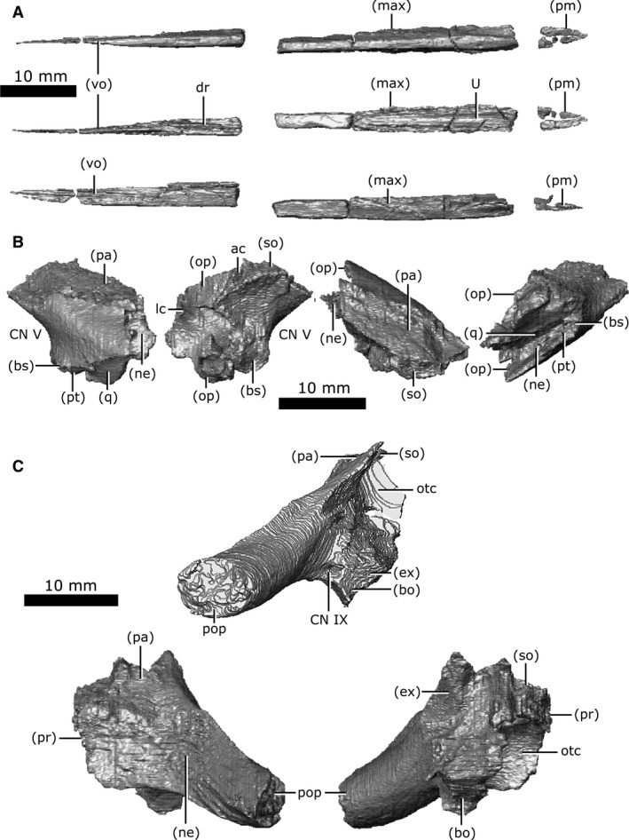
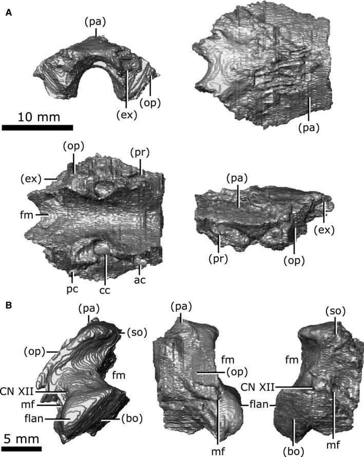
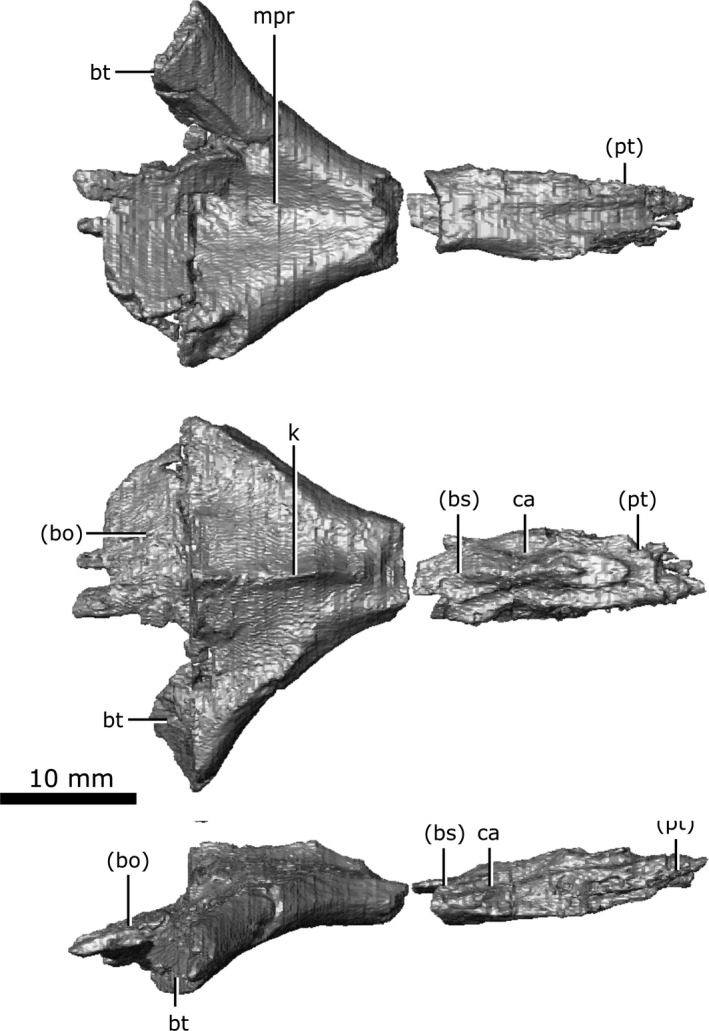
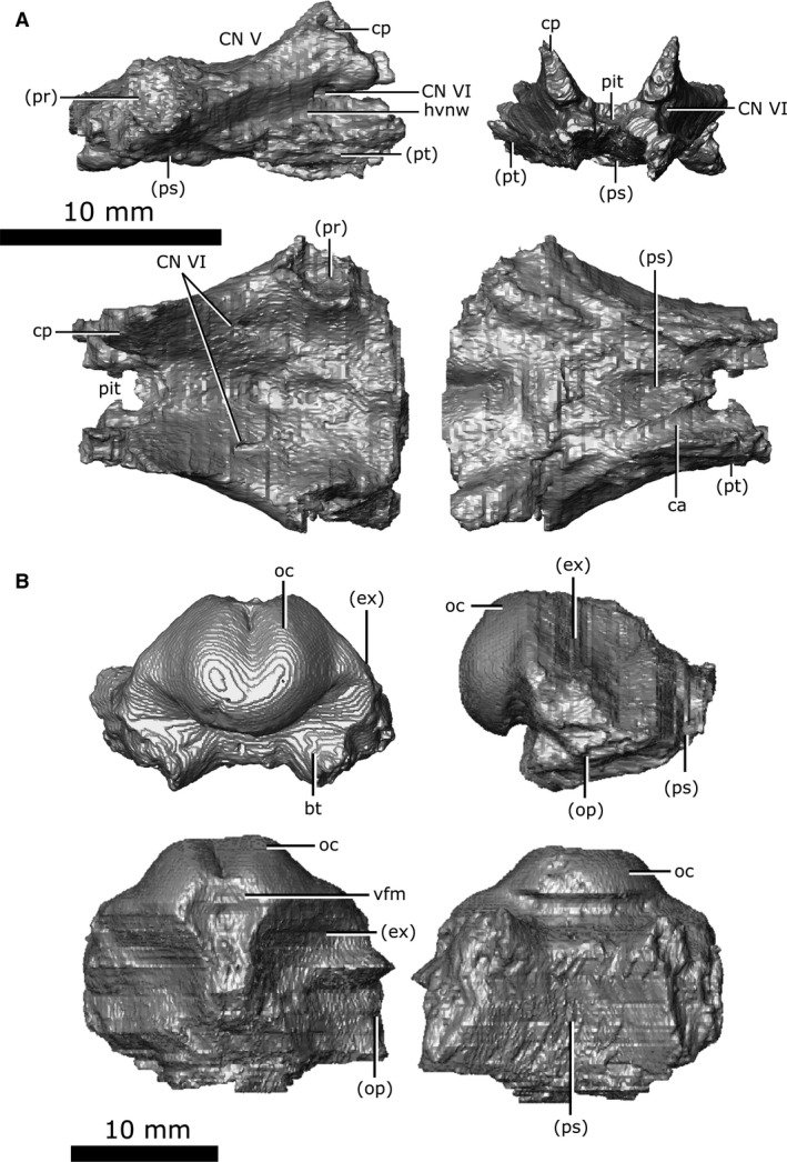
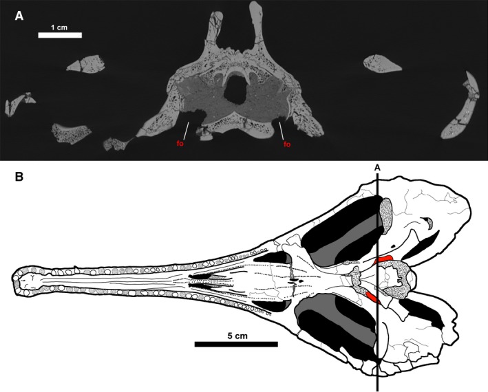
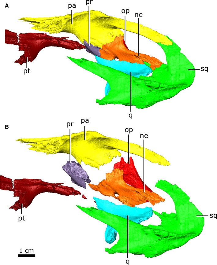
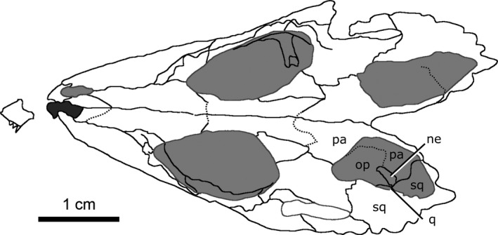
Choristoderes are extinct neodiapsid reptiles that are well known for their unusual cranial anatomy, possessing an elongated snout and expanded temporal arches. Although choristodere skulls are well described externally, their internal anatomy remains unknown. An internal description was needed to shed light on peculiarities of the choristodere skull, such as paired gaps on the ventral surface of the skull that may pertain to the fenestra ovalis, and a putative neomorphic ossification in the lateral wall of the braincase. Our goals were: (i) to describe the cranial elements of Champsosaurus lindoei in three dimensions; (ii) to describe paired gaps on the ventral surface of the skull to determine if these are indeed the fenestrae ovales; (iii) to illustrate the morphology of the putative neomorphic bone; and (iv) to consider the possible developmental and functional origins of the neomorph. We examined the cranial anatomy of the choristodere Champsosaurus lindoei (CMN 8920) using high-resolution micro-computed tomography scanning. We found that the paired gaps on the ventral surface of the skull do pertain to the fenestrae ovales, an unusual arrangement that may be convergent with some plesiosaurs, some aistopods, and some urodeles. The implications of this morphology in Champsosaurus are unknown and will be the subject of future work. We found that the neomorphic bone is a distinct ossification, but is not part of the wall of the brain cavity or the auditory capsule. Variation in the developmental pathways of cranial bones in living amniotes was surveyed to determine how the neomorphic bone may have developed. We found that the chondrocranium and splanchnocranium show little to no variation across amniotes, and the neomorphic bone is therefore most likely to have developed from the dermatocranium; however, the stapes is a pre-existing cranial element that is undescribed in choristoderes and may be homologous with the neomorphic bone. If the neomorphic bone is not homologous with the stapes, the neomorph likely developed from the dermatocranium through incomplete fusion of ossification centres from a pre-existing bone, most likely the parietal. Based on the apparent morphology of the neomorph in Coeruleodraco, the neomorph was probably too small to play a significant structural role in the skull of early choristoderes and it may have arisen through non-adaptive means. In neochoristoderes, such as Champsosaurus, the neomorph was likely recruited to support the expanded temporal arches.

Keywords: choristodera; computed tomography; development; evolution; skull anatomy.


Figures

Figure 1
Figure 1
Articulated skull of Champsosaurus lindoei (CMN 8920). (A) dorsal view; (B) ventral view; (C) lateral view.
Figure 2
Figure 2
Articulated skull of Champsosaurus lindoei (CMN 8920). (A) anterior view; (B) posterior view.
Figure 3
Figure 3
Major skull openings of Champsosaurus lindoei (CMN 8920). (A) dorsal view; (B) ventral view.
Figure 4
Figure 4
Skull measurements for Champsosaurus lindoei (CMN 8920). See Table 1 for corresponding measurement descriptions and values.
Figure 5
Figure 5
Isolated premaxilla and maxilla of Champsosaurus lindoei (CMN 8920). (A) left premaxilla in dorsal view (left), ventral view (middle), lateral view (top right), medial view (bottom right); (B) left maxilla in lateral view (top), medial view (second from top), dorsal view (second from bottom), ventral view (bottom).
Figure 6
Figure 6
Isolated nasal and lacrimal of Champsosaurus lindoei (CMN 8920). (A) nasal in dorsal view (top), ventral view (middle), right lateral view (bottom); (B) left lacrimal in dorsal view (top left), ventral view (top right), lateral view (bottom left), medial view (bottom right).
Figure 7
Figure 7
Isolated prefrontals and frontals of Champsosaurus lindoei (CMN 8920). (A) prefrontals in dorsal view (top), ventral view (middle), right lateral view (bottom); (B) frontals in dorsal view (top), ventral view (middle), right lateral view (bottom).
Figure 8
Figure 8
Isolated postfrontal and postorbital of Champsosaurus lindoei (CMN 8920). (A) left postfrontal in dorsal view (top left), ventral view (top right), lateral view (bottom left), medial view (bottom right); (B) left postorbital in dorsal view (top left), ventral view (top right), lateral view (bottom left), medial view (bottom right).
Figure 9
Figure 9
Isolated parietals of Champsosaurus lindoei (CMN 8920) in dorsal view (top), ventral view (middle), right lateral view (bottom).
Figure 10
Figure 10
Isolated left neomorphic bone of Champsosaurus lindoei (CMN 8920) in lateral view (top left), medial view (top right), dorsal view (middle left), ventral view (middle right), anterior view (bottom left), posterior view (bottom right).
Figure 11
Figure 11
Isolated jugal, quadratojugal, and squamosal of Champsosaurus lindoei (CMN 8920). (A) left jugal in lateral view (top left), medial view (top right), dorsal view (bottom left), ventral view (bottom right); (B) left quadratojugal in lateral view (top left), medial view (top right), dorsal view (bottom left), ventral view (bottom right); (C) left squamosal in lateral view (top left), medial view (top right), dorsal view (bottom left), ventral view (bottom right).
Figure 12
Figure 12
Isolated vomers, palatine, and ectopterygoid of Champsosaurus lindoei (CMN 8920). (A) vomers in ventral view (top), dorsal view (middle), right lateral view (bottom); (B) left palatine in ventral view (top), dorsal view (second from top), lateral view (second from bottom), medial view (bottom); (C) left ectopterygoid in ventral view (left), dorsal view (second from left), lateral view (second from right), medial view (right).
Figure 13
Figure 13
Isolated pterygoids of Champsosaurus lindoei (CMN 8920) in ventral view (top), dorsal view (middle), right lateral view (bottom).
Figure 14
Figure 14
Tooth morphology of Champsosaurus lindoei (CMN 8920). (A) Labial view of left maxillary tooth row; (B) Basal transverse cross section of the maxillary teeth; (C) Apical transverse cross section of the maxillary teeth; (D) Left palatal dentition. Red coloured areas mark the palatal teeth, light‐grey coloured area marks the nasopalatal trough.
Figure 15
Figure 15
Isolated left quadrate of Champsosaurus lindoei (CMN 8920) in ventral view (top left), dorsal view (top right), lateral view (bottom left), medial view (bottom right).
Figure 16
Figure 16
Isolated internarial, prootic, and opisthotic of Champsosaurus lindoei (CMN 8920). (A) internarial in ventral view (top), dorsal view (middle), right lateral view (bottom); (B) left prootic in lateral view (left), medial view (second from left), dorsal view (second from right), lateral view (right); (C) left opisthotic in posterior view (top), lateral view (bottom left), medial view (bottom right).
Figure 17
Figure 17
Isolated supraoccipital and exoccipital of Champsosaurus lindoei (CMN 8920). (A) supraoccipital in posterior view (top left), dorsal view (top right), ventral view (bottom left), left lateral view (bottom right); (B) left exoccipital in posterior view (left), lateral view (middle), medial view (right).
Figure 18
Figure 18
Isolated parasphenoid of Champsosaurus lindoei (CMN 8920) in ventral view (top), dorsal view (middle), right lateral view (bottom).
Figure 19
Figure 19
Isolated basisphenoid and basioccipital of Champsosaurus lindoei (CMN 8920). (A) basisphenoid in right lateral view (top left), anterior view (top right), dorsal view (bottom left), ventral view (bottom right); (B) basioccipital in posterior view (top left), right lateral view (top right), dorsal view (bottom left), ventral view (bottom right).
Figure 20
Figure 20
The fenestrae ovales of Champsosaurus lindoei. (A) coronal cross section of the braincase of CMN 8920; (B) ventral view of CMN 8920 with the fenestrae ovales coloured red. Black line labelled ‘A’ indicates the position of cross section A. Fine stippling indicates fractured surfaces exposing cancellous bone.
Figure 21
Figure 21
Left neomorphic bone and adjoining bones of Champsosaurus lindoei (CMN 8920) in left dorsolateral view with the elements (A) articulated, and (B) exploded.
Figure 22
Figure 22
Line drawing of the skull of Coeruleodraco jurassicus (IVPP V 23318) in dorsal view. Modified from Matsumoto et al. (2019).

References

    1. Abo‐Eleneen RE, Othman SI, Al‐Harbi HM, et al. (2017) Anatomical study of the skull of amphisbaenian Diplometopon zarudnyi (Squamata, Amphisbaenia). Saudi J Biol Sci 1, 1–11. - PMC - PubMed
    1. Atkins JB, Franz‐Odendaal TA (2016) The evolutionary and morphological history of the parasphenoid bone in vertebrates. Acta Zool 97, 255–263.
    1. Atkins JB, Reisz R, Maddin HC (2019) Braincase simplification and the origin of lissamphibians. PLoS ONE 14, e0213694. - PMC - PubMed
    1. Bailleul AM, Scannella JB, Horner JR, et al. (2016) Fusion patterns in the skulls of modern archosaurs reveal that sutures are ambiguous maturity indicators for the Dinosauria. PLoS ONE 11, 1–26. - PMC - PubMed
    1. de Beer G (1937) The Development of the Vertebrate Skull. Toronto: Oxford University Press.

Publication types