. 2019 Dec 30;25(1):144.
doi: 10.3390/molecules25010144.

Neuroprotective Effects of Tetrahydrocurcumin against Glutamate-Induced Oxidative Stress in Hippocampal HT22 Cells

Affiliations

Neuroprotective Effects of Tetrahydrocurcumin against Glutamate-Induced Oxidative Stress in Hippocampal HT22 Cells

Chang-Hyun Park et al. Molecules. .

Abstract

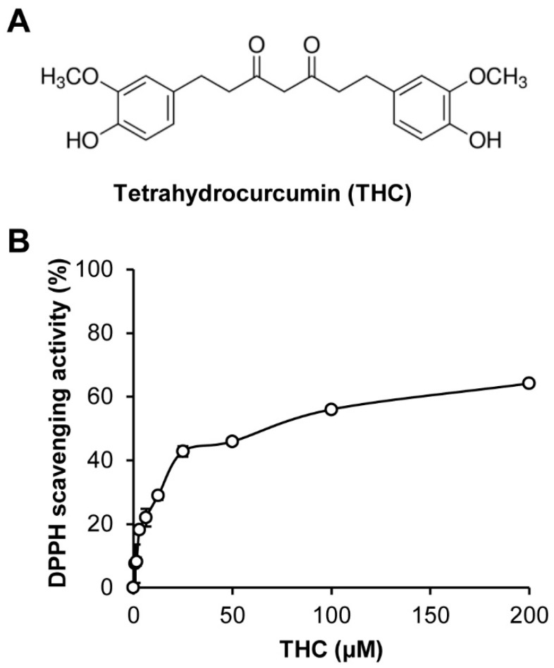
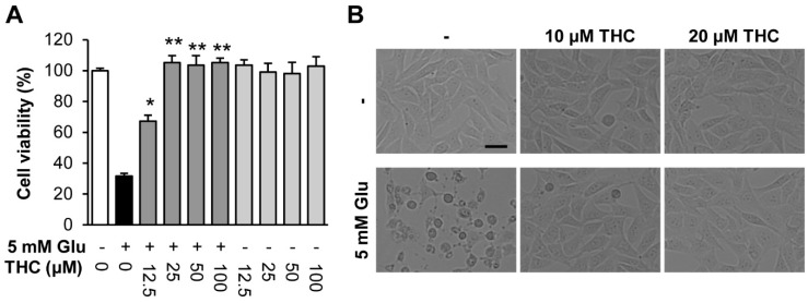
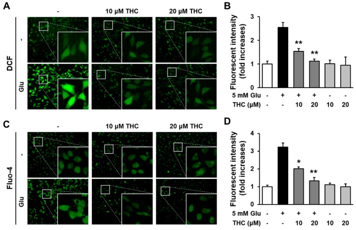
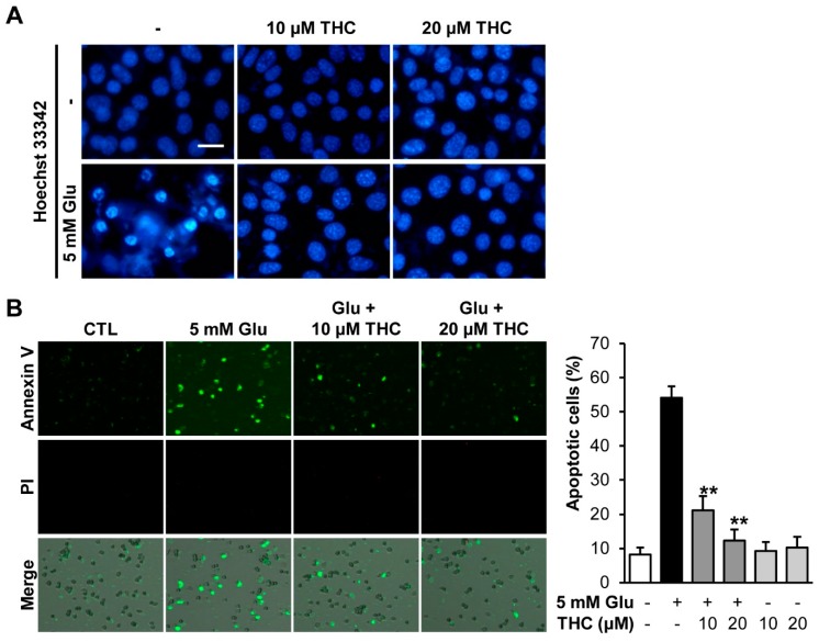
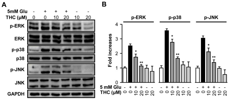
In the central nervous system, glutamate is a major excitable neurotransmitter responsible for many cellular functions. However, excessive levels of glutamate induce neuronal cell death via oxidative stress during acute brain injuries as well as chronic neurodegenerative diseases. The present study was conducted to examine the effect of tetrahydrocurcumin (THC), a major secondary metabolite of curcumin, and its possible mechanism against glutamate-induced cell death. We prepared THC using curcumin isolated from Curcuma longa (turmeric) and demonstrated the protective effect of THC against glutamate-induced oxidative stress in HT22 cells. THC abrogated glutamate-induced HT22 cell death and showed a strong antioxidant effect. THC also significantly reduced intracellular calcium ion increased by glutamate. Additionally, THC significantly reduced the accumulation of intracellular oxidative stress induced by glutamate. Furthermore, THC significantly diminished apoptotic cell death indicated by annexin V-positive in HT22 cells. Western blot analysis indicated that the phosphorylation of mitogen-activated protein kinases including c-Jun N-terminal kinase, extracellular signal-related kinases 1/2, and p38 by glutamate was significantly diminished by treatment with THC. In conclusion, THC is a potent neuroprotectant against glutamate-induced neuronal cell death by inhibiting the accumulation of oxidative stress and phosphorylation of mitogen-activated protein kinases.

Keywords: Ca2+; HT22 cells; glutamate; mitogen-activated protein kinase; oxidative stress; tetrahydrocurcumin.


Conflict of interest statement

The authors declare no conflict of interest.

Figures

Figure 1
Figure 1
Tetrahydrocurcumin (THC) possessed antioxidative activity. (A) Chemical structure of THC prepared from curcumin isolated from Curcuma longa (Turmeric). (B) The bar graph represents DPPH scavenging activity of THC.
Figure 2
Figure 2
Tetrahydrocurcumin (THC) prevented glutamate-induced HT22 cell death. (A) Cell viability was measured using a CyTox assay kit 24 h after treatment with 5 mM glutamate with or without THC. Bars denote the percentage of cell viability (mean ± S.E.M., * p < 0.05 and ** p < 0.001 compared to glutamate-treated cells). (B) Microscopic images were obtained after exposure of HT22 cells to glutamate for 24 h (scale bar, 50 μm).
Figure 3
Figure 3
THC diminished the increase in intracellular ROS and Ca2+ via its antioxidant activity. (A) Cells were treated with 5 mM glutamate with or without 10 or 20 μM THC for 8 h and stained with H2DCF-DA. Green indicates DCF fluorescence (20×). (B) Bars denote the ROS levels measured by fold-increases in the fluorescent intensity of DCF (** p < 0.001 compared to glutamate-treated cells). (C) Cells were treated with 5 mM glutamate with or without 10 or 20 μM THC for 8 h and stained with Fluo-4 AM for an additional 30 min. Green fluorescence indicates DCF active cells (20×). (D) Bars denote the levels of [Ca2+]i measured by fold-increases in the fluorescent intensity of Fluo-4 (* p < 0.05 and ** p < 0.001 compared to glutamate-treated cells).
Figure 4
Figure 4
THC blocked glutamate-induced apoptotic cell death in HT22 cells. (A) Cells were treated with 5 mM glutamate with or without 10 or 20 μM THC for 12 h and stained with Hoechst 33342. (B) We also performed Tali-image based analysis to identify the percentage of apoptotic cells. Merged images (bottom) were combined with bright field (gray; not shown), annexin V (green; top), propidium iodide (red; middle) images. The bars denote the percentages apoptotic cells determined by annexin V-positive cells (** p < 0.001 compared to glutamate-treated cells).
Figure 5
Figure 5
THC blocked phosphorylation of mitogen-activated protein kinases (MAPKs) induced by glutamate in HT22 cells. (A) HT22 cells were treated with 5 mM glutamate with or without 10 or 20 µM THC for 8 h. Western blot analysis were performed with specific antibodies for p38, p-p38, ERK, p-ERK, JNK, p-JNK, and GAPDH. (B) Immunoreactive bands were quantitatively determined using ImageJ software. The bars denote the fold increases compared with control (mean ± S.E.M, * p < 0.05 and ** p < 0.001 compared to glutamate-treated cells).

References

    1. Choi D.W. Glutamate neurotoxicity and diseases of the nervous system. Neuron. 1988;1:623–634. doi: 10.1016/0896-6273(88)90162-6. - DOI - PubMed
    1. Thoreson W.B., Witkovsky P. Glutamate receptors and circuits in the vertebrate retina. Prog. Retin. Eye Res. 1999;18:765–810. doi: 10.1016/S1350-9462(98)00031-7. - DOI - PubMed
    1. Coyle J.T., Puttfarcken P. Oxidative stress, glutamate, and neurodegenerative disorders. Science. 1993;262:689–695. doi: 10.1126/science.7901908. - DOI - PubMed
    1. Hwang O. Role of oxidative stress in Parkinson’s disease. Exp. Neurobiol. 2013;22:461–491. doi: 10.5607/en.2013.22.1.11. - DOI - PMC - PubMed
    1. Murphy T.H., Miyamoto M., Sastre A., Schnaar R.L., Coyle J.T. Glutamate toxicity in a neuronal cell line involves inhibition of cystine transport leading to oxidative stress. Neuron. 1989;2:1547–1558. doi: 10.1016/0896-6273(89)90043-3. - DOI - PubMed