

Morphometric Evaluation of Spermatogenic Cells and Seminiferous Tubules and Exploration of Luteinizing Hormone Beta Polypeptide in Testis of Datong Yak
- PMID: 31905946
- PMCID: PMC7022877
- DOI: 10.3390/ani10010066
Morphometric Evaluation of Spermatogenic Cells and Seminiferous Tubules and Exploration of Luteinizing Hormone Beta Polypeptide in Testis of Datong Yak
Abstract
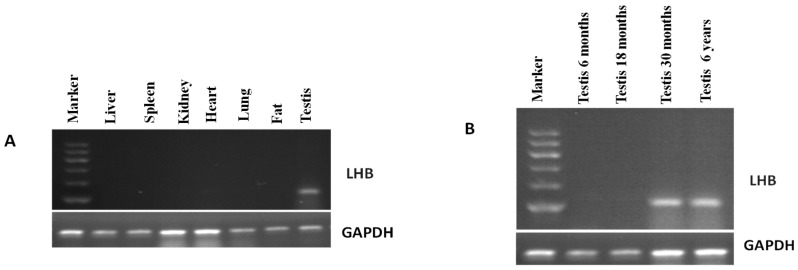
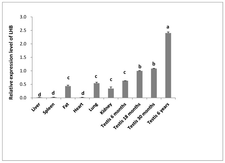
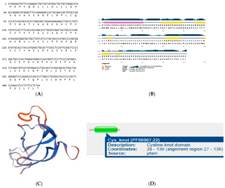
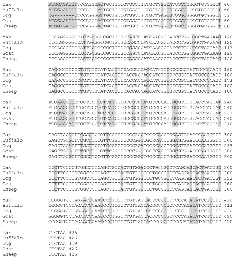
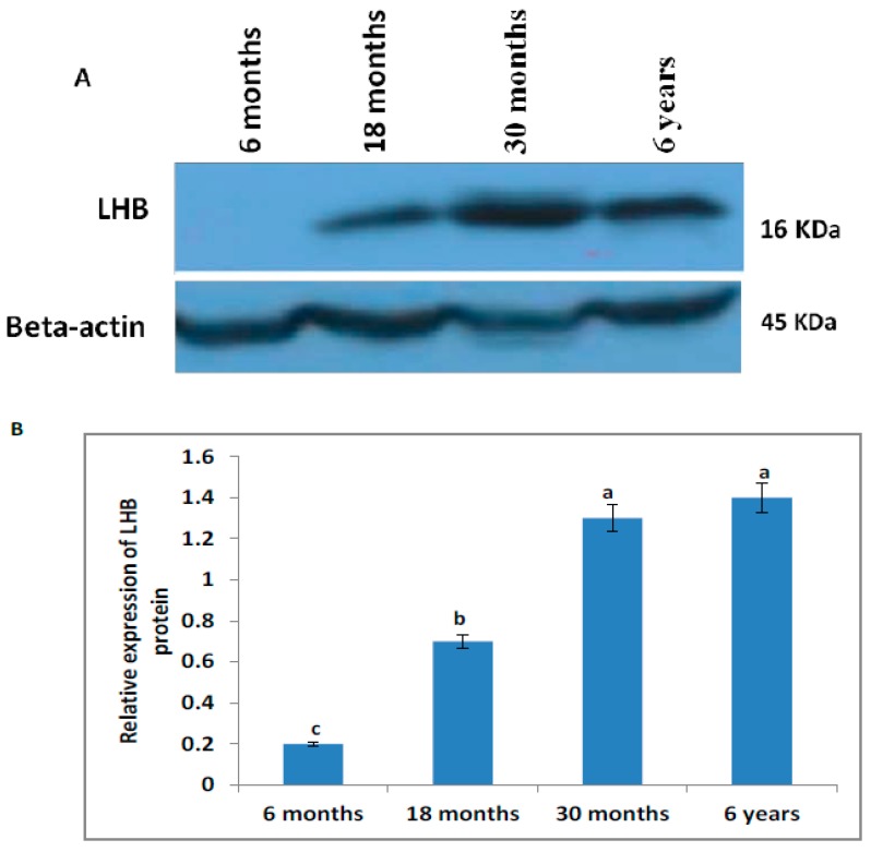
Histological examination of testes is essential for understanding infertility, sex development, and growth. Therefore, to understand the histomorphology of testes at different developmental stages, we performed hematoxylin and eosin staining of Yak testis. Our results revealed that the diameters of spermatogenic cells and their nuclei were significantly larger (p < 0.05) in the testis at six years compared to at six and 18 months. No significant difference was noted between 30 months and six years. The study was designed to compare the expression profile of LHB in Datong yak. The expression pattern of LHB was explored using quantitative PCR, semi-quantitative PCR, molecular bioinformatic, and Western blot analysis. Our observations indicated that expression of LHB was significantly higher (p < 0.05) in the testis of Datong yak. Western blotting indicated that the molecular mass of LHB protein was 16 kDa in yak. The protein encoded by yak LHB included conserved cysteine-knot domain regions. The high expression of LHB in testis indicated that LHB may be vital for the development of male gonads and the fertility of Datong yak.
Keywords: LHB; gene expression; histomorphology; testis; yak.
Conflict of interest statement
The authors declare no conflict of interest.
Figures

Similar articles
-
Expression Analysis of IZUMO1 Gene during Testicular Development of Datong Yak (Bos Grunniens).Animals (Basel). 2019 May 29;9(6):292. doi: 10.3390/ani9060292. Animals (Basel). 2019. PMID: 31146500 Free PMC article.
-
Molecular Cloning and Characterization of SYCP3 and TSEG2 Genes in the Testicles of Sexually Mature and Immature Yak.Genes (Basel). 2019 Oct 30;10(11):867. doi: 10.3390/genes10110867. Genes (Basel). 2019. PMID: 31671664 Free PMC article.
-
The low expression of Dmrt7 is associated with spermatogenic arrest in cattle-yak.Mol Biol Rep. 2014 Nov;41(11):7255-63. doi: 10.1007/s11033-014-3611-x. Epub 2014 Jul 23. Mol Biol Rep. 2014. PMID: 25052188
-
Regulation by Hsp27/P53 in testis development and sperm apoptosis of male cattle (cattle-yak and yak).J Cell Physiol. 2018 Jan;234(1):650-660. doi: 10.1002/jcp.26822. Epub 2018 Aug 21. J Cell Physiol. 2018. PMID: 30132847
-
The expression of epidermal growth factor (EGF) and its receptor (EGFR) during post-natal testes development in the yak.Reprod Domest Anim. 2014 Dec;49(6):970-6. doi: 10.1111/rda.12416. Epub 2014 Sep 26. Reprod Domest Anim. 2014. PMID: 25263304
Cited by
-
Age-Related Changes in the Testicular Morphophysiology of the Cane Rat (Thryonomys swinderianus).J Microsc Ultrastruct. 2021 Jul 9;10(3):118-126. doi: 10.4103/jmau.jmau_84_20. eCollection 2022 Jul-Sep. J Microsc Ultrastruct. 2021. PMID: 36504588 Free PMC article.
-
Enrichment of spermatogonial stem cells and staging of the testis cycle in a dasyurid marsupial, the fat-tailed dunnart.Stem Cells. 2025 Mar 10;43(3):sxaf007. doi: 10.1093/stmcls/sxaf007. Stem Cells. 2025. PMID: 39943734 Free PMC article.
-
Quantitative Proteomic Analysis Reveals Key Proteins Involved in Testicular Development of Yaks.Int J Mol Sci. 2024 Aug 2;25(15):8433. doi: 10.3390/ijms25158433. Int J Mol Sci. 2024. PMID: 39126002 Free PMC article.
-
Aging induced testicular damage: analyzing the ameliorative potential of Mucuna pruriens seed extract.3 Biotech. 2023 Jun;13(6):206. doi: 10.1007/s13205-023-03618-8. Epub 2023 May 22. 3 Biotech. 2023. PMID: 37229277 Free PMC article.
-
Comparative analysis of the testes from wild-type and Alkbh5-knockout mice using single-cell RNA sequencing.G3 (Bethesda). 2022 Jul 29;12(8):jkac130. doi: 10.1093/g3journal/jkac130. G3 (Bethesda). 2022. PMID: 35652742 Free PMC article.
References
-
- Davis J.R., Langford G.A., Kirby P.J. The testicular capsule. In: Johnson A.D., Gomes R., Vandemark N.L., editors. The Testis. Development, Anatomy and Physiology. Academic Press; London, UK: 1970. pp. 281–337.
-
- Leal M.C., Becker-Silva S.C., Chiarini-Garcia H., Franca L.R. Sertoli cell efficiency and daily sperm production in goats (Capra hircus) Anim. Reprod. 2004;1:122128.
LinkOut - more resources
Full Text Sources
