

Transcriptional Differences of Coding and Non-Coding Genes Related to the Absence of Melanocyte in Skins of Bama Pig
- PMID: 31905971
- PMCID: PMC7017308
- DOI: 10.3390/genes11010047
Transcriptional Differences of Coding and Non-Coding Genes Related to the Absence of Melanocyte in Skins of Bama Pig
Abstract
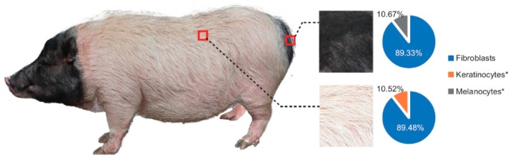
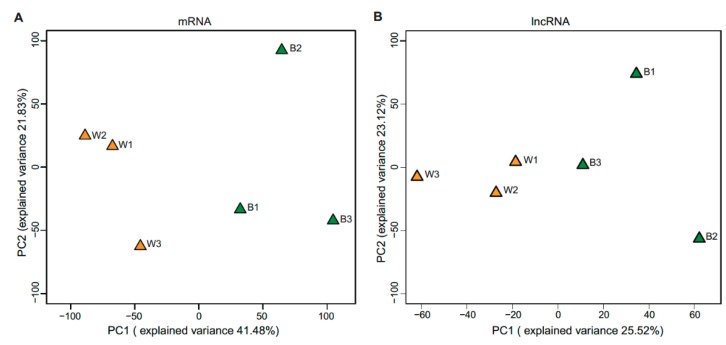
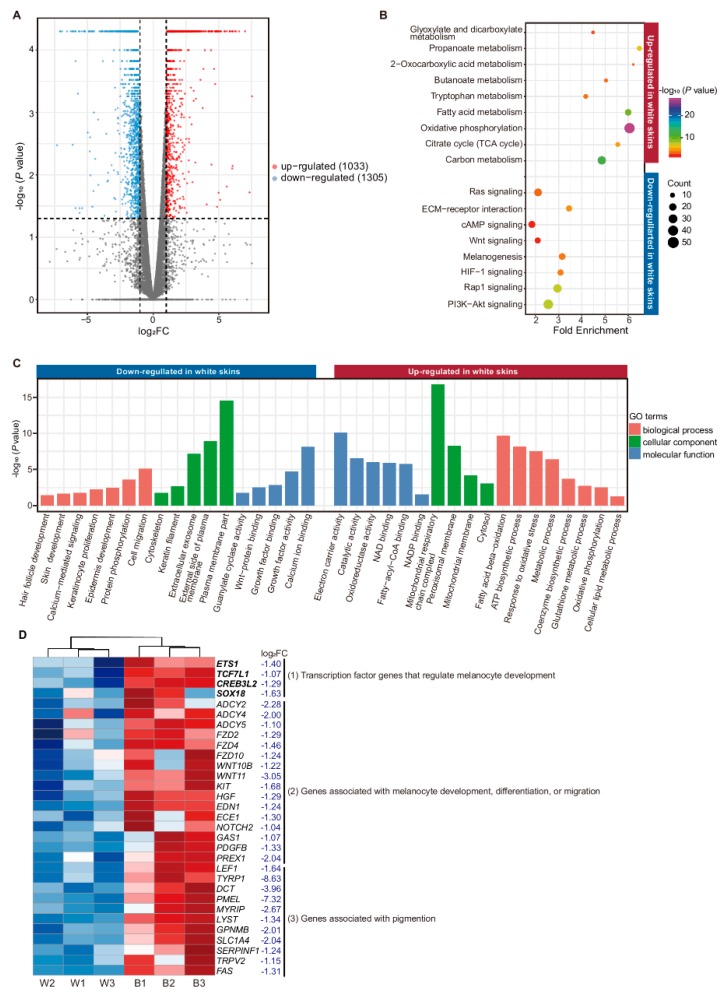
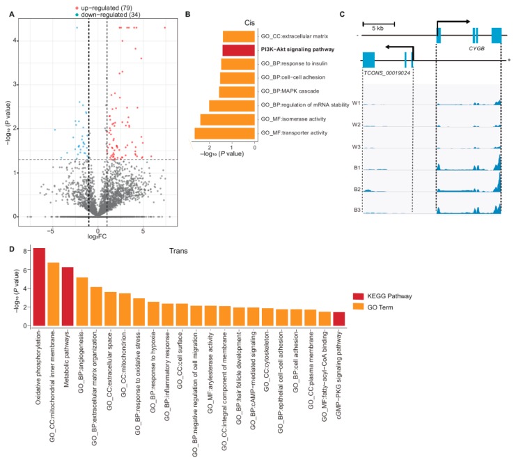
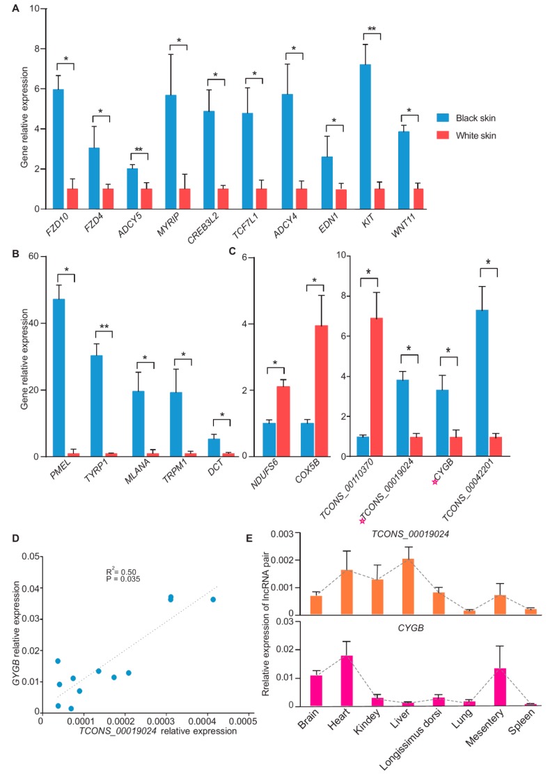
Skin is the body's largest organ, and the main function of skin is to protect underlying organs from possible external damage. Melanocytes play an important role in skin pigmentation. The Bama pig has a "two-end-black" phenotype with different coat colors across skin regions, e.g., white skin (without melanocytes) and black skin (with melanocytes), which could be a model to investigate skin-related disorders, specifically loss of melanocytes. Here, we generated expression profiles of mRNAs and long noncoding RNAs in Bama pig skins with different coat colors. In total, 14,900 mRNAs and 7549 lncRNAs were expressed. Overall, 2338 mRNAs/113 lncRNAs with FDR-adjusted p-value ≤ 0.05 were considered to be differentially expressed (DE) mRNAs/lncRNAs, with 1305 down-regulated mRNAs and 1033 up-regulated mRNAs in white skin with|log2(fold change)| > 1. The genes down-regulated in white skin were associated with pigmentation, melanocyte-keratinocyte interaction, and keratin, while up-regulated ones were mainly associated with cellular energy metabolisms. Furthermore, those DE lncRNAs were predicted to be implicated in pigmentation, keratin synthesis and cellular energy metabolism. In general, this study provides insight into the transcriptional difference involved in melanocyte-loss-induced keratinocyte changes and promotes the Bama pig as a biomedical model in skin research.
Keywords: Bama pig; melanocyte deficiency; model; transcriptome.
Conflict of interest statement
The authors declare no conflict of interest.
Figures
Similar articles
-
Transcriptomic Analysis of Coding Genes and Non-Coding RNAs Reveals Complex Regulatory Networks Underlying the Black Back and White Belly Coat Phenotype in Chinese Wuzhishan Pigs.Genes (Basel). 2019 Mar 7;10(3):201. doi: 10.3390/genes10030201. Genes (Basel). 2019. PMID: 30866582 Free PMC article.
-
The comprehensive detection of miRNA, lncRNA, and circRNA in regulation of mouse melanocyte and skin development.Biol Res. 2020 Feb 3;53(1):4. doi: 10.1186/s40659-020-0272-1. Biol Res. 2020. PMID: 32014065 Free PMC article.
-
Identification and comparison of long non-conding RNA in Jinhua and Landrace pigs.Biochem Biophys Res Commun. 2018 Nov 30;506(3):765-771. doi: 10.1016/j.bbrc.2018.06.028. Epub 2018 Jun 23. Biochem Biophys Res Commun. 2018. PMID: 29890140
-
Comprehensive transcriptional profiling of porcine brain aging.Gene. 2019 Apr 20;693:1-9. doi: 10.1016/j.gene.2019.01.019. Epub 2019 Jan 26. Gene. 2019. PMID: 30695714
-
Long non-coding RNA expression profile in Cdk5-knockdown mouse skin.Gene. 2018 Sep 25;672:195-201. doi: 10.1016/j.gene.2018.05.120. Epub 2018 Jun 8. Gene. 2018. PMID: 29890311 Review.
Cited by
-
Exploring the regulatory role of long non-coding RNAs in pigmentation in juvenile Plectropomus leopardus.Sci Rep. 2025 Jul 31;15(1):27977. doi: 10.1038/s41598-025-13347-y. Sci Rep. 2025. PMID: 40745010 Free PMC article.
-
Transcriptome profiling analysis of uterus during chicken laying periods.BMC Genomics. 2023 Aug 3;24(1):433. doi: 10.1186/s12864-023-09521-z. BMC Genomics. 2023. PMID: 37537566 Free PMC article.
-
Grade follicles transcriptional profiling analysis in different laying stages in chicken.BMC Genomics. 2022 Jul 7;23(1):492. doi: 10.1186/s12864-022-08728-w. BMC Genomics. 2022. PMID: 35794517 Free PMC article.
-
Comprehensive analysis of lncRNAs modified by m6A methylation in sheep skin.Anim Biosci. 2024 Nov;37(11):1887-1990. doi: 10.5713/ab.24.0039. Epub 2024 May 7. Anim Biosci. 2024. PMID: 38754841 Free PMC article.
-
Polygenic risk scores of fasting insulin and insulin-related traits in a Taiwanese Han population.Cell Biosci. 2025 Aug 5;15(1):115. doi: 10.1186/s13578-025-01454-2. Cell Biosci. 2025. PMID: 40765060 Free PMC article.
References
-
- Kanitakis J. Anatomy, histology and immunohistochemistry of normal human skin. Eur. J. Dermatol. 2002;12:390–399. - PubMed
Publication types
MeSH terms
Substances
LinkOut - more resources
Full Text Sources
Molecular Biology Databases
