Review
. 2019 Dec 30;21(1):266.
doi: 10.3390/ijms21010266.

Cell-to-Cell Communication in Learning and Memory: From Neuro- and Glio-Transmission to Information Exchange Mediated by Extracellular Vesicles

Affiliations
Review

Cell-to-Cell Communication in Learning and Memory: From Neuro- and Glio-Transmission to Information Exchange Mediated by Extracellular Vesicles

Gabriella Schiera et al. Int J Mol Sci. .

Abstract

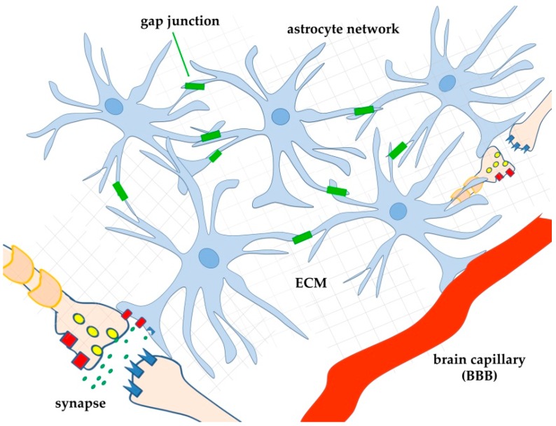
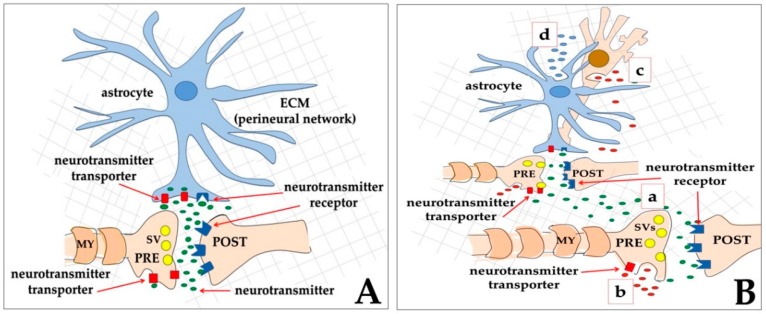
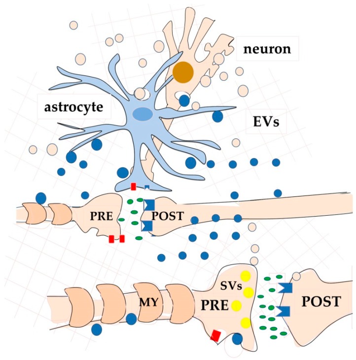
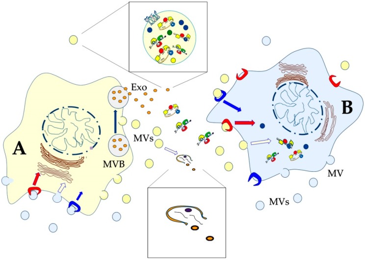
Most aspects of nervous system development and function rely on the continuous crosstalk between neurons and the variegated universe of non-neuronal cells surrounding them. The most extraordinary property of this cellular community is its ability to undergo adaptive modifications in response to environmental cues originating from inside or outside the body. Such ability, known as neuronal plasticity, allows long-lasting modifications of the strength, composition and efficacy of the connections between neurons, which constitutes the biochemical base for learning and memory. Nerve cells communicate with each other through both wiring (synaptic) and volume transmission of signals. It is by now clear that glial cells, and in particular astrocytes, also play critical roles in both modes by releasing different kinds of molecules (e.g., D-serine secreted by astrocytes). On the other hand, neurons produce factors that can regulate the activity of glial cells, including their ability to release regulatory molecules. In the last fifteen years it has been demonstrated that both neurons and glial cells release extracellular vesicles (EVs) of different kinds, both in physiologic and pathological conditions. Here we discuss the possible involvement of EVs in the events underlying learning and memory, in both physiologic and pathological conditions.

Keywords: extracellular vesicles; glial cells; learning; memory; synaptic plasticity; tetrapartite synapse; tripartite synapsis; volume transmission; wiring transmission.


Conflict of interest statement

The authors declare no conflict of interest.

Figures

Figure 1
Figure 1
Schematic drawing of astrocyte interconnection by gap junctions (green rectangles). By forming a network, astrocytes can influence synapses far away from the active ones, allowing different (and distant) synapses to interact with each other also in the absence of proximity (lateral regulation of synaptic transmission by astrocytes, [86]). For simplicity, only two distant astrocytes, each interacting with one synapse (model of the tripartite synapse), and one astrocyte interacting with one brain capillary are shown. Thanks to the network, astrocytes can probably also transfer metabolites (for example, glucose) from blood to distant neurons. BBB: blood-brain-barrier; ECM: extracellular matrix.
Figure 2
Figure 2
Schematic drawing of two types of neurotransmission, according to the initial proposal of the model [109,110]. (A) In the wiring transmission (WT), the presynaptic neuron (PRE) releases neurotransmitter molecules (small green ovals) that bind to their receptors (dark blue) on the post-synaptic element (POST). Excess neurotransmitters are then taken back by the presynaptic neuron through neurotransmitter transporters (red rectangles); In (B), a few ways to obtain volume transmission (VT) are represented: (a), the presynaptic neuron (PRE) releases a neurotransmitter (small green ovals) that not only binds to its receptors on the post-synaptic element (POST), but also diffuses at different distances, thus reaching other faraway synapses that will be activated; (b) extrasynaptic release from the axon of signal molecules (small red ovals) into the extracellular matrix, outside the synaptic cleft; (c) extrasynaptic release of signal molecules (small red ovals) from the soma of a neuron; (d) gliotransmitter release from an astrocyte (small blue ovals). In both WT and VT, astrocytes play a central role since they can express on their membranes both neurotransmitter receptors and neurotransmitter transporters, and, in addition, they can release gliotransmitters.
Figure 3
Figure 3
Schematic drawings of one further aspect of VT-neurotransmission [114]. A variety of molecules and metabolites can be exchanged both among neurons, and among neurons and other brain cells, via extracellular vesicles (EVs) of different size and composition. Neurons release EVs (probably for most exosomes) mainly from the soma and dendrites. As an example of a glial cell, an astrocyte is drawn. For clarity, vesicles have been depicted the same colour as the producing cell: light pink produced by the neuron and blue produced by the astrocyte.
Figure 4
Figure 4
Schematic drawing of two cells that communicate with each other by exchanging extracellular vesicles of different sizes and origins. Some vesicles directly bud from the plasma membrane (microvesicles, MVs), while exosomes (Exo) derive from the multivesicular body (MVB). For clarity, vesicles have been depicted in the same colour as the producing cell: yellow produced by the yellow cell (A), and light blue produced by the light blue cell (B). After release, some EVs are quickly lysed and release their content into the extracellular space (lower enlarged view). Some of them contain matrix metalloproteases and other hydrolytic enzymes responsible for the digestion of various ECM components. Alternatively, intact EVs can interact with the target cells that can internalize them through a variety of pathways [145]. The vesicles can also directly fuse with the plasma membrane of the target cell. Both MVs and exosomes are endowed with proteins, lipids, and nucleic acids that can influence different physiological and pathological functions of the target cell (upper enlarged view).
Figure 5
Figure 5
Neurons are highly polarized cells with only one nucleus and highly differentiated peripheries. Polarization depends on the cytoskeleton-dependent trafficking of organelles and vesicle/molecule complexes in both anterograde- and retrograde-direction. Among the transported complexes, ribonucleoprotein complexes (RNPs) have been also described. RNPs contain a variety of RNAs and RNA-binding regulatory proteins (RBPs). During their trip to the periphery mRNAs are repressed. It has been reported that, at post-synaptic sites, upon synapse activation, and largely in response to calcium waves, some RBPs undergo post-translational modifications that allow the release and translation of mRNAs. Some of the newly synthesized proteins can accumulate at the synapse, while others can shuttle back to the nucleus to modify chromatin structure and expression. At the same time, microtubules ensure transport of organelles such as mitochondria and synaptic vesicles.
Figure 6
Figure 6
All the cell types in the CNS release EVs of different size, origin, and composition. For clarity, both larger and smaller vesicles have been coloured the same colour as the producing cell. A few components of the vesicles are reported in the inserts with the relevant references [27,31,179,182,183,184,189,197,198,199,200,201]. Abbreviations: ApoD, apoprotein D; BBB, blood–brain barrier; CNP, 2′,3′-Cyclic Nucleotide 3′ Phosphodiesterase; EAAT, excitatory amino acid transporter; FGF, fibroblast growth factor; GS, glutamine synthetase; MAG, myelin-associated glycoprotein; MBP, myelin basic protein; MCT1, monocarboxylate transporter 1; MMPs, matrix metallopeptidases; MOG, myelin oligodendrocyte glycoprotein; MVB: multivescicular body; PLP, myelin proteolipid protein; PRPC, cellular prion protein; VEGF, vascular endothelial growth factor.
Figure 7
Figure 7
Schematic drawing of a procedure for purifying neuron-derived EVs, according to [250]. A few molecules that have been found increased (upward arrows) or decreased (downward arrows) in EVs purified from the blood of patients are reported in the boxes, together with some relevant references [30,31,200].

Similar articles

Cited by

References

    1. Bliss T.V., Lømo T. Long-lasting potentiation of synaptic transmission in the dentate area of the anaesthetized rabbit following stimulation of the perforant path. J. Physiol. 1973;232:331–356. doi: 10.1113/jphysiol.1973.sp010273. - DOI - PMC - PubMed
    1. Collingridge G.L., Kehl S.J., McLennan H. Excitatory amino acids in synaptic transmission in the Schaffer collateral-commissural pathway of the rat hippocampus. J. Physiol. 1983;334:33–46. doi: 10.1113/jphysiol.1983.sp014478. - DOI - PMC - PubMed
    1. Morris R.G., Anderson E., Lynch G.S., Baudry M. Selective impairment of learning and blockade of long-term potentiation by an N-methyl-D-aspartate receptor antagonist, AP5. Nature. 1986;319:774–776. doi: 10.1038/319774a0. - DOI - PubMed
    1. Morris R.G. Synaptic plasticity and learning: Selective impairment of learning rats and blockade of long-term potentiation in vivo by the N-methyl-D-aspartate receptor antagonist AP5. J. Neurosci. 1989;9:3040–3057. doi: 10.1523/JNEUROSCI.09-09-03040.1989. - DOI - PMC - PubMed
    1. Bailey C.H., Bartsch D., Kandel E.R. Toward a molecular definition of long-term memory storage. Proc. Natl. Acad. Sci. USA. 1996;93:13445–13452. doi: 10.1073/pnas.93.24.13445. - DOI - PMC - PubMed