Review
. 2020 Jan 1;21(1):302.
doi: 10.3390/ijms21010302.

A Single Cell but Many Different Transcripts: A Journey into the World of Long Non-Coding RNAs

Affiliations
Review

A Single Cell but Many Different Transcripts: A Journey into the World of Long Non-Coding RNAs

Enrico Alessio et al. Int J Mol Sci. .

Abstract

In late 2012 it was evidenced that most of the human genome is transcribed but only a small percentage of the transcripts are translated. This observation supported the importance of non-coding RNAs and it was confirmed in several organisms. The most abundant non-translated transcripts are long non-coding RNAs (lncRNAs). In contrast to protein-coding RNAs, they show a more cell-specific expression. To understand the function of lncRNAs, it is fundamental to investigate in which cells they are preferentially expressed and to detect their subcellular localization. Recent improvements of techniques that localize single RNA molecules in tissues like single-cell RNA sequencing and fluorescence amplification methods have given a considerable boost in the knowledge of the lncRNA functions. In recent years, single-cell transcription variability was associated with non-coding RNA expression, revealing this class of RNAs as important transcripts in the cell lineage specification. The purpose of this review is to collect updated information about lncRNA classification and new findings on their function derived from single-cell analysis. We also retained useful for all researchers to describe the methods available for single-cell analysis and the databases collecting single-cell and lncRNA data. Tables are included to schematize, describe, and compare exposed concepts.

Keywords: lncRNA database; lncRNAs; long non-coding RNAs; non-coding RNAs; single-cell; single-cell database; single-cell expression; single-cell sequencing.


Conflict of interest statement

The authors declare no conflict of interest. The funders had no role in the design of the study; in the collection, analyses, or interpretation of data; in the writing of the manuscript, or in the decision to publish the results.

Figures

Figure 1
Figure 1
Different RNA types in cells. RNAs are divided in coding (if they are translated; blue) and non-coding (if they are not translated; yellow). Non-coding RNAs can be distinguished in different RNA types depending on their size or function. Long non-coding RNAs are longer than 200 nt and all displayed categories are discussed in the manuscript. Transcripts presented can be localized in the cell nucleus, cytoplasm, or in both compartments, in mitochondria or chloroplasts.
Figure 2
Figure 2
Scheme of different types of long non-coding RNAs (lncRNAs). (A) licRNAs are localized between coding genes and do not overlap any of them. (B) Genic lncRNAs are overlapped to coding sequences. (C) Splice variants. (D) Ribosomal RNAs are components of ribosomes. (E) Chromatin interacting lncRNAs allow chromatin modifications permitting the correct localization of modifying enzymes. (F) miRNA sponging lncRNAs reduce the availability of miRNAs to modulate the expression of their mRNA target. (G) Enhancer lncRNAs are transcribed from DNA regions with enhancing functions. (H) SINEUPs are lncRNAs that associating to mRNAs are able to enhance their translation. (I) lncRNAs can also code for micropeptides. (J) lncRNAs are distinguished according to the position of their targets. Green arrows indicate protein (E), miRNA (F), or lncRNA (J) targets. AA is for amino acids.
Figure 3
Figure 3
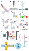
Methods to isolate single cells. (A) Micropipette isolation method consists of diluting the sample until obtaining a single cell on each well. (B) Laser capture microdissection utilizes a laser linked to a microscope to collect single cells from solid tissues. (C) Fluorescent activated cell sorting can sort cells tagged with fluorescent markers. (D) Capillary-based technology employs mechanical suction to isolate the cells. (E) Punching technologies are based on the principle of capturing cells on microwells and “punch” them on collection vials. (F) The dielectrophoresis system uses the electrokinetic properties of cells to move them on a chip. (G) Microfluidic platforms can capture single cells and barcoded beads into droplets of water in an oil phase. Each bead contains several primers composed by PCR handles, cell barcodes, and unique molecular identifiers (UMIs) sequences. PCR handles are common in all beads and allows PCR amplification after cDNA synthesis. Cell barcode sequences, identical across all the primers of the same microparticle but different from those on different beads, allow recovery of the cells’ origin. UMIs, different on each primer, allow mRNA transcripts to be digitally counted and to identify PCR duplicates. An oligod(T) sequence is present at the end of all primers for capturing polyadenylated RNAs and priming reverse transcription.
Figure 4
Figure 4
(A) Single-cell RNA sequencing of 8-cell embryos revealed that, contrary to what was previously believed, each cell has a different set of expressed genes and that the expression of XIST is not equal in the eight cells. (B) A scheme of an inactivated X chromosome with lncRNA XIST represented by red rectangles. Some genes are transcribed from this chromosome even if it is considered inactivated. Expressed lncRNAs are represented by purple semicircles while mRNAs are shown in yellow semicircles. In green, it is marked the position of ATRX, one of the mRNAs that escape the inactivation of the X chromosome. Microscope images show the expression of XIST (red), ATRX (green) and the position of the two X chromosomes (blue). Moreover, it is represented an enlargement of a single nucleus (white rectangle) for each staining. FISH images are modified from [155]. (C) Differences in lncRNA expression between cells at different stages of reprogramming. Some lncRNAs seem to be specifically expressed in the less specialized cells, suggesting that they might be involved with the maintenance of the pluripotency of stem cells and that might be used as tools to induce specialized cells to transition into less specialized cells (like iPSCs). Heat map is modified from [156].

References

    1. Li J., Liu C. Coding or noncoding, the converging concepts of RNAs. Front. Genet. 2019;10:496. doi: 10.3389/fgene.2019.00496. - DOI - PMC - PubMed
    1. Consortium E.P. An integrated encyclopedia of DNA elements in the human genome. Nature. 2012;489:57–74. doi: 10.1038/nature11247. - DOI - PMC - PubMed
    1. Vandevenne M., Delmarcelle M., Galleni M. RNA regulatory networks as a control of stochasticity in biological systems. Front. Genet. 2019;10:403. doi: 10.3389/fgene.2019.00403. - DOI - PMC - PubMed
    1. Vaquerizas J.M., Kummerfeld S.K., Teichmann S.A., Luscombe N.M. A census of human transcription factors: Function, expression and evolution. Nat. Rev. Genet. 2009;10:252–263. doi: 10.1038/nrg2538. - DOI - PubMed
    1. Pertea M. The human transcriptome: An unfinished story. Genes. 2012;3:344–360. doi: 10.3390/genes3030344. - DOI - PMC - PubMed

LinkOut - more resources