

Survival Time of Visual Gains after Diabetic Vitrectomy and Its Relationship with Ischemic Heart Disease
- PMID: 31906417
- PMCID: PMC6981366
- DOI: 10.3390/ijerph17010310
Survival Time of Visual Gains after Diabetic Vitrectomy and Its Relationship with Ischemic Heart Disease
Abstract
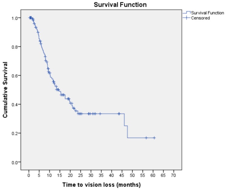
Vitrectomy surgery in proliferative diabetic retinopathy improves the vision-related quality of life. However, there is lack of data on the duration of maintenance of visual gains post vitrectomy. This study thus aimed to determine the survival time of visual gains and the prognostic factors of vision loss after vitrectomy surgery for complications of proliferative diabetic retinopathy. A retrospective cohort study was conducted in an ophthalmology clinic in Malaysia. We included 134 patients with type 2 diabetes mellitus on follow-up after vitrectomy for proliferative diabetic retinopathy. Visual acuity was measured using the log of minimum angle of resolution (LogMar). A gain of ≥0.3 LogMar sustained on two subsequent visits was considered evidence of visual improvement post vitrectomy. Subjects were considered to have vision loss when their post-operative visual acuity subsequently dropped by ≥0.3 LogMar. Kaplan-Meier analysis was used to determine the survival time of visual gains. Cox Proportional Hazard regression was used to determine the prognostic factors of vision loss. The median age of patients was 56.00 years (IQR ± 10.00). The median duration of diabetes mellitus was 14.00 years (IQR ± 10.00). Approximately 50% of patients with initial improvement post vitrectomy subsequently experienced vision loss. The survival time, i.e., the median time from surgery until the number of patients with vision loss formed half of the original cohort, was 14.63 months (95% CI: 9.95, 19.32). Ischemic heart disease was a significant prognostic factor of vision loss. Patients with underlying ischemic heart disease (adjusted HR: 1.97, 95% CI: 1.18, 3.33) had a higher risk of vision loss post vitrectomy, after adjusting for other factors. Approximately half the patients with initial visual gains post vitrectomy maintained their vision for at least one year. Ischemic heart disease was a poor prognostic factor for preservation of visual gains post vitrectomy.
Keywords: LogMar; ischemic heart disease; post vitrectomy; prognostic factors; type 2 diabetes mellitus.
Conflict of interest statement
The authors declare no conflict of interest.
Figures
References
-
- Letchuman G.R., Wan Nazaimoon W.M., Wan Mohamad W.B., Chandran L.R., Tee G.H., Jamaiyah H., Isa M.R., Zanariah H., Fatanah I., Ahmad Faudzi Y. Prevalence of diabetes in the Malaysian national health morbidity survey III 2006. Med. J. Malays. 2010;65:180–186. - PubMed
-
- Institute for Public Health (IPH) National Health and Morbidity Survey 2015 (NHMS 2015), Non-Communicable Diseases, Risk Factors Other Health Problems. Volume II. Ministry of Health Malaysia; Kuala Lumpur, Malaysia: 2015.
Publication types
MeSH terms
LinkOut - more resources
Full Text Sources
Medical
