

Immunohistochemical Analysis of a Vitreous Membrane Removed from a Patient with Incontinentia Pigmenti-Related Retinal Detachment
- PMID: 31906444
- PMCID: PMC7158695
- DOI: 10.3390/vision4010005
Immunohistochemical Analysis of a Vitreous Membrane Removed from a Patient with Incontinentia Pigmenti-Related Retinal Detachment
Abstract
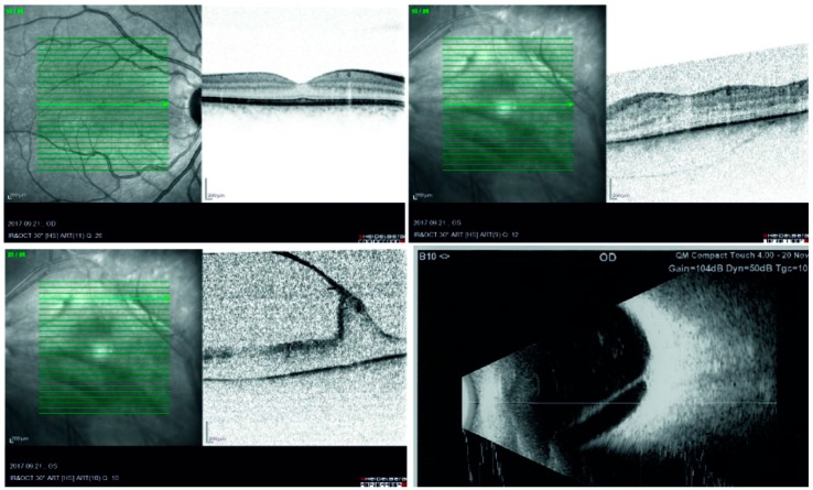
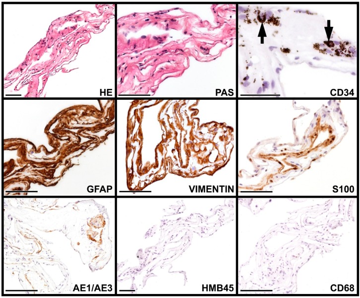
This is a case history of a 23-year-old woman suffering from incontinentia pigmenti (IP). The patient's vision in the left eye started to deteriorate due to cataract progression at the age of 22, and by the age of 23, it dropped from 0.9 to 0.04. Ultrasound examination confirmed tractional vitreoretinal membranes. Vitrectomy was performed, therefore, on her left eye. The histological evaluation of vitreous membrane revealed a complex immunophenotype (positivity for glial fibrillary acidic protein (GFAP), vimentin, S-100, anti-pan cytokeratin antibody (AE/AE3), and smooth muscle-specific actin (SMA) to various extents). The right eye remained unsymptomatic throughout this course. Besides being the first to analyze the tractional vitreoretinal membrane in IP with immunohistochemical methods, this case study points out that extreme cases of asymmetric side involvement in IP do exist, even to the point of one eye being completely unsymptomatic.
Keywords: epiretinal membrane; hereditary diseases; immunohistochemistry; incontinentia pigmenti.
Conflict of interest statement
The authors declare no conflict of interest.
Figures
Similar articles
-
[Treatment of retinopathy of incontinentia pigmenti by anti-vascular endothelial growth factor].Zhonghua Yan Ke Za Zhi. 2019 Apr 11;55(4):294-301. doi: 10.3760/cma.j.issn.0412-4081.2019.04.012. Zhonghua Yan Ke Za Zhi. 2019. PMID: 30982292 Chinese.
-
Incontinentia pigmenti-associated ocular anomalies of paediatric incontinentia pigmenti patients in China.Acta Ophthalmol. 2019 May;97(3):265-272. doi: 10.1111/aos.13781. Epub 2018 Aug 3. Acta Ophthalmol. 2019. PMID: 30073775
-
Multifocal hypopigmented retinal pigment epithelial lesions in incontinentia pigmenti.Retina. 2006 Mar;26(3):328-33. doi: 10.1097/00006982-200603000-00012. Retina. 2006. PMID: 16508434
-
[Paediatric retinal detachment and hereditary vitreoretinal disorders].Klin Monbl Augenheilkd. 2013 Sep;230(9):914-9. doi: 10.1055/s-0033-1350759. Epub 2013 Aug 28. Klin Monbl Augenheilkd. 2013. PMID: 23986190 Review. German.
-
Incontinentia pigmenti: a case report and literature review.Turk J Pediatr. 2009 Mar-Apr;51(2):190-4. Turk J Pediatr. 2009. PMID: 19480336 Review.
References
-
- Aradhya S., Woffendin H., Jakins T., Bardaro T., Esposito T., Smahi A., Shaw C., Levy M., Munnich A., D’Urso M., et al. A recurrent deletion in the ubiquitously expressed NEMO (IKK-gamma) gene accounts for the vast majority of incontinentia pigmenti mutations. Hum. Mol. Genet. 2001;10:2171–2179. doi: 10.1093/hmg/10.19.2171. - DOI - PubMed
-
- Nishimura M., Oka Y., Takagi I., Yamana T., Kitano A. The clinical features and treatment of the retinopathy in Bloch-Sulzberger syndrome (incontinentia pigmenti) Jpn. J. Ophthalmol. 1980;24:310–319.
Publication types
LinkOut - more resources
Full Text Sources
Miscellaneous
