Review
. 2020 Jan 2;9(1):110.
doi: 10.3390/cells9010110.

Dissecting the Heterogeneity in T-Cell Mediated Inflammation in IBD

Affiliations
Review

Dissecting the Heterogeneity in T-Cell Mediated Inflammation in IBD

Irma Tindemans et al. Cells. .

Abstract

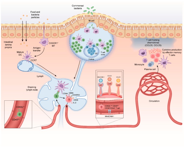
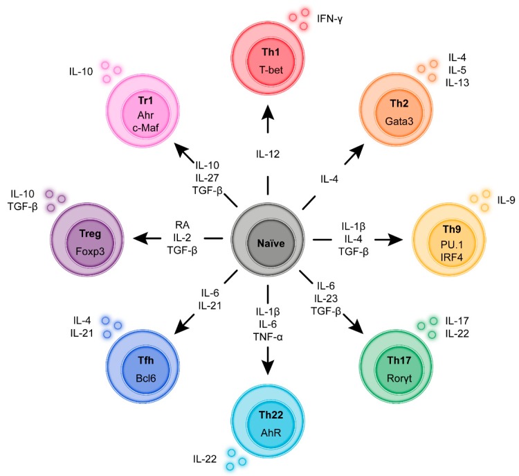
Infiltration of the lamina propria by inflammatory CD4+ T-cell populations is a key characteristic of chronic intestinal inflammation. Memory-phenotype CD4+ T-cell frequencies are increased in inflamed intestinal tissue of IBD patients compared to tissue of healthy controls and are associated with disease flares and a more complicated disease course. Therefore, a tightly controlled balance between regulatory and inflammatory CD4+ T-cell populations is crucial to prevent uncontrolled CD4+ T-cell responses and subsequent intestinal tissue damage. While at steady state, T-cells display mainly a regulatory phenotype, increased in Th1, Th2, Th9, Th17, and Th17.1 responses, and reduced Treg and Tr1 responses have all been suggested to play a role in IBD pathophysiology. However, it is highly unlikely that all these responses are altered in each individual patient. With the rapidly expanding plethora of therapeutic options to inhibit inflammatory T-cell responses and stimulate regulatory T-cell responses, a crucial need is emerging for a robust set of immunological assays to predict and monitor therapeutic success at an individual level. Consequently, it is crucial to differentiate dominant inflammatory and regulatory CD4+ T helper responses in patients and relate these to disease course and therapy response. In this review, we provide an overview of how intestinal CD4+ T-cell responses arise, discuss the main phenotypes of CD4+ T helper responses, and review how they are implicated in IBD.

Keywords: CD4+ T-cell; IBD; T helper cell; intestine.


Conflict of interest statement

The authors have no conflicts of interest.

Figures

Figure 1
Figure 1
Mechanisms of CD4+ T-cell differentiation in the healthy intestine. (i) Antigens can be taken up by specialized microfold cells (M-cells) that can transport microbial antigens across the mucosal epithelium from the lumen to the subepithelial dome in the Peyer’s patch (PP); (ii) soluble antigens can diffuse through epithelial tight junctions and can be transferred across epithelial cells by transcellular routes and be ingested by phagocytes such as macrophages and dendritic cells (DC); and (iii) luminal antigens can also be captured by transepithelial projecting dendrites from macrophage-like CX3CR1+ APCs in the lamina propria. M-cells and CX3CR1+ APCs can transfer antigen to migratory dendritic cells that migrate from the lamina propria to inductive sites to interact with naïve CD4+ T-cells. Naïve CD4+ CD45RA chemokine receptor C-C motif receptor 7+ (CCR7) T-cells migrate from the peripheral blood into gut-draining lymph nodes and PP. The DC-mediated T-cell activation requires three signals; (i) T-cell receptor (TCR) stimulation with antigenic peptides in the context of major histocompatibility complex class II (MHCII), (ii) co-stimulation via CD28-CD80/CD86 engagement and (iii) cytokine signals mostly provided by APCs. Upon activation, T-cells acquire CD69, CD25 (IL-2 receptor-alpha chain) CD44 and CD45RO. T-cells down-regulate expression of CD62L, CCR7, and CD45RA and start secreting IL-2, a cytokine needed for proliferation. After a 3–4-day proliferation period the differentiated T-cells egress from the lymph node into the blood. A subpopulation of these CD45ROCD44+CD4+ T-cells will re-express CD62Lhi and CCR7 and provide central memory in lymphoid tissues. The remainder of the CD62LnegCD45ROCD44+CD4+ T-cells will migrate to the intestinal lamina propria where they reside as long-lived memory CD4+ T-cells. Crucially, differentiation of T-cells in the MLN is associated with imprinting of higher levels of retinoic acid-induced CD38. As this CD62LnegCD38+ effector T-cell phenotype is induced regardless of regulatory or inflammatory T-cell function it can be used to monitor gut-homing T-cells in human peripheral blood. Homing of memory-phenotype CD62LloCD45ROCD44+CD4+ T-cells to the small and large intestine requires expression of the integrin α4β7 which binds to mucosal vascular addressin cell adhesion molecule 1 (MADCAM1) on endothelial cells in blood vessels. In the small intestine, epithelial cells produce the chemokine CCL25 which attracts memory T-cells expressing the receptor CCR9 Chemokine receptors involved in regulation of CD4+ T-cell homing to the colon at steady state are less well defined, although recently, it was discovered that G protein coupled receptor 15 (GPR15) directs CD4+ T-cells to the colon. During intestinal inflammation, CCR6 and its ligand CCL20 also contribute to CD4+ T-cell recruitment to the inflamed small and large intestine, demonstrating that CD4+ T-cells can use alternative homing receptors during inflammation when compared to steady state.
Figure 2
Figure 2
Induction and transcriptional program of the main regulatory and Inflammatory CD4+ T-cell sub-populations. During antigen-specific activation via the T-cell receptor (TCR) and subsequent proliferation in the lymph node naïve CD4+ T-cells undergo a functional programming termed differentiation. Depending on the strength of the TCR signal and in response to cytokine co-stimulation by the APC the T-cells express specific transcription factors that skew differentiation to a range of functionalities such as host defense against intracellular pathogens, defense against extracellular pathogens, defense against helminths or immune suppression. Although these differentiation programs are not irreversible classification according to these “subsets” based on dominant cytokine profiles and transcription factors is commonly used to better describe and characterize immune responses.

Similar articles

Cited by

References

    1. Preza G.C., Yang O.O., Elliott J., Anton P.A., Ochoa M.T. T lymphocyte density and distribution in human colorectal mucosa, and inefficiency of current cell isolation protocols. PLoS ONE. 2015;10:e0122723. doi: 10.1371/journal.pone.0122723. - DOI - PMC - PubMed
    1. Mowat A.M. To respond or not to respond—A personal perspective of intestinal tolerance. Nat. Rev. Immunol. 2018;18:405–415. doi: 10.1038/s41577-018-0002-x. - DOI - PubMed
    1. Duchmann R., Kaiser I., Hermann E., Mayet W., Ewe K., Meyer zum Buschenfelde K.H. Tolerance exists towards resident intestinal flora but is broken in active inflammatory bowel disease (IBD) Clin. Exp. Immunol. 1995;102:448–455. doi: 10.1111/j.1365-2249.1995.tb03836.x. - DOI - PMC - PubMed
    1. Louis E., Van Kemseke C., Reenaers C. Necessity of phenotypic classification of inflammatory bowel disease. Best Pract Res. Clin. Gastroenterol. 2011;25(Suppl. 1):S2–S7. doi: 10.1016/S1521-6918(11)70003-8. - DOI - PubMed
    1. Levine A., Griffiths A., Markowitz J., Wilson D.C., Turner D., Russell R.K., Fell J., Ruemmele F.M., Walters T., Sherlock M., et al. Pediatric Modification of the Montreal Classification for Inflammatory Bowel Disease: The Paris Classification. Inflamm. Bowel Dis. 2011;17:1314–1321. doi: 10.1002/ibd.21493. - DOI - PubMed

Publication types