Review
. 2020 Jan 2;9(1):112.
doi: 10.3390/cells9010112.

Context-Dependent Strategies for Enhanced Genome Editing of Genodermatoses

Affiliations
Review

Context-Dependent Strategies for Enhanced Genome Editing of Genodermatoses

Oliver Patrick March et al. Cells. .

Abstract

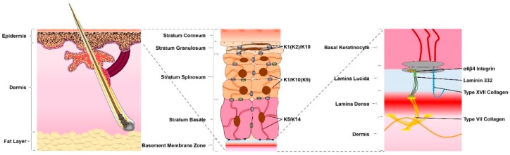
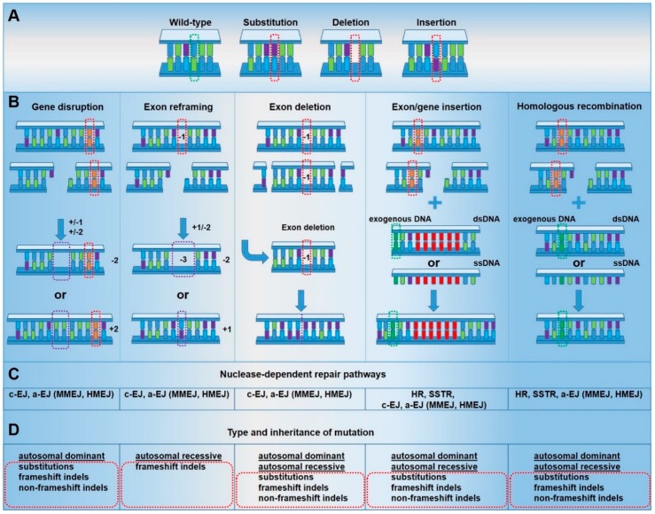
The skin provides direct protection to the human body from assault by the harsh external environment. The crucial function of this organ is significantly disrupted in genodermatoses patients. Genodermatoses comprise a heterogeneous group of largely monogenetic skin disorders, typically involving mutations in genes encoding structural proteins. Therapeutic options for this debilitating group of diseases, including epidermolysis bullosa, primarily consist of wound management. Genome editing approaches co-opt double-strand break repair pathways to introduce desired sequence alterations at specific loci. Rapid advances in genome editing technologies have the potential to propel novel genetic therapies into the clinic. However, the associated phenotypes of many mutations may be treated via several genome editing strategies. Therefore, for potential clinical applications, implementation of efficient approaches based upon mutation, gene and disease context is necessary. Here, we describe current genome editing approaches for the treatment of genodermatoses, along with a discussion of the optimal strategy for each genetic context, in order to achieve enhanced genome editing approaches.

Keywords: DNA repair pathways; designer nucleases; epidermolysis bullosa; genodermatoses; genome editing; keratinopathies.


Conflict of interest statement

The authors declare no conflict of interest.

Figures

Figure 1
Figure 1
The anatomy of the skin. The skin consists of three layers: fat, dermis and epidermis. The epidermis is typically composed of four layers, situated above the basement membrane. From inner to outer, these are: the stratum basale, stratum spinosum, stratum granulosum and stratum corneum—associated with different expression profiles with respect to keratins and intracellular adhesions. Keratins 5 and 14 (K5/K14) are expressed in basal cells. Suprabasal cells demonstrate a downregulation of K5/K14 in favor of K1/K10 in normal interfollicular epidermis and K1/K9 in the palms and soles. Cells of the stratum granulosum additionally express K2. The basal keratinocytes of the stratum basale connect the epidermis to the basement membrane zone, comprised of the lamina lucida and lamina densa, which provides connection between the epidermis and the underlying dermis. Structural proteins within the basement membrane zone, such as α6β4 integrin, laminin 332, type XVII collagen and type VII collagen, are crucial for the integrity and stability of the skin.
Figure 2
Figure 2
Context-dependent gene repair strategies. Genome editing technologies rely on the formation of specific double-strand breaks (DSBs) within a gene and their resolution via DSB repair pathways. The targeting strategy, nature, and context of the DSB determine the repair outcome of the different genome editing approaches. Therefore, the choice of an efficient targeting strategy for genome editing of genodermatoses is highly dependent on the type and inheritance of the mutation. (A) Types of mutations. (B) Gene repair strategies. (C) Nuclease-dependent DNA repair pathways. (D) Type and inheritance of mutation, which can be targeted by the respective gene repair strategy. (Orange bars and red dotted rectangles indicate the mutation site; dark green bars and dotted rectangles indicate wild-type (wt) sequence; purple dotted rectangles indicate wanted repair outcomes; red bars indicate exon/gene insertion.)

References

    1. Rognoni E., Watt F.M. Skin Cell Heterogeneity in Development, Wound Healing, and Cancer. Trends Cell Biol. 2018;28:709–722. doi: 10.1016/j.tcb.2018.05.002. - DOI - PMC - PubMed
    1. Hsu Y.C., Li L., Fuchs E. Emerging interactions between skin stem cells and their niches. Nat. Med. 2014;20:847–856. doi: 10.1038/nm.3643. - DOI - PMC - PubMed
    1. Fuchs E. Cell biology: More than skin deep. J. Cell Biol. 2015;209:629–631. doi: 10.1083/jcb.201503129. - DOI - PMC - PubMed
    1. Has C., Bruckner-Tuderman L. Molecular and diagnostic aspects of genetic skin fragility. J. Dermatol. Sci. 2006;44:129–144. doi: 10.1016/j.jdermsci.2006.08.003. - DOI - PubMed
    1. Simpson C.L., Patel D.M., Green K.J. Deconstructing the skin: Cytoarchitectural determinants of epidermal morphogenesis. Nat. Rev. Mol. Cell Biol. 2011;12:565–580. doi: 10.1038/nrm3175. - DOI - PMC - PubMed

Publication types