

Synergistic Effects on Incorporation of β-Tricalcium Phosphate and Graphene Oxide Nanoparticles to Silk Fibroin/Soy Protein Isolate Scaffolds for Bone Tissue Engineering
- PMID: 31906498
- PMCID: PMC7023539
- DOI: 10.3390/polym12010069
Synergistic Effects on Incorporation of β-Tricalcium Phosphate and Graphene Oxide Nanoparticles to Silk Fibroin/Soy Protein Isolate Scaffolds for Bone Tissue Engineering
Abstract
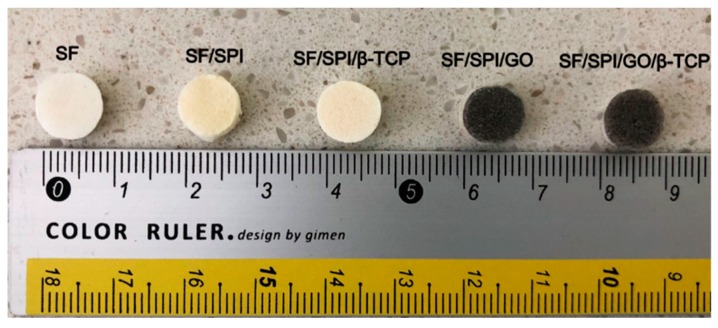
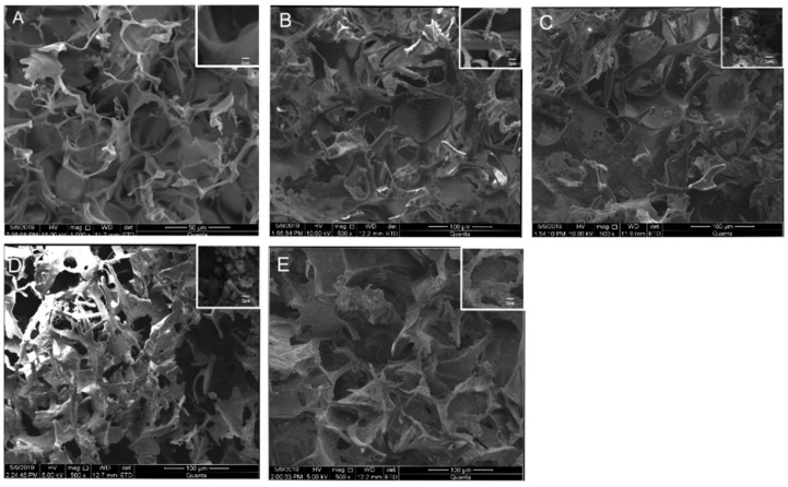
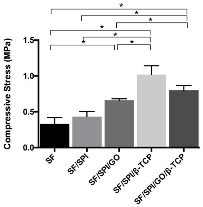
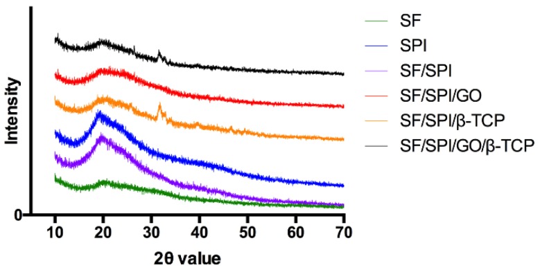
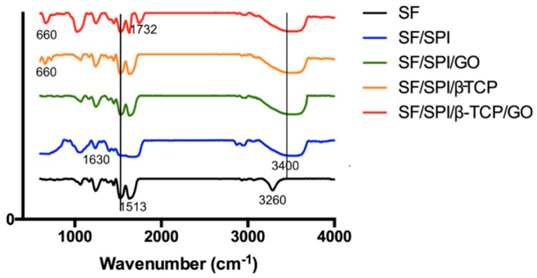
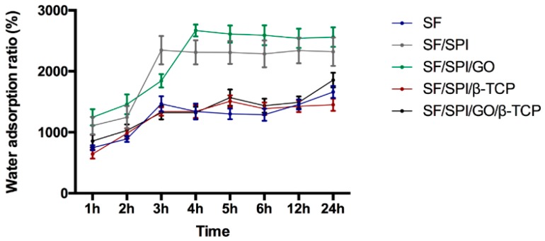
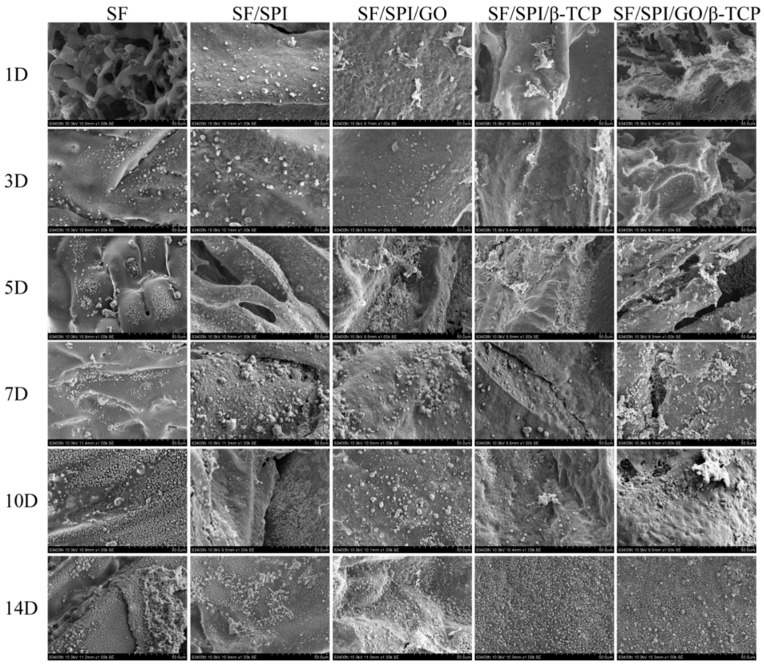
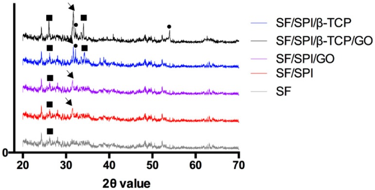
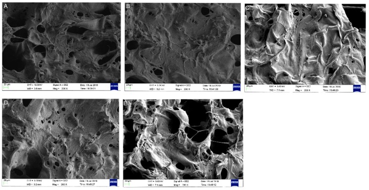
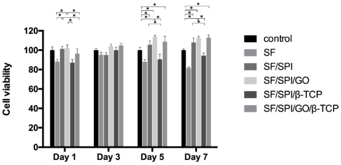
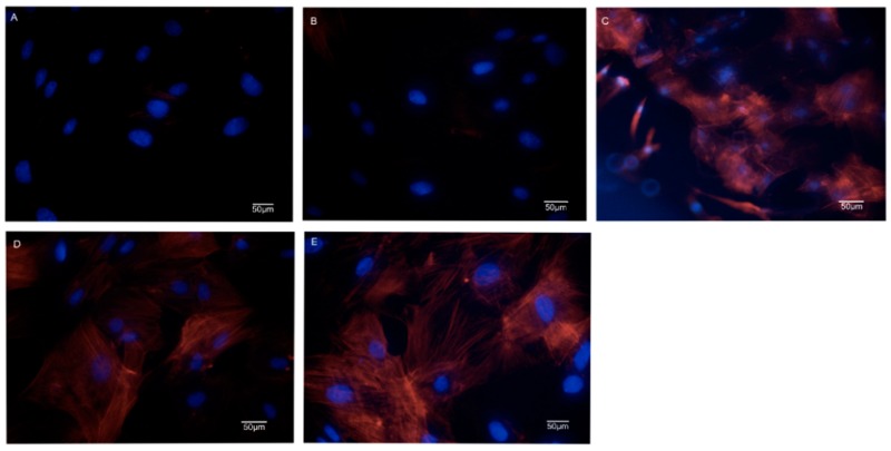
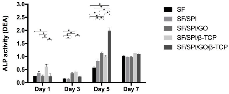
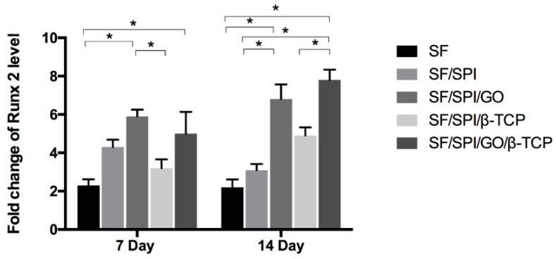
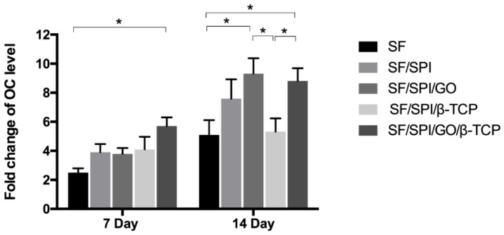
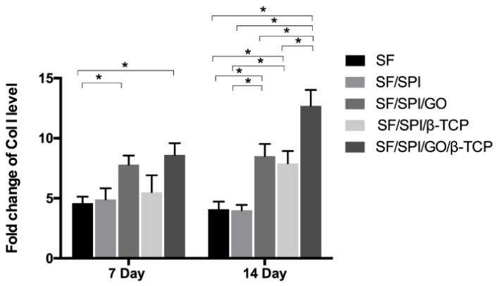
In bone tissue engineering, an ideal scaffold is required to have favorable physical, chemical (or physicochemical), and biological (or biochemical) properties to promote osteogenesis. Although silk fibroin (SF) and/or soy protein isolate (SPI) scaffolds have been widely used as an alternative to autologous and heterologous bone grafts, the poor mechanical property and insufficient osteoinductive capability has become an obstacle for their in vivo applications. Herein, β-tricalcium phosphate (β-TCP) and graphene oxide (GO) nanoparticles are incorporated into SF/SPI scaffolds simultaneously or individually. Physical and chemical properties of these composite scaffolds are evaluated using field emission scanning electron microscope (FESEM), X-ray diffraction (XRD) and attenuated total reflectance Fourier transformed infrared spectroscopy (ATR-FTIR). Biocompatibility and osteogenesis of the composite scaffolds are evaluated using bone marrow mesenchymal stem cells (BMSCs). All the composite scaffolds have a complex porous structure with proper pore sizes and porosities. Physicochemical properties of the scaffolds can be significantly increased through the incorporation of β-TCP and GO nanoparticles. Alkaline phosphatase activity (ALP) and osteogenesis-related gene expression of the BMSCs are significantly enhanced in the presence of β-TCP and GO nanoparticles. Especially, β-TCP and GO nanoparticles have a synergistic effect on promoting osteogenesis. These results suggest that the β-TCP and GO enhanced SF/SPI scaffolds are promising candidates for bone tissue regeneration.
Keywords: graphene oxide; osteogenesis; scaffold; silk fibroin; soy protein isolate; β-tricalcium phosphate.
Conflict of interest statement
The authors declare no conflict of interest. The founding sponsors had no role in the design of the study; in the collection, analyses, or interpretation of data; in the writing of the manuscript, and in the decision to publish the results.
Figures
Similar articles
-
The incorporation of β-tricalcium phosphate nanoparticles within silk fibroin composite scaffolds for enhanced bone regeneration: An in vitro and in vivo study.J Biomater Appl. 2022 Apr;36(9):1567-1578. doi: 10.1177/08853282211065621. Epub 2022 Feb 8. J Biomater Appl. 2022. PMID: 35135370
-
Enhanced osteogenesis of β-tricalcium phosphate reinforced silk fibroin scaffold for bone tissue biofabrication.Int J Biol Macromol. 2017 Feb;95:14-23. doi: 10.1016/j.ijbiomac.2016.11.002. Epub 2016 Nov 3. Int J Biol Macromol. 2017. PMID: 27818295
-
Graphene oxide-modified silk fibroin/nanohydroxyapatite scaffold loaded with urine-derived stem cells for immunomodulation and bone regeneration.Stem Cell Res Ther. 2021 Dec 4;12(1):591. doi: 10.1186/s13287-021-02634-w. Stem Cell Res Ther. 2021. PMID: 34863288 Free PMC article.
-
Mimicking bone remodeling scaffolds of polyvinylalcohol/silk fibroin with phytoactive compound of soy protein isolate as surgical supporting biomaterials for tissue formation at defect area in osteoporosis; characterization, morphology, andin-vitrotesting.Biomed Mater. 2025 Mar 13;20(2). doi: 10.1088/1748-605X/adb66f. Biomed Mater. 2025. PMID: 39951896
-
Biological Scaffolds Assembled with Magnetic Nanoparticles for Bone Tissue Engineering: A Review.Materials (Basel). 2023 Feb 8;16(4):1429. doi: 10.3390/ma16041429. Materials (Basel). 2023. PMID: 36837058 Free PMC article. Review.
Cited by
-
Clinical Efficacy of Polycaprolactone β-Calcium Triphosphate Composite for Osteoconduction in Rabbit Bone Defect Model.Polymers (Basel). 2021 Jul 31;13(15):2552. doi: 10.3390/polym13152552. Polymers (Basel). 2021. PMID: 34372155 Free PMC article.
-
Three-Dimensional Printing Strategies for Irregularly Shaped Cartilage Tissue Engineering: Current State and Challenges.Front Bioeng Biotechnol. 2022 Jan 5;9:777039. doi: 10.3389/fbioe.2021.777039. eCollection 2021. Front Bioeng Biotechnol. 2022. PMID: 35071199 Free PMC article. Review.
-
The Future of Graphene: Preparation from Biomass Waste and Sports Applications.Molecules. 2024 Apr 17;29(8):1825. doi: 10.3390/molecules29081825. Molecules. 2024. PMID: 38675644 Free PMC article. Review.
-
A Comprehensive Review of Nanoparticles: From Classification to Application and Toxicity.Molecules. 2024 Jul 25;29(15):3482. doi: 10.3390/molecules29153482. Molecules. 2024. PMID: 39124888 Free PMC article. Review.
-
A Novel Zwitterionic Hydrogel Incorporated with Graphene Oxide for Bone Tissue Engineering: Synthesis, Characterization, and Promotion of Osteogenic Differentiation of Bone Mesenchymal Stem Cells.Int J Mol Sci. 2023 Jan 31;24(3):2691. doi: 10.3390/ijms24032691. Int J Mol Sci. 2023. PMID: 36769013 Free PMC article.
References
-
- Thibaudeau L., Taubenberger A.V., Holzapfel B.M., Quent V.M., Fuehrmann T., Hesami P., Brown T.D., Dalton P.D., Power C.A., Hollier B.G., et al. A tissue-engineered humanized xenograft model of human breast cancer metastasis to bone. Dis. Model. Mech. 2014;7:299–309. doi: 10.1242/dmm.014076. - DOI - PMC - PubMed
Grants and funding
LinkOut - more resources
Full Text Sources
Miscellaneous
