Review
. 2020 Jan 2;9(1):113.
doi: 10.3390/cells9010113.

MicroRNAs: Biological Regulators in Pathogen-Host Interactions

Affiliations
Review

MicroRNAs: Biological Regulators in Pathogen-Host Interactions

Stephanie Maia Acuña et al. Cells. .

Abstract

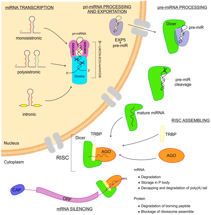
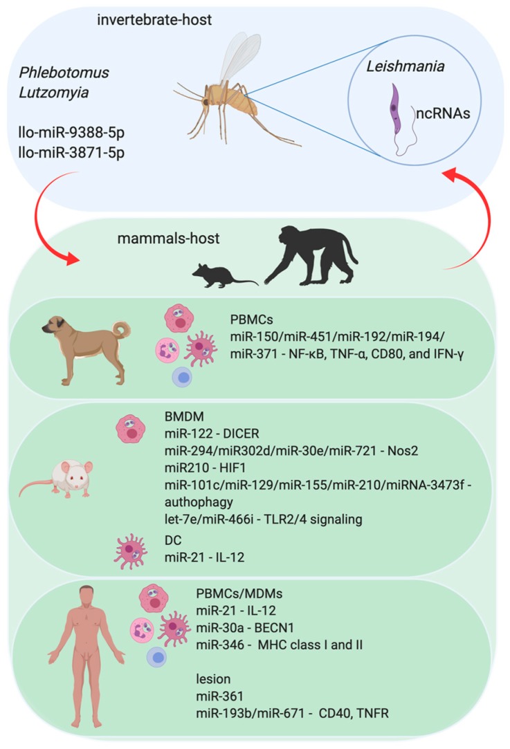
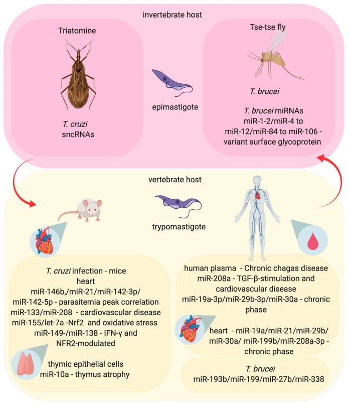
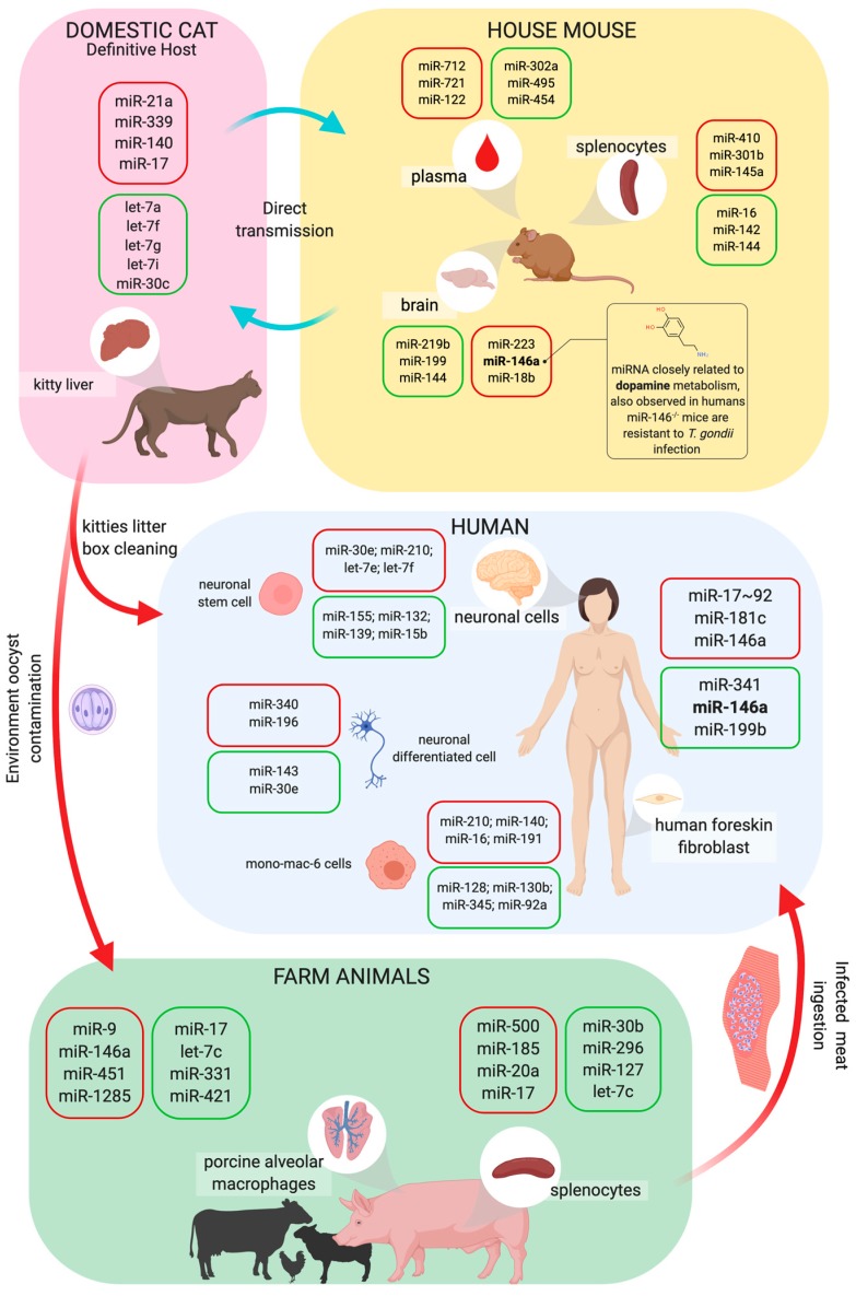
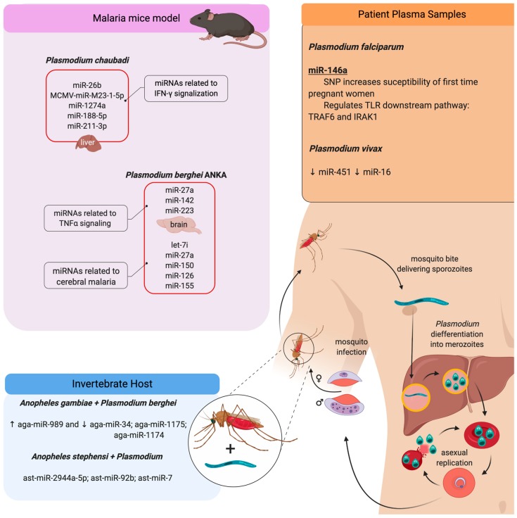
An inflammatory response is essential for combating invading pathogens. Several effector components, as well as immune cell populations, are involved in mounting an immune response, thereby destroying pathogenic organisms such as bacteria, fungi, viruses, and parasites. In the past decade, microRNAs (miRNAs), a group of noncoding small RNAs, have emerged as functionally significant regulatory molecules with the significant capability of fine-tuning biological processes. The important role of miRNAs in inflammation and immune responses is highlighted by studies in which the regulation of miRNAs in the host was shown to be related to infectious diseases and associated with the eradication or susceptibility of the infection. Here, we review the biological aspects of microRNAs, focusing on their roles as regulators of gene expression during pathogen-host interactions and their implications in the immune response against Leishmania, Trypanosoma, Toxoplasma, and Plasmodium infectious diseases.

Keywords: gene expression; host; immune response; microRNAs; parasite; pathogen; post-transcriptional.


Conflict of interest statement

The authors declare no conflict of interest.

Figures

Figure 1
Figure 1
Biogenesis of microRNAs (miRNAs). The miRNAs are small non-coding RNAs transcribed from DNA sequences which can be monocistronic or polycistronic, comprised within exons, introns, or a unique host gene. Right after the transcription, this new RNA sequence is called primary-miRNA (pri-miRNA), which is folded in a hairpin conformation and coupled with the microprocessor: a combination of DGCR8 and Drosha RNases which cut the pri-miRNA, making it a pre-miRNA. Afterward, the pre-miRNA is coupled to Exportin 5 and then exported to the cytoplasm where this complex is found by Dicer, which cuts the pre-miRNA, releasing two mature miRNA arms. The mature miRNA complexed to the Dicer can now couple with the Argonaute and TRBP proteins, thus assembling the RNA Induced Silencing Complex–RISC. When the RISC is done, it can find the messenger RNAs that are the targets to the miRNA complexed. Once found, the message is now silenced and the gene expression regulated.
Figure 2
Figure 2
The role of miRNA in the Leishmania parasite and parasite–host interaction. The protozoan infection caused by Leishmania parasite leads to Leishmaniasis in mammalian hosts. The infection begins with the transmission during the blood meal of infected female sandflies (Phlebotomus and/or Lutzomyia) and the consequent injection of infective promastigotes into the host’s epidermis. Flagellated and mobile extracellular promastigote form of Leishmania proliferate in the gut of sandflies. Upon penetration, promastigotes are identified by immune phagocytic cells, such as macrophages, neutrophils, and dendritic cells (DC). Once the parasites are phagocytized by these immune cells, they are differentiated into amastigote forms and begin to multiply within those cells. Amastigotes have a reduced flagellum and ovaleted morphology and survive inside the phagolysosomal compartment. Parasites in promastigote form express noncoding RNAs (ncRNAs) that can regulate the invertebrate host–sandfly–metabolic pathways. However, sandflies also express miRNAs, which can affect the parasite biology, influencing its success in the colonization of sandflies’ digestive tract and differentiation to the infective forms and the vertebrate host. Not only do the invertebrate hosts express miRNAs, but the vertebrate one also does. The experimental models using human Monocytes Derived-Macrophages (MDM) or murine Bone Marrow-Derived Macrophages (BMDM), and also in the Peripheral Blood Cells (PBMCs) from human and dogs show that infection of mammalian hosts can interfere with the profile of host miRNAs. Also, the plasma samples or the cutaneous lesion site from leishmaniasis patients show modifications in the miRNA profile.
Figure 3
Figure 3
The influences of miRNA in the interaction between the Trypanosoma, vector, and mammalian host in Chagas disease and Sleep sickness. The Trypanosoma cruzi causes a Chagas disease and is transmitted during the Triatomine bug blood-meal, when the bug defecates and transfers trypomastigotes form to the vertebrate host. The itchy skin, due to the bug’s bite, allows the trypomastigote form to enter in the dermis, where the parasite infects any cell, differentiates into amastigote form, and replicates inside the infected cells, causing the clinical symptoms of the disease. T. cruzi express small noncoding RNAs (sncRNAs) that can change the regulation of gene expression inside the invertebrate host (Triatomine). Also, T. cruzi infection can modify miRNAs expression in the human and murine heart-infected cells, in the murine thymic epithelial infected cells or circulating miRNAs in human plasma samples, impacting cardiomyopathy, parasitemia and the immune response. On the other hand, sleep sickness is caused by T. brucei. The parasite is transmitted during the blood-meal of infected tsetse flies, which inject trypomastigote form into the vertebrate host. Different from the T. cruzi infection, T. brucei trypomastigotes circulate in the blood, lymph, and spinal fluid and can cross the blood brain barrier to infect the central nervous system. This parasite expresses miRNAs correlated with the modulation of the expression of virulence factors, such as variant surface glycoproteins. The miRNA profile is altered in plasma samples of patients infected with T. brucei.
Figure 4
Figure 4
Life cycle of Toxoplasma and host miRNA interaction. Toxoplasma gondii has a complex life cycle, in which the parasite can infect a large range of animals, such as domestic cats, farm animals, mice, and even humans. Cats are the T. gondii definitive host. Oocysts are released from infected cats’ feces, sporulate in the environment, and become infective. Samples of kittens’ liver present modifications in miRNA expression. Humans can also become infected by eating undercooked meat of infected animals or while nursing infected cats. In the human host, this infection can lead to toxoplasmosis, a disease that affects various organs’ tissues such as skeletal muscle, myocardium, and the brain. As a result, neuronal cells, monocyte/macrophage, and fibroblasts from infected patients present modifications in miRNA expression. Also, porcine alveolar macrophages and splenocytes samples have altered miRNA profiles in infected pigs. To study toxoplasmosis in vivo, mice models are commonly used because mice can naturally be infected by the parasites and affect multiple organs while changing their miRNA profile. Mice are largely used to study the disease outcomes and treatments, they can also be naturally infected and have multiple organs affected with the infection, and it is already known that the spleen, plasma, and brain have their miRNA profile changed during the infection. The box color represents the increase (red) or decrease (green) of miRNAs expression.
Figure 5
Figure 5
Plasmodium induces miRNA profile modifications during pathogen–host interaction. Human malaria infection begins with the bite of infected female mosquito Anopheles (invertebrate host), delivering sporozoite forms that migrate to the liver and differentiate into merozoites. These forms leave the liver and start the blood-stage asexual replication. In this stage, Plasmodium can differentiate into gametocytes, which can be ingested by the mosquito and develop the sexual phase of the life cycle. Afterward, the mosquito can infect another individual. The species P. falciparum and P. vivax are anthropophilic, infecting only humans; both of them can induce modifications in the miRNA profile in human plasma samples. Besides, mice infections with P. chaubadi and P. beghei have been used as malaria experimental models. In the murine infection, the parasite changes the circulating miRNA profile and it is seen in the same way in the liver of infected mice with P. chaubadi as it is seen in the brain of mice infected with P. berghei ANKA. Although, the invertebrate host presents modifications in the miRNA profile when infected with Plasmodium. The red boxes represent the upregulation of miRNAs related to these models. The arrows represent the upregulation (↑) or downregulation (↓) of miRNA.

References

    1. Fernandes J.C.R., Acuna S.M., Aoki J.I., Floeter-Winter L.M., Muxel S.M. Long non-coding RNAs in the regulation of gene expression: Physiology and disease. Non-Coding RNA. 2019;5:17. doi: 10.3390/ncrna5010017. - DOI - PMC - PubMed
    1. Long Y., Wang X., Youmans D.T., Cech T.R. How do lncRNAs regulate transcription? Sci. Adv. 2017;3:eaao2110. doi: 10.1126/sciadv.aao2110. - DOI - PMC - PubMed
    1. Derrien T., Johnson R., Bussotti G., Tanzer A., Djebali S., Tilgner H., Guernec G., Martin D., Merkel A., Knowles D.G., et al. The gencode v7 catalog of human long noncoding RNAs: Analysis of their gene structure, evolution, and expression. Genome Res. 2012;22:1775–1789. doi: 10.1101/gr.132159.111. - DOI - PMC - PubMed
    1. Djebali S., Davis C.A., Merkel A., Dobin A., Lassmann T., Mortazavi A., Tanzer A., Lagarde J., Lin W., Schlesinger F., et al. Landscape of transcription in human cells. Nature. 2012;489:101–108. doi: 10.1038/nature11233. - DOI - PMC - PubMed
    1. Ulitsky I., Bartel D.P. LincRNAs: Genomics, evolution, and mechanisms. Cell. 2013;154:26–46. doi: 10.1016/j.cell.2013.06.020. - DOI - PMC - PubMed

Publication types

LinkOut - more resources