. 2020 Jan 2;8(1):70.
doi: 10.3390/microorganisms8010070.

Role of FAD-I in Fusobacterial Interspecies Interaction and Biofilm Formation

Affiliations

Role of FAD-I in Fusobacterial Interspecies Interaction and Biofilm Formation

Bhumika Shokeen et al. Microorganisms. .

Erratum in

Abstract

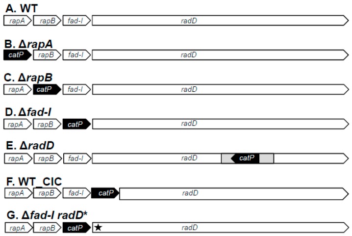
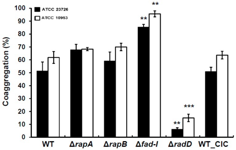
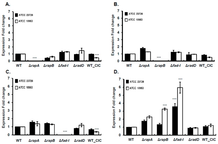
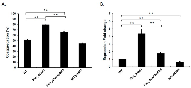
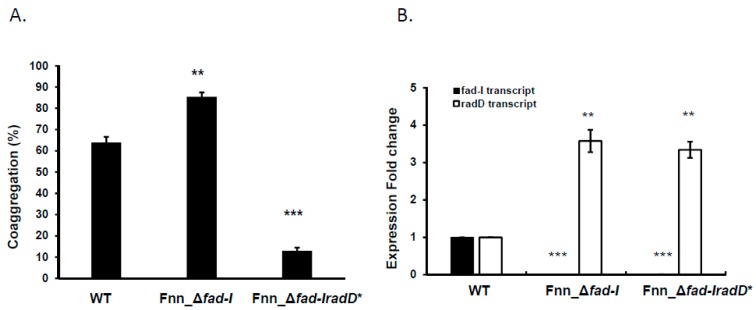
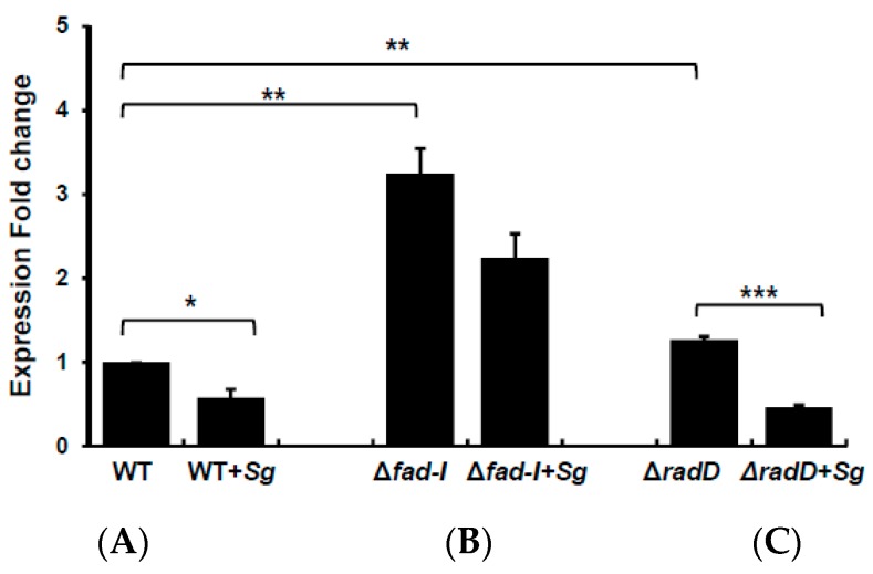
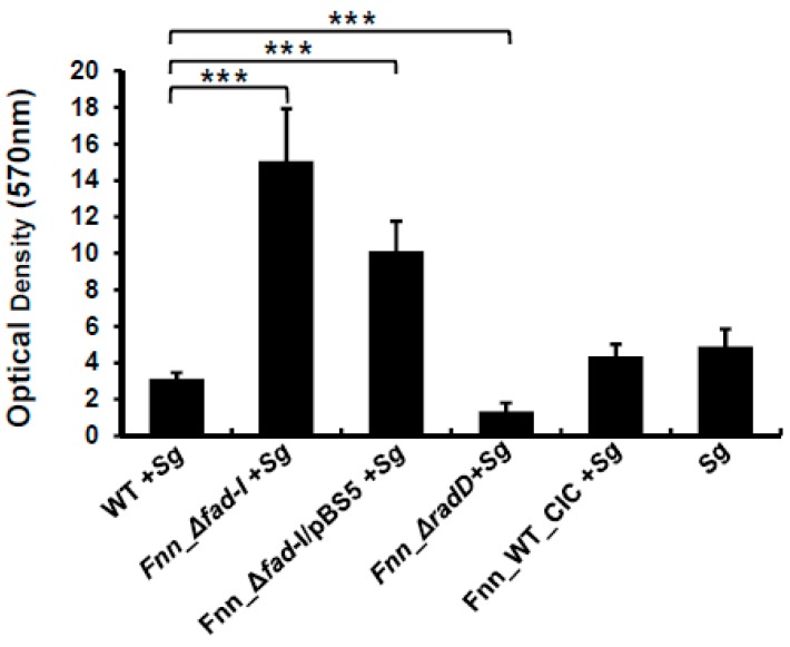
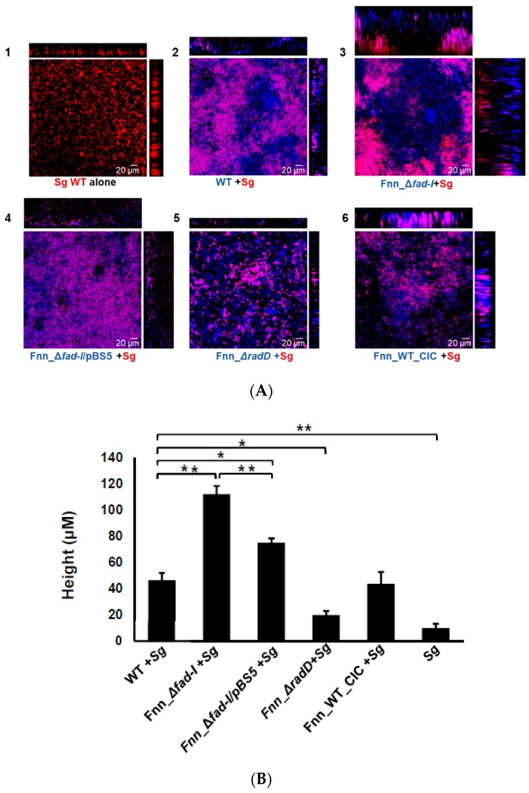
: RadD, a major adhesin of oral fusobacteria, is part of a four-gene operon encoding the small lipoprotein FAD-I and two currently uncharacterized small proteins encoded by the rapA and rapB genes. Previously, we described a role for FAD-I in the induction of human B-defensin 2 (hBD2) upon contact with oral epithelial cells. Here, we investigated potential roles for fad-I, rapA, and rapB in interspecies interaction and biofilm formation. Gene inactivation mutants were generated for each of these genes in the nucleatum and polymorphum subspecies of Fusobacterium nucleatum and characterized for their adherence to partner species, biofilm formation, and operon transcription. Binding to Streptococcus gordonii was increased in all mutant strains with Δfad-I having the most significant effect. This increased adherence was directly proportional to elevated radD transcript levels and resulted in significantly different architecture and height of the biofilms formed by Δfad-I and S. gordonii compared to the wild-type parent. In conclusion, FAD-I is important for fusobacterial interspecies interaction as its lack leads to increased production of the RadD adhesin suggesting a role of FAD-I in its regulation. This regulatory effect does not require the presence of functional RadD.

Keywords: Fusobacterium nucleatum; RadD; biofilm; fad-I; interspecies interaction.


Conflict of interest statement

The authors have no conflicts of interest to disclose.

Figures

Figure 1
Figure 1
Schematic representation of gene inactivation mutants of the radD-encoding four-gene operon and controls (A) wild-type (WT), (B) ΔrapA, (C) ΔrapB, (D) Δfad-I, (E) ΔradD (F), and wild-type-catP insertion control (WT_CIC) in F. nucleatum subspecies nucleatum and polymorphum. (G) Δfad-I radD*: F. nucleatum ssp. nucleatum ATCC 23726 radD frameshift mutant in the Δfad-I background.
Figure 2
Figure 2
Quantitative Coaggregation of wild-type F. nucleatum ssp. nucleatum (ATCC 23726) and F. nucleatum ssp. polymorphum (ATCC 10953) and their various mutant derivatives: ΔrapA, ΔrapB, Δfad-I, ΔradD, and control (WT_CIC) with S. gordonii. Data are represented as mean of percentage coaggregation and standard error of mean of three independent experiments. (** p ≤ 0.01 and *** p ≤ 0.001 compared to the wild-type control).
Figure 3
Figure 3
Transcriptional analysis of radD-operon genes in wild-type F. nucleatum ssp. nucleatum (ATCC 23726) and F. nucleatum ssp. polymorphum (ATCC 10953) and their mutant derivatives. Expression fold changes compared to wild-type are shown for (A) rapA, (B) rapB, (C) fad-I, and (D) radD for mutant derivatives and the WT_CIC control strains. Data are presented as the mean and standard error of mean of three independent experiments. (*** represents p ≤ 0.001 compared to the wild-type control).
Figure 4
Figure 4
Characterization of the fad-I complement of F. nucleatum ssp. nucleatum. (A) Quantitative coaggregation of the mutant derivatives of wild-type ATCC 23726 (WT), Fnn_Δfad-I, and Fnn_Δfad-I/pBS5 with S. gordonii is shown as percentage coaggregation. (B) Transcriptional levels of radD in the Fnn_Δfad-I mutant and the Fnn_Δfad-I/pBS5 complement along with a vector only control WT/pHS58 are shown as fold change in comparison to the wild-type parent ATCC 23726. All data are presented as the mean and standard error of mean of three independent experiments (** represents p ≤ 0.01).
Figure 5
Figure 5
Quantitative coaggregation and transcriptional analysis of F. nucleatum ssp. nucleatum, Fnn_Δfad-I, and Fnn_Δfad-IradD*. (A) Coaggregation of wild-type ATCC 23726 (WT), Fnn_Δfad-I, and Fnn_Δfad-IradD* with S. gordonii is represented as mean of percentage coaggregation and standard error of mean of three independent experiments. (B) fad-I and radD transcript levels are represented as expression fold change compared to the wild-type parent ATCC 23726. The data represent the mean and standard error of mean of three independent experiments. (** represents p ≤ 0.01 compared to the WT control, *** represents p ≤ 0.001 than the WT).
Figure 6
Figure 6
Transcriptional analysis of radD levels in the presence of S. gordonii. Presented are radD levels in (A) wild-type ATCC 23726 (WT), (B) Fnn_Δfad-I, and (C) Fnn_ΔradD alone or in the presence of the partner species. Data are presented as the mean and standard error of mean of three independent experiments (* represents p ≤ 0.05, ** represents p ≤ 0.01, *** represents p ≤ 0.001).
Figure 7
Figure 7
Biomass of dual-species biofilms formed by F. nucleatum ssp nucleatum ATCC 23726 and its radD-operon mutant derivatives with S. gordonii. The biomass was assessed via the crystal violet assay. Data are presented as the mean and standard error of mean of three independent experiments (*** represents p ≤ 0.001).
Figure 8
Figure 8
(A). Visualization of dual-species biofilm formed between wild-type and mutant strains of F. nucleatum ssp nucleatum ATCC 23726 and S. gordonii (mCherry) by CLSM. The biofilm was fluorescently labeled with SYTO9. The S. gordonii (Sg) cells constitutively express mCherry and appear red on the images. Wild-type (WT) F. nucleatum (Fn) and its mutants (fad-I, fad-I/pBS5, radD, WT-CIC) are stained by syto9-only which are pseudo-colored as blue in the Zen software. Association of F. nucleatum and S. gordonii in the biofilm is observed as purple color in the confocal images. Each image panel is represented by x-z axis view on top and y-z axis view on the right side of the x-y view. The various panels show the biofilm formed by: (1) S. gordonii (Sg) alone; (2) wild-type ATCC 23726 with S. gordonii; (3) Fnn_Δfad-I with S. gordonii; (4) Fnn_Δfad-I/pBS5 with S. gordonii; (5) Fnn_ΔradD with S. gordonii; (6) Fnn_WT_CIC with S. gordonii. (B) Comparison of the height of the dual species biofilm of the wild-type and mutant strains of F. nucleatum ssp nucleatum ATCC 23726 with S. gordonii, as observed from the confocal images. The data represents mean of the height and standard error of mean of biofilm as observed in three independent experiments with height measurements captured in five randomly chosen locations in each experiment (n = 15). The single species biofilm of S. gordonii is also included as control. (* represents p ≤ 0.05, ** represents p ≤ 0.01).

Similar articles

Cited by

References

    1. Handley P., Rickard A. Coaggregation—Is it a universal biofilm phenomenon? In: Gilbert P., Allison D., editors. Biofilm Community Interactions: Chance or Necessity? Bioline; London, UK: 2001. pp. 1–10.
    1. Kolenbrander P.E. Intergeneric coaggregation among human oral bacteria and ecology of dental plaque. Annu. Rev. Microbiol. 1988;42:627–656. doi: 10.1146/annurev.mi.42.100188.003211. - DOI - PubMed
    1. Ellen R.P., Lepine G., Nghiem P.M. In vitro models that support adhesion specificity in biofilms of oral bacteria. Adv. Dent. Res. 1997;11:33–42. doi: 10.1177/08959374970110011401. - DOI - PubMed
    1. Kolenbrander P.E., Andersen R.N., Kazmerzak K., Wu R., Palmer R.J., Jr. Spatial organization of oral bacteria in biofilms. Methods Enzymol. 1999;310:322–332. doi: 10.1016/s0076-6879(99)10026-0. - DOI - PubMed
    1. Kolenbrander P.E. Oral microbial communities: Biofilms, interactions, and genetic systems. Annu. Rev. Microbiol. 2000;54:413–437. doi: 10.1146/annurev.micro.54.1.413. - DOI - PubMed