. 2020 Jan 2;21(1):319.
doi: 10.3390/ijms21010319.

Extended cleavage specificities of two mast cell chymase-related proteases and one granzyme B-like protease from the platypus, a monotreme

Affiliations

Extended cleavage specificities of two mast cell chymase-related proteases and one granzyme B-like protease from the platypus, a monotreme

Zhirong Fu et al. Int J Mol Sci. .

Abstract

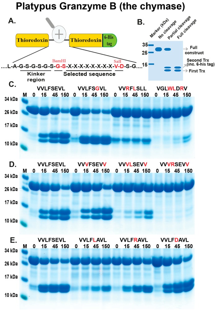
Mast cells (MCs) are inflammatory cells primarily found in tissues in close contact with the external environment, such as the skin and the intestinal mucosa. They store large amounts of active components in cytoplasmic granules, ready for rapid release. The major protein content of these granules is proteases, which can account for up to 35 % of the total cellular protein. Depending on their primary cleavage specificity, they can generally be subdivided into chymases and tryptases. Here we present the extended cleavage specificities of two such proteases from the platypus. Both of them show an extended chymotrypsin-like specificity almost identical to other mammalian MC chymases. This suggests that MC chymotryptic enzymes have been conserved, both in structure and extended cleavage specificity, for more than 200 million years, indicating major functions in MC-dependent physiological processes. We have also studied a third closely related protease, originating from the same chymase locus whose cleavage specificity is closely related to the apoptosis-inducing protease from cytotoxic T cells, granzyme B. The presence of both a chymase and granzyme B in all studied mammals indicates that these two proteases bordering the locus are the founding members of this locus.

Keywords: animal model; chymase; cleavage specificity; human chymase; mast cell; monotremes; platypus.


Conflict of interest statement

The authors declare no conflict of interest.

Figures

Figure 1
Figure 1
A phylogenetic tree of chymase loci encoded serine proteases. The amino acid sequences of a panel of chymase loci encoded proteases were analysed for sequence relatedness with the program MrBase. A bootstrap tree based on 1000 replicates was generated and the bootstrap values are depicted at each branch of the tree. The different subfamilies of chymase loci genes were colour-coded for easy identification and the genes of primary interest, monotreme and marsupial enzymes, are marked by red arrows and the related alligator and Xenopus proteases are marked with green arrows. The granzyme B related in dark green and the cathepsin G related in light green.
Figure 2
Figure 2
The chymase locus. The chymase locus encodes a number of hematopoietic serine proteases, including the α-chymases, β-chymases, cathepsin G, and several granzymes [12]. Genes are colour-coded: the α-chymase-related genes are marked in light blue, the β-chymases in slightly darker blue, cathepsin G in green, the M8 family in light green, the granzymes in dark blue.
Figure 3
Figure 3
Analysis of the recombinant platypus enzymes used in the determination of their extended cleavage specificities. Three different recombinant platypus enzymes were produced as proenzymes, containing an N-terminal His6-tag and an enterokinase site, in the HEK-293 EBNA cells with the episomal vector pCEP-Pu2. These proenzymes were first purified on Ni-NTA beads (-EK) and then activated by removal of the His-6 tag by enterokinase digestion (+EK). After purification and activation, the three enzymes were analysed by separation on SDS-PAGE and visualized with Coomassie Brilliant Blue staining. The proteases before and after enterokinase cleavage are marked with small red arrow heads. The few additional bands seen on the gel are originating from the conditioned cell medium, and the major band of a size of approximately 69 kDa is bovine serum albumin from the serum that represent the major protein of the cell medium.
Figure 4
Figure 4
Analysis of the cleavage specificity of platypus GzmB (the chymase) by the use of recombinant protein substrates. Panel (A) shows the overall structure of the recombinant protein substrates used for analysis of the efficiency in cleavage by the three different enzymes (analysed in Figure 4, Figure 5 and Figure 6). In these substrates, two thioredoxin molecules are positioned in tandem and the proteins have a His-6 tag positioned in their C-termini of the second thioredoxin. The different cleavable sequences are inserted in a linker region between the two thioredoxin molecules by the use of two unique restriction sites, one Bam HI and one SalI site, which are indicated in the bottom of panel (A). In panel (B), an example cleavage is shown to highlight possible cleavage patterns. Panels (CE) show the cleavage of a number of substrates by the platypus granzyme B, the chymase. The sequences of the different substrates are indicated above the images of the gels. The red residues highlight the major difference between this substrate and the reference substrate positioned at the beginning (Left side) of this panel. The time of cleavage, in minutes, is also indicated above the corresponding lanes of the different gels. The un-cleaved substrates have a molecular weight of approximately 25 kDa and the cleaved substrates appear as two closely-located bands with a size of 12–13 kDa.
Figure 5
Figure 5
Analysis of the cleavage specificity of platypus DDN1-like (also a chymase) by the use of recombinant protein substrates. The different panels show the cleavage of a number of substrates by the platypus DDN1-like. The sequences of the different substrates are indicated above the pictures of the gels. The red residues highlight the major difference between this substrate and the reference substrate positioned at the beginning (Left side) of this panel. The time of cleavage, in minutes, is also indicated above the corresponding lanes of the different gels. The un-cleaved substrates have a molecular weight of approximately 25 kDa and the cleaved substrates appear as two closely-located bands with a size of 12–13 kDa.
Figure 6
Figure 6
Analysis of the cleavage specificity of platypus GzmBGH (the granzyme B equivalent) by the use of recombinant protein substrates. The different panels show the cleavage of a number of substrates by the platypus GzmBGH. The sequences of the different substrates are indicated above the pictures of the gels. The red residues highlight the major difference between this substrate and the reference substrate positioned at the beginning (Left side) of this panel. The time of cleavage, in minutes, is also indicated above the corresponding lanes of the different gels. The un-cleaved substrates have a molecular weight of approximately 25 kDa and the cleaved substrates appear as two closely-located bands with a size of 12–13 kDa.

Similar articles

Cited by

References

    1. Galli S.J. Mast cells and basophils. Curr. Opin. Hematol. 2000;7:32–39. doi: 10.1097/00062752-200001000-00007. - DOI - PubMed
    1. Galli S.J., Grimbaldeston M., Tsai M. Immunomodulatory mast cells: Negative, as well as positive, regulators of immunity. Nat. Rev. Immunol. 2008;8:478–486. doi: 10.1038/nri2327. - DOI - PMC - PubMed
    1. Hellman L.T., Akula S., Thorpe M., Fu Z. Tracing the Origins of IgE, Mast Cells, and Allergies by Studies of Wild Animals. Front. Immunol. 2017;8:1749. doi: 10.3389/fimmu.2017.01749. - DOI - PMC - PubMed
    1. Abe T., Swieter M., Imai T., Hollander N.D., Befus A.D. Mast cell heterogeneity: Two-dimensional gel electrophoretic analyses of rat peritoneal and intestinal mucosal mast cell. Eur. J. Immunol. 1990;20:1941–1947. doi: 10.1002/eji.1830200911. - DOI - PubMed
    1. Lützelschwab C., Aveskogh M., Pejler G., Hellman L. Secretory Granule Proteases in Rat Mast Cells. Cloning of 10 Different Serine Proteases and a Carboxypeptidase A from Various Rat Mast Cell Populations. J. Exp. Med. 1997;185:13–30. doi: 10.1084/jem.185.1.13. - DOI - PMC - PubMed

LinkOut - more resources