. 2020 Jan 6;20(1):7.
doi: 10.1186/s12903-019-0987-y.

Cyclic compression emerged dual effects on the osteogenic and osteoclastic status of LPS-induced inflammatory human periodontal ligament cells according to loading force

Affiliations

Cyclic compression emerged dual effects on the osteogenic and osteoclastic status of LPS-induced inflammatory human periodontal ligament cells according to loading force

Ru Jia et al. BMC Oral Health. .

Abstract

Background: Appropriate mechanical stimulation is essential for bone homeostasis in healthy periodontal tissues. While the osteogenesis and osteoclast differentiation of inflammatory periodontal ligament cells under different dynamic loading has not been yet clear. The aim of this study is to clarify the inflammatory, osteogenic and pro-osteoclastic effects of different cyclic stress loading on the inflammatory human periodontal ligament cells (hPDLCs).

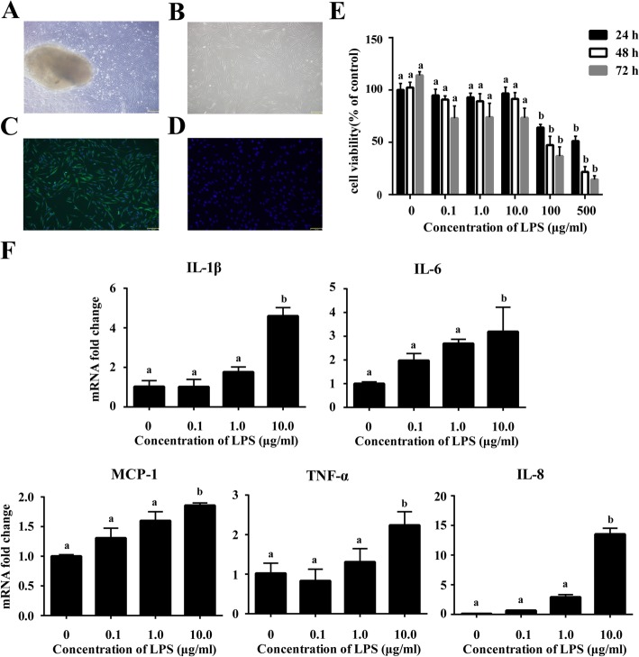
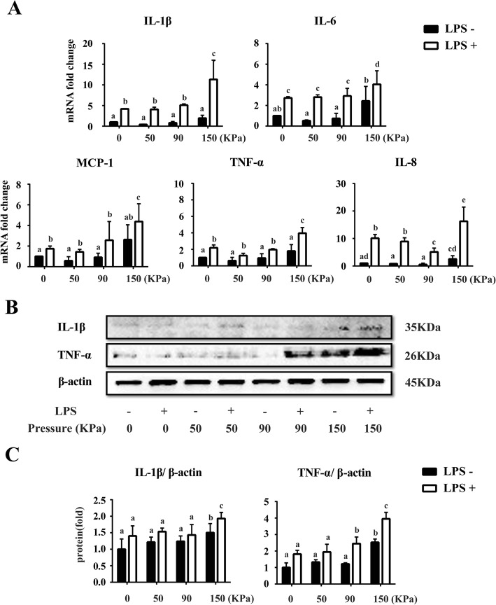
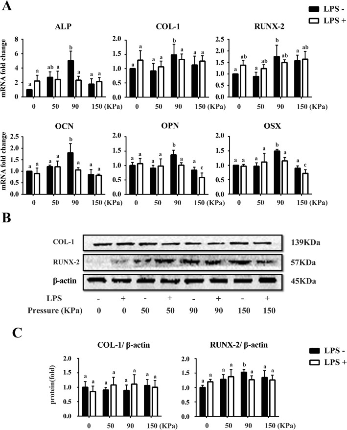
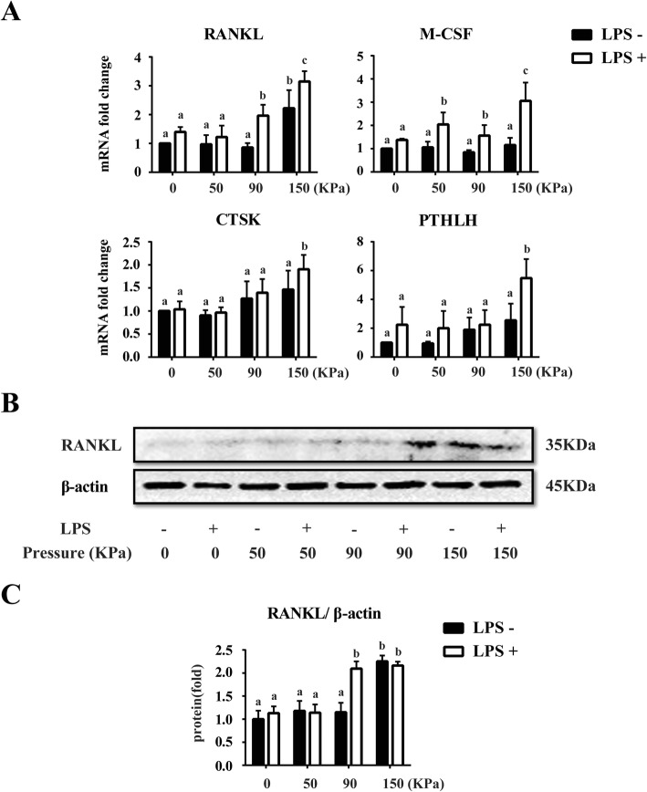
Methods: hPDLCs were isolated from healthy premolars and cultured in alpha minimum Eagle's medium (α-MEM). Lipopolysaccharides (LPS) were used to induce the inflammation state of hPDLCs in vitro. Determination of LPS concentration for the model of inflammatory periodontium was based on MTT and genes expression analysis. Then the cyclic stress of 0, 0-50, 0-90 and 0-150 kPa was applied to the inflammatory hPDLCs for 5 days respectively. mRNA and protein levels of osteogenic, osteoclastic and inflammation-related markers were examined after the treatment.

Results: MTT and RT-PCR results showed that 10 μg/ml LPS up-regulated TNF-α, IL-1β, IL-6, IL-8 and MCP-1 mRNA levels (P < 0.05) and did not affect the cell viability (P > 0.05). The excessive loading of stress (150 kPa) with or without LPS strongly increased the expression of inflammatory-related markers TNF-α, IL-1β, IL-6, IL-8, MCP-1 (P < 0.05) and osteoclastic markers RANKL, M-CSF, PTHLH and CTSK compared with other groups (P < 0.05), but had no significant effect on osteogenic genes. While 0-90 kPa cyclic pressure could up-regulate the expression of osteogenic genes ALP, COL-1, RUNX2, OCN, OPN and OSX in the healthy hPDLSCs.

Conclusions: Collectively, it could be concluded that 0-150 kPa was an excessive stress loading which accelerated both inflammatory and osteoclastic effects, while 0-90 kPa may be a positive factor for the osteogenic differentiation of hPDLCs in vitro.

Keywords: Dynamic loading; LPS; Osteogenic differentiation; Periodontitis; hPDLCs.


Conflict of interest statement

The authors declare that they have no competing interests.

Figures

Fig. 1
Fig. 1
Cell characterization and LPS-induced inflammation of hPDLCs in vitro. Primary cells grew out from the tissue explants (a) and were spindle shaped (b). Immunocytochemistry staining showed that cells were vimentin positive (c) and cytokeratin negative (d) (magnification: 100×, scale bar: 150 μm). After treated by different concentrations of LPS (range = 0–500 μg/ml) for 24, 48 and 72 h respectively, cell viability of hPDLCs was evaluated with MTT assay (e). mRNA expressions of pro-inflammatory cytokines IL-1β, IL-6, IL-8, MCP-1 and TNF-α in hPDLCs after 0.1, 1.0 and 10 μg/ml LPS treatment were detected using real-time PCR (f). Data were represented as means ± SEM, n = 6 (hPDLCs from six donors). The bars with different lowercase letters were significantly different from each other (P < 0.05), and those with the same letter exhibited no significant difference
Fig. 2
Fig. 2
Expression of mRNA and protein levels of the inflammatory markers in hPDLCs after different dynamic cyclic stress loading for 5 days. a Real-time PCR results of pro-inflammatory markers, including IL-1β, IL-6, IL-8, TNF-α and MCP-1 mRNA expression in hPDLCs after different cyclic stress loading for 5 days with LPS or not. b Western blotting analysis for IL-1β and TNF-α using total protein isolated from different groups of hPDLCs. c Quantification of Western blotting analysis. Protein content was expressed relative to the control and represented three similar independent experiments with triplicate observations in each experiment. Data were represented as the means ± SEM, n = 6 (hPDLCs from six donors). The bars with different lowercase letters were significantly different from each other (P < 0.05), and those with the same letter exhibited no significant difference
Fig. 3
Fig. 3
Expression of mRNA and protein levels of the osteoblastic markers in hPDLCs after different dynamic cyclic stress loading for 5 days. a Real-time PCR results of osteoblastic markers ALP, COL-1, RUNX-2, OCN, OPN and OSX mRNA expression in hPDLCs after different cyclic stress loading for 5 days with LPS or not. b Western blotting analysis for COL-1 and RUNX-2 using total protein isolated from different groups of hPDLCs. c Quantification of Western blotting analysis. Protein content was expressed relative to the control and represented three similar independent experiments with triplicate observations in each experiment. Data were represented as means ± SEM, n = 6 (hPDLCs from six donors). The bars with different lowercase letters were significantly different from each other (P < 0.05), and those with the same letter exhibited no significant difference
Fig. 4
Fig. 4
Expression of mRNA and protein levels of the pro-osteoclastic markers in hPDLCs after different dynamic cyclic stress loading for 5 days. a Real-time PCR results of pro-osteoclastic markers RANKL, M-CSF, CTSK and PTHLH mRNA expression in hPDLCs after different cyclic stress loading for 5 days with LPS or not. b Western blotting analysis for RANKL using total protein isolated from different groups of hPDLCs. c Quantification of Western blotting analysis. Protein content was expressed relative to the control and represented three similar independent experiments with triplicate observations in each experiment. Data were represented as means ± SEM, n = 6 (hPDLCs from six donors). The bars with different lowercase letters were significantly different from each other (P < 0.05), and those with the same letter exhibited no significant difference

References

    1. Golz L, Memmert S, Rath-Deschner B, Jager A, Appel T, Baumgarten G, Gotz W, Frede S. LPS from P gingivalis and hypoxia increases oxidative stress in periodontal ligament fibroblasts and contributes to periodontitis. Mediators Inflamm. 2014;2014:986264. doi: 10.1155/2014/986264. - DOI - PMC - PubMed
    1. Chi XP, Ouyang XY, Wang YX. Hydrogen sulfide synergistically upregulates Porphyromonas gingivalis lipopolysaccharide-induced expression of IL-6 and IL-8 via NF-kappaB signalling in periodontal fibroblasts. Arch Oral Biol. 2014;59(9):954–961. doi: 10.1016/j.archoralbio.2014.05.022. - DOI - PubMed
    1. Baker PJ, Dixon M, Evans RT, Dufour L, Johnson E, Roopenian DC. CD4(+) T cells and the proinflammatory cytokines gamma interferon and interleukin-6 contribute to alveolar bone loss in mice. Infect Immun. 1999;67(6):2804–2809. doi: 10.1128/IAI.67.6.2804-2809.1999. - DOI - PMC - PubMed
    1. Ulevitch RJ, Tobias PS. Receptor-dependent mechanisms of cell stimulation by bacterial endotoxin. Annu Rev Immunol. 1995;13:437–457. doi: 10.1146/annurev.iy.13.040195.002253. - DOI - PubMed
    1. Kato H, Taguchi Y, Tominaga K, Umeda M, Tanaka A. Porphyromonas gingivalis LPS inhibits osteoblastic differentiation and promotes pro-inflammatory cytokine production in human periodontal ligament stem cells. Arch Oral Biol. 2014;59(2):167–175. doi: 10.1016/j.archoralbio.2013.11.008. - DOI - PubMed

Publication types

Substances