. 2020 Feb 21;64(3):e01889-19.
doi: 10.1128/AAC.01889-19. Print 2020 Feb 21.

Hydroxylamine and Carboxymethoxylamine Can Inhibit Toxoplasma gondii Growth through an Aspartate Aminotransferase-Independent Pathway

Affiliations

Hydroxylamine and Carboxymethoxylamine Can Inhibit Toxoplasma gondii Growth through an Aspartate Aminotransferase-Independent Pathway

Jixu Li et al. Antimicrob Agents Chemother. .

Abstract

Toxoplasma gondii is an obligate intracellular protozoan parasite and a successful parasitic pathogen in diverse organisms and host cell types. Hydroxylamine (HYD) and carboxymethoxylamine (CAR) have been reported as inhibitors of aspartate aminotransferases (AATs) and interfere with the proliferation in Plasmodium falciparum Therefore, AATs are suggested as drug targets against Plasmodium The T. gondii genome encodes only one predicted AAT in both T. gondii type I strain RH and type II strain PLK. However, the effects of HYD and CAR, as well as their relationship with AAT, on T. gondii remain unclear. In this study, we found that HYD and CAR impaired the lytic cycle of T. gondiiin vitro, including the inhibition of invasion or reinvasion, intracellular replication, and egress. Importantly, HYD and CAR could control acute toxoplasmosis in vivo Further studies showed that HYD and CAR could inhibit the transamination activity of rTgAAT in vitro However, our results confirmed that deficiency of AAT in both RH and PLK did not reduce the virulence in mice, although the growth ability of the parasites was affected in vitro HYD and CAR could still inhibit the growth of AAT-deficient parasites. These findings indicated that HYD and CAR inhibition of T. gondii growth and control of toxoplasmosis can occur in an AAT-independent pathway. Overall, further studies focusing on the elucidation of the mechanism of inhibition are warranted. Our study hints at new substrates of HYD and CAR as potential drug targets to inhibit T. gondii growth.

Keywords: Toxoplasma gondii; aspartate aminotransferase; carboxymethoxylamine; hydroxylamine; toxoplasmosis.


Figures

FIG 1
FIG 1
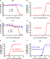
HYD and CAR were able to inhibit parasite growth. (A) HFF cell viability upon treatment with HYD and CAR. (B) Inhibition of HFF growth with HYD treatment. The 50% inhibitory concentration (IC50) was examined. (C) Vero cell viability upon treatment with HYD and CAR. (D) Inhibition of Vero cell growth by HYD treatment. (E and F) Inhibition of T. gondii type I parasites (RH-GFP) after HYD (E) and CAR (F) treatment. The IC50 and selectivity index values were determined.
FIG 2
FIG 2
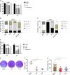
T. gondii lytic cycle is impaired by HYD and CAR in vitro. (A) Invasion assay. Purified tachyzoites were pretreated with either of the two compounds of 2- or 4-fold IC50 values, sulfadiazine (1 mg/ml), or DMSO for 1 h at 37°C, followed by invasion for 2 h, and dual staining was used to determine the percentages of invaded parasites. (B and C) Intracellular replication assay. HYD and CAR 2-fold (B) or 4-fold (C) IC50 values were allowed to treat infected RH parasites for 24 h, and the numbers of parasites in 100 random vacuoles were counted and plotted. (D) Egress assay. Infected cells were treated with compounds at 2- and 4-fold IC50s for 10 min before incubation with 3 μM A23187. After incubation, mouse anti-SAG1 and rabbit anti-GRA7 were used to measure the percentage of staining egressed PVs. At least 300 vacuoles were counted per slip. (E) Plaque formation assay. A total of 150 fresh RH strain tachyzoites were used to infect the HFF cell monolayer and allowed to grow for 8 days with HYD or CAR at an 2-fold IC50 value, and then 0.1% crystal violet was used for staining. (F and G) Relative plaque numbers (F) and plaque sizes (G) from panel D. The data are presented as the means ± the SEM of at least three independent experiments. *, P < 0.05; **, P < 0.01; ***, P < 0.001; ****, P < 0.0001 compared to DMSO treatment, determined by chi-square test (invasion and egress assay), Tukey’s multiple-comparison test (replication assay, a heavy asterisk represents HYD versus vehicle, and a light asterisk represents CAR versus vehicle), and one-way ANOVA plus Tukey-Kramer post hoc analysis (plaque assay).
FIG 3
FIG 3
HYD and CAR control acute Toxoplasma infection in mice. Female BALB/c mice were infected i.p. with an acute dose of 50,000 PLK tachyzoites and treated i.p. from day 1 to day 7 postinfection with 5 or 20 mg/kg HYD; 10, 25, or 50 mg/kg CAR; or PBS once daily. The body weight, morbidity, mortality, and clinical signs were noted. (A) Body weight (%). (B) Mean clinical scores. The scores varied from 0 (no signs) to 10 (all signs). (C) Survival rates. *, P < 0.05, log-rank (Mantel-Cox) test. (D) Parasite burdens of survival mice brains. At day 30 postinfection, the brains were collected, and DNA was extracted, 800 ng of DNA was used to detect the number of parasites. ***, P < 0.001, one-way ANOVA plus Tukey-Kramer post hoc analysis.
FIG 4
FIG 4
HYD and CAR impair rTgAAT catalytic activity. (A) Soluble rTgRHAAT expression. The concentration of soluble rTgAAT-fused GST was determined. (B) Generation of an enzyme-coupled assay. Expressed GST was used as a control. (C) Enzyme reaction. The enzyme activities of catalysis aspartate and α-ketoglutarate into glutamate were determined using 1 μg of rTgAAT. (D) Inhibitor assay. The enzyme reaction was inhibited with increasing amounts of HYD and CAR.
FIG 5
FIG 5
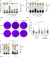
AAT deficiency slows down RH and PLK parasite growth in vitro. (A) Plaque assay. The growth of 150 Δaat or ComAAT tachyzoites in vitro was compared to that of parental RH strains. Plaques were visualized by staining with 0.1% crystal violet. (B) Relative size of the plaques in panel A. The data are presented as means ± the SEM of three independent experiments. (C) Plaque formation of the ΔPLKaat tachyzoites in vitro. Vero cells were infected by 300 tachyzoites and cultured for 12 days. (D) Relative size of plaques in panel C. The data are presented as means ± the SEM of three independent experiments. ***, P < 0.001, one-way ANOVA plus Tukey-Kramer post hoc analysis. (E and F) Invasion, replication, and egress assays of mutants compared to parental and complemented strains in RH (E) and PLK (F) lines. The data are presented as means ± the SEM of at least three independent experiments. *, P < 0.05; **, P < 0.01; ***, P < 0.001; ****, P < 0.0001, determined by chi-square test (invasion and egress assay) and Tukey’s multiple-comparison test (replication assay).
FIG 6
FIG 6
Supplementation with α-ketoglutarate (α-Keto) rescues growth defects in Δaat tachyzoites. (A) Supplementation assay with α-ketoglutarate (400 μM) on type 1 strain RH. Tachyzoites were grown with or without α-ketoglutarate 24 h postinfection, and the numbers of parasites in each PV were then determined. (B) PLK replication rescued by α-ketoglutarate (400 μM). Infected tachyzoites were cultured in Vero cells with or without α-ketoglutarate for 24 h, and then the numbers of parasites in each PV were determined. (C) Plaque formation of ΔPLKaat tachyzoites under α-ketoglutarate supplementation conditions. (D) Relative size of plaques in panel C. (E) The high concentration of α-ketoglutarate (2 mM) led to faster replication of AAT-deficient parasites compared to parental PLK in vitro. The data are presented as means ± the SEM of three independent experiments. –, no treatment with α-ketoglutarate; +, treatment by a low concentration (400 μM); ++, treatment by high a concentration (2 mM). *, P < 0.05; **, P < 0.01; ***, P < 0.001, one-way ANOVA plus Dunnett’s multiple-comparison test (replication assay) and one-way ANOVA plus Tukey-Kramer post hoc analysis (plaque assay).
FIG 7
FIG 7
Virulence tests of Δaat tachyzoites in mice. (A and B) Body weight (%) (A) and survival rate (B) during ΔRHaat infection. Mice were infected by i.p. injection with 100 RH (n = 11), ΔRHaat (n = 11), or ComRHAAT (n = 6) tachyzoites. (C) Body weight during ΔPLKaat infection. Six mice were infected with 10,000, 30,000, and 50,000 tachyzoites by i.p. injection. (D) Survival rate of PLK AAT deficiency parasite infection in mice. Survival curves of mice infected with ΔPLKaat tachyzoites were noted until day 30. *, P < 0.05; **, P < 0.01, log-rank (Mantel-Cox) test.
FIG 8
FIG 8
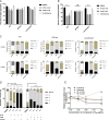
HYD and CAR inhibit T. gondii growth through an AAT-independent pathway. (A and B) Effects of AAT-deficient parasite invasion treated with HYD and CAR compared to parental and complemented. The data show the means ± the SEM of three independent experiments. ***, P < 0.001, determined by chi-square test. (C) Effects of HYD and CAR treatment for AAT-deficient parasite replication. The concentration of the 2- or 4-fold IC50 values of HYD and CAR were used to treat parasite replication by three strains. *, P < 0.05; **, P < 0.01; ***, P < 0.001; ****, P < 0.0001. A heavy asterisk stands for HYD versus vehicle, and a light asterisk stands for CAR versus vehicle, determined by Tukey’s multiple-comparison test. (D) α-Ketoglutarate (α-Keto) assay. α-Ketoglutarate was supplemented to the HYD and CAR treatment medium, and then replication was determined at 24 h postinfection. ***, P < 0.001; ****, P < 0.0001, determined by Tukey’s multiple-comparison test. (E) Effects of HYD and CAR on mitochondrial genome and apicoplast genome. A total of 2 × 107 tachyzoites were treated by 0.25-, 0.5-, and 1-fold IC50 values of HYD, CAR, or sulfadiazine (1 mg/ml) for 5 days to investigate the expression of CytB and the EF-Tu gene by qPCR. *, P < 0.05; ***, P < 0.001, one-way ANOVA plus Dunnett’s multiple-comparison test.

Similar articles

Cited by

References

    1. Jacot D, Waller RF, Soldati-Favre D, MacPherson DA, MacRae JI. 2016. Apicomplexan energy metabolism: carbon source promiscuity and the quiescence hyperbole. Trends Parasitol 32:56–70. doi:10.1016/j.pt.2015.09.001. - DOI - PubMed
    1. Weiss LM, Dubey JP. 2009. Toxoplasmosis: a history of clinical observations. Int J Parasitol 9:895–901. doi:10.1016/j.ijpara.2009.02.004. - DOI - PMC - PubMed
    1. Guo H, Gao Y, Jia H, Moumouni PFA, Masatani T, Liu M, Lee SH, Galon EM, Li J, Li Y, Tumwebaze MA, Benedicto B, Xuan X. 2019. Characterization of strain-specific phenotypes associated with knockout of dense granule protein 9 in Toxoplasma gondii. Mol Biochem Parasitol 229:53–61. doi:10.1016/j.molbiopara.2019.01.003. - DOI - PubMed
    1. Howe DK, Sibley LD. 1995. Toxoplasma gondii comprises three clonal lineages: correlation of parasite genotype with human disease. J Infect Dis 172:1561–1566. doi:10.1093/infdis/172.6.1561. - DOI - PubMed
    1. Sibley LD, Ajioka JW. 2008. Population structure of Toxoplasma gondii: clonal expansion driven by infrequent recombination and selective sweeps. Annu Rev Microbiol 62:329–351. doi:10.1146/annurev.micro.62.081307.162925. - DOI - PubMed

Publication types

MeSH terms