Review
. 2020 Jan 15;204(2):294-305.
doi: 10.4049/jimmunol.1900821.

Brain Parenchymal and Extraparenchymal Macrophages in Development, Homeostasis, and Disease

Affiliations
Review

Brain Parenchymal and Extraparenchymal Macrophages in Development, Homeostasis, and Disease

Simone Brioschi et al. J Immunol. .

Abstract

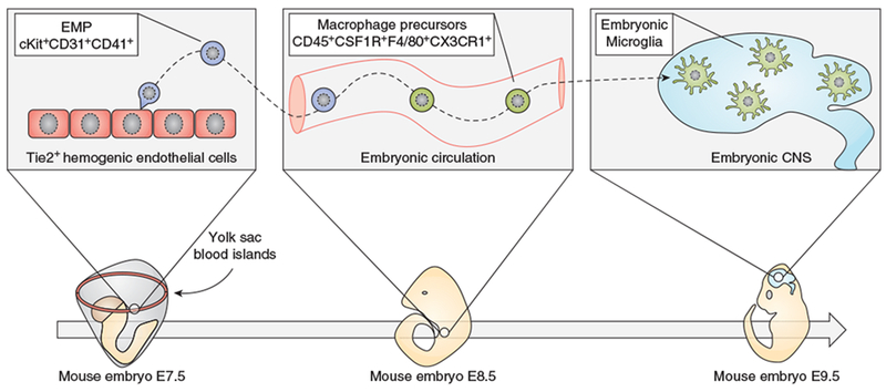
Microglia are parenchymal macrophages of the CNS; as professional phagocytes they are important for maintenance of the brain's physiology. These cells are generated through primitive hematopoiesis in the yolk sac and migrate into the brain rudiment after establishment of embryonic circulation. Thereafter, microglia develop in a stepwise fashion, reaching complete maturity after birth. In the CNS, microglia self-renew without input from blood monocytes. Recent RNA-sequencing studies have defined a molecular signature for microglia under homeostasis. However, during disease, microglia undergo remarkable phenotypic changes, which reflect the acquisition of specialized functions tailored to the pathological context. In addition to microglia, the brain-border regions host populations of extraparenchymal macrophages with disparate origins and phenotypes that have recently been delineated. In this review we outline recent findings that provide a deeper understanding of both parenchymal microglia and extraparenchymal brain macrophages in homeostasis and during disease.


Figures

Figure 1:
Figure 1:
Key steps in the development of embryonic microglia
Figure 2:
Figure 2:
Possible mechanisms involving the DAM signature genes.
Figure 3:
Figure 3:
Molecular signature of microglia and BAMs under homeostasis.
Figure 4:
Figure 4:
Two alternative hypotheses for the origin of microglia and BAMs. a) Hypothesis 1 - EMPs arise from YS-blood islands around E7.5 (1) and at E8.5 migrate into the embryo proper where they differentiate into macrophage precursors (pre-Macs) (2). These cells seed all embryonic tissues including the brain (3). Depending on the local environment, they differentiate into either microglia (4) or BAMs (5). In this model, microglia and BAMs originate from common progenitors and the local environment plays a major role in determining their fate. The genes indicated in boxes 4 and 5 refer to transcriptional signatures identified in the adult mouse brain. b) Hypothesis 2 - EMPs arise from YS-blood islands around E7.5 (1) and at E8.5 migrate into the embryo proper where they differentiate into macrophage precursors (pre-Macs) (2). These cells generate two separate lineages (x and y) giving rise to either pre-microglia (3) or pre-BAMs (4), which respectively colonize the CNS parenchyma and the brain-blood interfaces. The local environment dictates the final maturation into microglia (5) and BAMs (6). In this model, microglia and BAMs share a common ancestor cell (EMP), but eventually develop through distinct lineages. Therefore, a combination of ontogeny and environment is critical for the fate of both cell types. The genes indicated in boxes 5 and 6 refer to transcriptional signatures identified in the adult mouse brain.

References

    1. Sieweke MH, and Allen JE. 2013. Beyond stem cells: self-renewal of differentiated macrophages. Science 342: 1242974. - PubMed
    1. Ginhoux F, and Jung S. 2014. Monocytes and macrophages: developmental pathways and tissue homeostasis. Nat. Rev. Immunol 14: 392–404. - PubMed
    1. Gomez Perdiguero E, Klapproth K, Schulz C, Busch K, Azzoni E, Crozet L, Garner H, Trouillet C, de Bruijn MF, Geissmann F, and Rodewald H-R. 2014. Tissue-resident macrophages originate from yolk-sac-derived erythro-myeloid progenitors. Nature 518: 547–551. - PMC - PubMed
    1. McGrath KE, Frame JM, Fegan KH, Bowen JR, Conway SJ, Catherman SC, Kingsley PD, Koniski AD, and Palis J. 2015. Distinct Sources of Hematopoietic Progenitors Emerge before HSCs and Provide Functional Blood Cells in the Mammalian Embryo. Cell Rep. 11: 1892–1904. - PMC - PubMed
    1. Ginhoux F, and Guilliams M. 2016. Tissue-Resident Macrophage Ontogeny and Homeostasis. Immunity 44: 439–449. - PubMed

Publication types